Far-field optical ultrathin lamella imaging system and method

An imaging method and imaging system technology, applied in chemical instruments and methods, material analysis through optical means, scientific instruments, etc., can solve the problems of low layer-by-layer imaging speed, long time, slow moving speed, etc.

Pending Publication Date: 2021-10-08
NORTHWEST UNIV(CN)
View PDF0 Cites 3 Cited by
  • Summary
  • Abstract
  • Description
  • Claims
  • Application Information

AI Technical Summary

Problems solved by technology

However, this technology usually generates a light sheet from the side of the imaging lens. In order to be compatible with generating a thin light sheet and maintain a long working distance, a specially designed lens is often used, and the thickness of the light sheet is generally on the order of microns. Still low resolution
In addition, the movement of biological samples requires the use of mechanical or piezoelectric translation stages, the movement speed is relatively slow, resulting in low layer-by-layer imaging speed, and 3D imaging takes a long time, which is not suitable for biomedical testing

Method used


Image

  • Far-field optical ultrathin lamella imaging system and method
  • Far-field optical ultrathin lamella imaging system and method
  • Far-field optical ultrathin lamella imaging system and method

Examples


Embodiment 1

[0057] A far-field optical ultra-thin slice imaging method, comprising the following steps:

[0058] Step 1. The laser emits a single laser beam and enters the beam modulation module;

[0059] Step 2, the beam modulation module modulates the incident single laser beam, and injects it into the rear aperture plane of the objective lens;

[0060] Step 3: The objective lens focuses the light beam incident on the rear aperture plane, and gathers it into the channel of the microfluidic chip to generate an ultra-thin light sheet;

[0061] Step 4, the microfluidic chip controls the flow of the fluorescent dye through the microfluidic chip control box; the microfluidic chip control box collects the fluorescence to the camera through the narrow band filter and the imaging lens.

[0062] Further, in step 2, the incident single laser beam is modulated, including the following steps:

[0063] Step 201, the spatial light filter, the first aperture diaphragm and the single lens sequentiall...

Embodiment 2

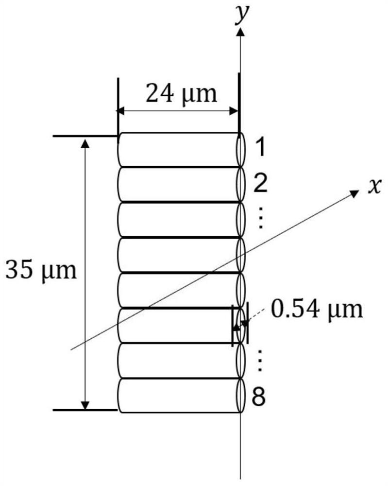
[0086] Such as Figure 6 Shown is a schematic structural diagram of the mid-to-far field optical ultra-thin slice imaging system of the present invention, including: a laser 1, a beam modulation module K2, an objective lens 12, a microfluidic chip control box 13, a microfluidic chip 14, a narrowband filter 15, Imaging lens 16 and camera 17;

[0087]The laser 1 emits a single laser beam and enters the beam modulation module K2; the beam modulation module K2 modulates the incident single laser beam, and enters the rear aperture plane of the objective lens 12; the objective lens 12 focuses the beam incident on the rear aperture plane, Converging to the inside of the channel of the microfluidic chip 14 to generate an ultra-thin light sheet; the microfluidic chip 14 controls the flow of fluorescently stained cells through the microfluidic chip control box 13; the fluorescent light emitted by the cells then passes through the narrowband filter 15 and imaging A lens 16 collects the ...


PUM

PropertyMeasurementUnit
thicknessaaaaaaaaaa
widthaaaaaaaaaa
heightaaaaaaaaaa

Abstract

The invention discloses a far-field optical ultrathin lamella imaging system and method, and relates to the technical field of optical and biomedical equipment. The imaging system comprises a laser, a light beam modulation module, an objective lens, a micro-fluidic chip control box, a micro-fluidic chip, a narrow-band filter, an imaging lens and an imaging device; the light beam modulation module modulates a single laser beam emitted by the laser, then the modulated single laser beam enters a rear aperture plane of the objective lens, and then the modulated single laser beam converges into a channel of the micro-fluidic chip to generate an ultrathin light sheet; the micro-fluidic chip controls the flow of a cell sample stained with fluorescence through the micro-fluidic chip control box; fluorescent light emitted by the cells is collected to the imaging device through the narrow-band filter and the imaging lens. Each Bessel light spot in the focusing area of the objective lens can be independently controlled, when the focusing light spots are close to each other, the interference influence is small, and continuous, high-resolution and high-flux lamellar imaging can be carried out on the fluorescence sample.

Description

technical field [0001] The invention relates to the technical field of optics and biomedical equipment, and more specifically relates to a far-field optical ultra-thin slice imaging system and method. Background technique [0002] The 3D structure of biological samples directly determines its biomechanical properties and functions in life phenomena. Rapid and high-resolution measurement of the 3D structure of biological samples is the frontier and hotspot of research in the fields of biomedicine and optical technology. At present, optical technologies capable of high-resolution measurement of the 3D structure of biological samples mainly include: Confocal Microscope, Stimulated Emission Depletion Microscope, Saturated Structured Illumination Microscopy, Random Positioning Imaging Microscopy, light sheet imaging technology, etc. [0003] When using a scanning microscope for biological imaging, such as using a confocal microscope and a stimulated radiation super-resolution mi...

Claims


Application Information

Patent Timeline
Patent Type & Authority Applications(China)
IPC IPC(8): G01N21/84G01N21/64G01N21/01B01L3/00
CPCG01N21/84G01N21/6428G01N21/6486G01N21/6458G01N21/01B01L3/5027G01N2021/0112B01L2200/12
Inventor 赵伟张策朱月强
Owner NORTHWEST UNIV(CN)
Why Eureka
Try Eureka