Fluorescent staining method for target protein of DNA nano-structure labeled cell

A nanostructure, fluorescent dyeing technology, applied in the field of optical microscopy, can solve problems such as loss of fluorescence signal

Active Publication Date: 2021-10-19
FUDAN UNIV +1
View PDF7 Cites 2 Cited by
  • Summary
  • Abstract
  • Description
  • Claims
  • Application Information

AI Technical Summary

Problems solved by technology

This method uses streptavidin and biotin as the medium, and uses the advantages of DNA nanostructures with multiple terminals (free radicals) to modify multiple fluorescent dyes and the characteristics of not being digested by proteases to label the target protein, which can not only overcome In the e

Method used


Image

  • Fluorescent staining method for target protein of DNA nano-structure labeled cell
  • Fluorescent staining method for target protein of DNA nano-structure labeled cell
  • Fluorescent staining method for target protein of DNA nano-structure labeled cell

Examples


Embodiment Construction

[0029] In order to illustrate the present invention more clearly, the present invention will be further described below in conjunction with preferred embodiments and accompanying drawings. Similar parts in the figures are denoted by the same reference numerals. Those skilled in the art should understand that the content specifically described below is illustrative rather than restrictive, and should not limit the protection scope of the present invention.

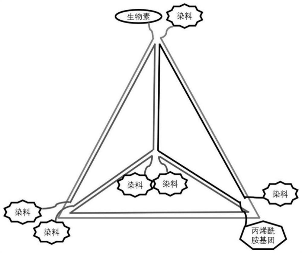
[0030] 1. Design and sequence synthesis of DNA tetrahedral nanostructures

[0031] The four strands of the DNA tetrahedral nanostructure were synthesized by Sangon Bioengineering (Shanghai) Co., Ltd., and biotin, fluorescent dyes and acrylamide groups were modified during synthesis. The 4 chain sequences are as follows:

[0032] A:

[0033] 5'-ACATTCCTAAGTCTGAAACATTACAGCTTGCTACACGAGAAGAGCCGCCATAGTA-3'

[0034] (SEQ ID NO:1), the 5' and 3' ends are modified with Alexa Fluor 488;

[0035] B:

[0036] 5'-TATCACCAGGCAGTTG...


PUM


Abstract

The invention belongs to the technical field of super-resolution microscopic imaging, and particularly relates to a fluorescent staining method for target protein of a DNA nano-structure labeled cell. Streptavidin is used as an intermediate, and a DNA nanostructure and an antibody of a biotin-modified target protein are connected together, so that the target protein is marked; and biotin, a group anchored on the hydrogel and more than two fluorescent dye molecules are simultaneously modified on the DNA nano structure. When the DNA nanostructure modifies the same dye, the brightness is very high, and the quality of the obtained microscopic picture can be greatly improved. When the dominant dye of the STED super-resolution microtechnique is modified, the expansion microtechnique can be well combined with the STED super-resolution technique. When a fluorescence resonance energy transfer dye pair is modified, the DNA nanostructure can have a flickering property, so that an expansion microtechnique is more conveniently combined with STORM and SOFI super-resolution techniques.

Description

technical field [0001] The invention belongs to the technical field of optical microscopy, and in particular relates to a fluorescent staining method for marking cell target proteins with DNA nanostructures. Background technique [0002] Expansion microscopy imaging technology is to specifically mark the target with fluorescence, such as protein, DNA, RNA, etc., and anchor the fluorescent group on the hydrogel. Fluorescent molecules that are indistinguishable from each other expand with the glue, increasing their distance from each other, allowing higher-resolution images to be acquired with common microscopy imaging equipment. Due to the simplicity of operation and readily available raw materials, expansion microscopy has a wide range of applications in single-celled animals, cell lines, tissues, and even animal body weight. [0003] The isotropic and uniform expansion of the sample is an important condition to ensure that the sample structure will not be distorted. To en...

Claims


Application Information

Patent Timeline
IPC IPC(8): G01N21/64
CPCG01N21/6486G01N21/6428G01N2021/6439
Inventor 马炯姚龙芳糜岚张丽陈丽雯王保举
Owner FUDAN UNIV
Why Eureka
Try Eureka