
Endogastric biopsy Raman image auxiliary diagnosis method and system based on artificial intelligence

An auxiliary diagnosis and artificial intelligence technology, applied in medical images, image enhancement, image analysis, etc., can solve the problems of complex picture and spectral data, difficult clinical interpretation, and lack of mature large sample data accumulation and summary.

Pending Publication Date: 2021-10-22
FUDAN UNIV
View PDF6 Cites 4 Cited by
  • Summary
  • Abstract
  • Description
  • Claims
  • Application Information

AI Technical Summary

Problems solved by technology

[0006] Because the pictures and spectral data of Raman imaging are relatively complex and there is no data accumulation and summary of mature large samples, clinical interpretation is very difficult

Method used


Image

  • Endogastric biopsy Raman image auxiliary diagnosis method and system based on artificial intelligence
  • Endogastric biopsy Raman image auxiliary diagnosis method and system based on artificial intelligence
  • Endogastric biopsy Raman image auxiliary diagnosis method and system based on artificial intelligence

Examples


Embodiment 1

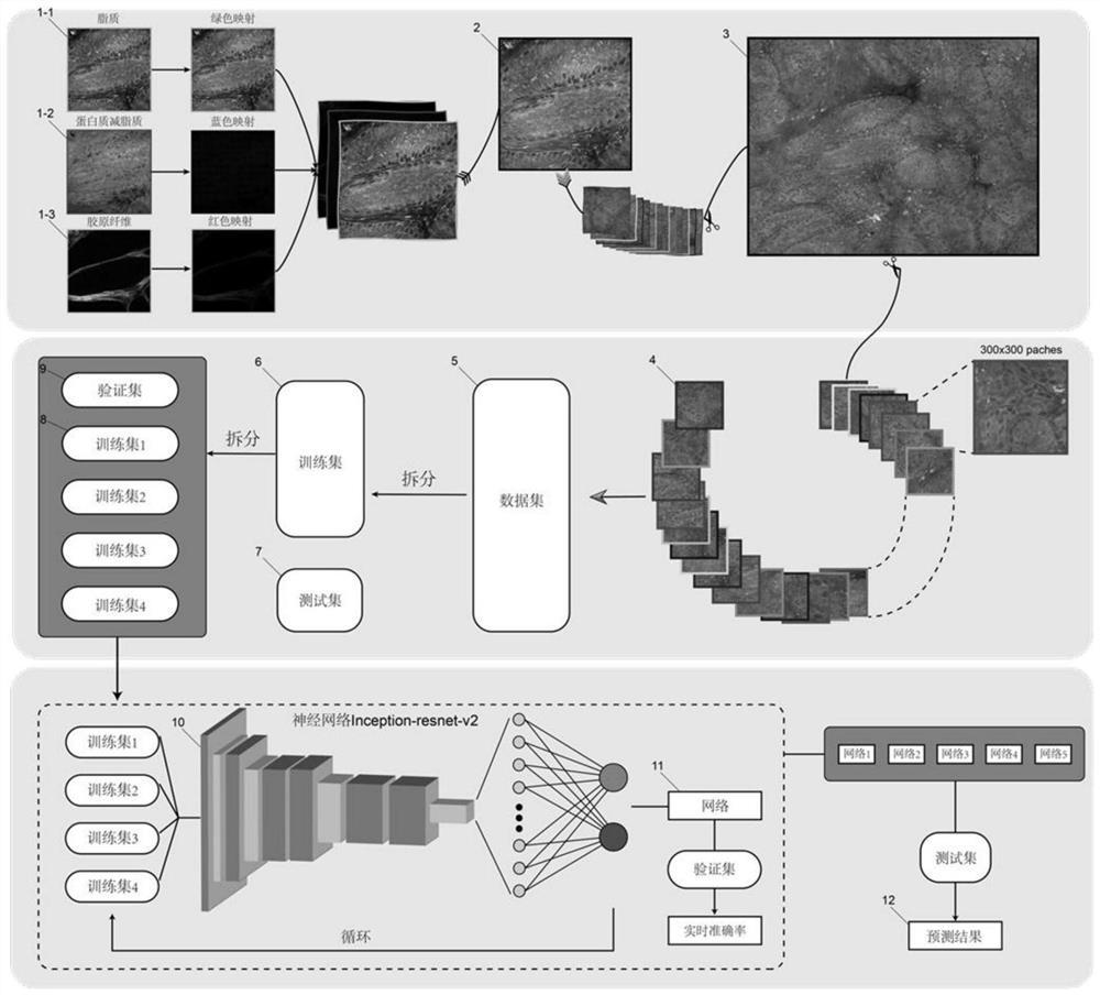
[0026] The invention provides an artificial intelligence-based rapid diagnosis method of gastric endoscopic biopsy Raman images. In a specific implementation, the figure 1 The processing flow shown. In this process, firstly, the two channel images formed by the stimulated Raman imaging system, namely lipid 1-1 and protein-reduced lipid 1-2, and the second harmonic channel image collagen 1-3 are respectively mapped to the stomach The different colors are then superimposed to form a stimulated Raman histopathology image 2, and then a stitching algorithm is used to generate a complete sample Raman pathology image 3 , and use the segmentation algorithm to segment all sample images into small images that conform to the input size of the neural network4, such as figure 2As shown, the input size of the network inception-resnet-v2 used in the present invention is 300x300x3, so the sample picture is cut to 300x300, and then data enhancement operations are performed by blurring, rot...

Embodiment 2

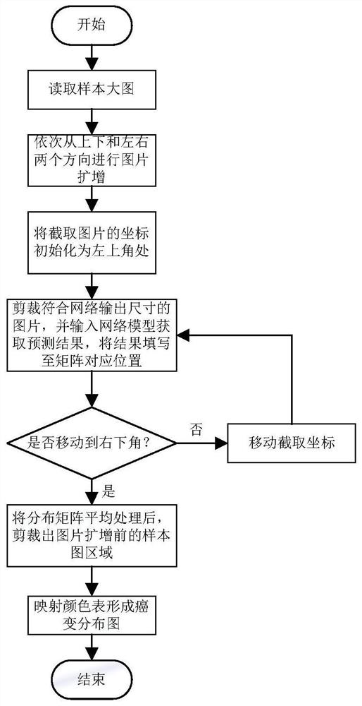
[0029] In this embodiment, an appropriate classification test algorithm is selected. In combination with Example 1, the algorithm for generating cancer distribution in fresh gastric biopsy tissue includes the following steps:

[0030] S1. Since the direct use of the cancer distribution generation algorithm will result in fewer calculations of the edge of the picture, the picture is first flipped and amplified according to the input size of the convolutional neural network, such as Figure 5 As shown in , first flip and amplify the upper and lower parts of the picture, and then flip and amplify the left and right parts. After the amplification, the size of the picture will be extended in four directions;

[0031] S2, the picture is divided into large pictures according to the movement steps selected each time by the small picture of the network input image size;

[0032] S3. Input the sequence of small images into the network, and generate the classification of the sequence of...


PUM


Abstract

The invention belongs to the technical field of medical equipment, and particularly relates to an endogastric biopsy Raman image auxiliary diagnosis method and system based on artificial intelligence. The artificial intelligence technology is applied to gastroscope stimulated Raman scattering endoscopic biopsy tissue image auxiliary diagnosis for the first time, and the method comprises the step: after histopathology image information is obtained by means of the stimulated Raman scattering microimaging technology, adopting the image classification and image omics data analysis based on a deep learning neural network and machine learning, and constructing an endogastric biopsy Raman image auxiliary diagnosis system. The system comprises a stomach tissue Raman image data preprocessing module, a neural network model, an algorithm module for training the neural network model, a neural network fine tuning module and a test module; compared with an existing traditional endoscope diagnosis and treatment system, the system has the advantages that real-time, rapid and intelligent diagnosis support in the endoscope examination process is achieved, and pathologists do not need to conduct explanation.

Description

technical field [0001] The invention belongs to the technical field of medical equipment, and in particular relates to an artificial intelligence-based Raman image-aided diagnosis method and system for gastric endoscopic biopsy. Background technique [0002] Gastric cancer is a major disease affecting human health. According to the latest annual report of the National Cancer Registry Center, the number of new cases of gastric cancer exceeds 680,000 each year, with about 500,000 deaths, making it the second most deadly tumor in my country. The key to the prevention and control of gastric cancer is early diagnosis and early treatment. Timely detection of early cancer, delay and reversal of precancerous lesions can effectively reduce the incidence and mortality of gastric cancer. Early gastric cancer and precancerous lesions cannot be diagnosed solely by endoscopic observation. Currently, pathological examination must be relied on, that is, the "endoscopy + biopsy" mode is ad...

Claims


Application Information

Patent Timeline
Patent Type & Authority Applications(China)
IPC IPC(8): G16H50/20G16H30/00G06T7/00G06T7/11G06K9/62
CPCG16H50/20G16H30/00G06T7/0012G06T7/11G06T2207/10068G06T2207/20081G06T2207/20084G06T2207/30096G06T2207/30092G06F18/24G06F18/214
Inventor 季敏标刘至杰胡皓敖建鹏周平红
Owner FUDAN UNIV
Learn More