
Preparation method of mouse meningeal single-cell suspension

A single-cell suspension and meningeal technology, applied in the field of genetic testing, can solve the problems that restrict the establishment of cell culture cell lines and functional gene verification research, and the inability to obtain single-cell suspensions of cell viability and tissue, achieving low-cost and high-efficiency acquisition , the effect of good vitality

Pending Publication Date: 2021-11-09
SHANGHAI PASSION BIOTECHNOLOGY CO LTD
View PDF3 Cites 3 Cited by
  • Summary
  • Abstract
  • Description
  • Claims
  • Application Information

AI Technical Summary

Problems solved by technology

Existing experimental techniques are still unable to obtain tissue single-cell suspensions with good cell viability, which restricts research work on tissue cell culture, establishment of cell lines, and functional gene verification.

Method used


Image

  • Preparation method of mouse meningeal single-cell suspension
  • Preparation method of mouse meningeal single-cell suspension
  • Preparation method of mouse meningeal single-cell suspension

Examples


preparation example Construction

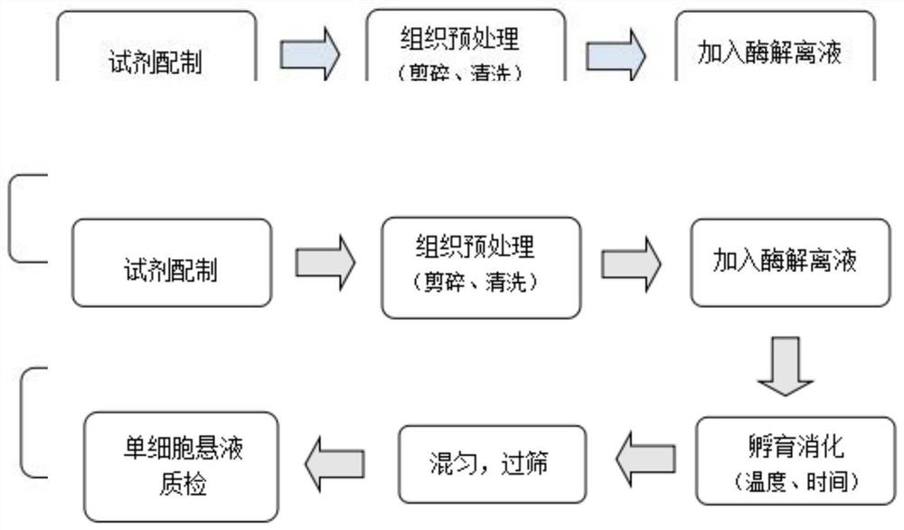
[0029] A preparation method of a single-cell suspension of mouse meninges, comprising the following steps:

[0030] 1) Preparation of reagents and medium, including serum-free medium and complete medium, preparation of cell cleaning solution, preparation of enzyme dissociation stock solution;

[0031] 2) Tissue pretreatment, add an appropriate amount of cell cleaning solution to a sterile RNase-free cell culture dish, and cut the tissue into 0.5-1mm pieces 2 After the small pieces, inhale into the 15ml test tube containing the cell cleaning solution, and rinse with the cell cleaning solution for 1-2 times until the excess tissue is removed;

[0032] 3) Digest and dissociate the tissue, suck off the cell cleaning solution, add 3-5ml enzyme dissociation stock solution, digest and incubate in a 37°C water bath for 20-30 minutes;

[0033] 4) To terminate the digestion, add an equal volume of the complete medium solution in step 1), gently pipette and mix well, no tissue fragments...

Embodiment 1

[0047] Step 1: Configure reagents and media

[0048] 1. Prepare the following two media respectively

[0049] (1) Serum-free medium: aliquoted Neurobasal Plus Medium contains 1% Penicillin-Streptomycin; add 1ml Penicillin-Streptomycin to 99ml Neurobasal Plus Medium.

[0050] (2) Complete medium: the divided Neurobasal Plus Medium contains 10% FBS and 1% Penicillin-Streptomycin; add 10ml of FBS and 1ml of Penicillin-Streptomycin to 89ml Neurobasal Plus Medium;

[0051] 2. Prepare cell washing solution: 1*HBSS-5% FBS solution: mix 95ml 1*HBSS solution with 5ml FBS solution;

[0052] 3. Prepare enzyme dissociation stock solution: Dissolve 13 mg of papain freeze-dried powder (Sigma, product number D4693-1G) in 10 ml of serum-free Neurobasal Plus Medium (Gibco, product number A3582901), and after it is completely dissolved, filter and remove bacteria. Store in a -20°C freezer to avoid repeated freezing and thawing.

[0053] Step Two: Tissue Pretreatment

[0054] Add an appropri...


PUM

PropertyMeasurementUnit
Number of living cellsaaaaaaaaaa

Abstract

The invention discloses a preparation method of a mouse meningeal single-cell suspension. The preparation method comprises the following steps of: 1) preparing a reagent and a culture medium; 2) carrying out tissue pretreatment; 3) carrying out tissue digestion and dissociation; 4) terminating digestion; 5) filtering by a micron cell filter; 6) obtaining a target tissue single-cell suspension; and 7) measuring the yield of living cells. The preparation method of the mouse meningeal single-cell suspension can be used for quickly and efficiently obtaining the mouse meningeal single-cell suspension at low cost and ensuring that dissociated cells keep good activity, can be applied to dissociation, separation and purification of mouse meningeal cells in a large range and establishment of a cell line, and provides important reference data for research of mouse nerve tissue single cells, gene functions and genetic breeding.

Description

technical field [0001] The invention relates to the technical field of gene detection, in particular to a method for preparing mouse meninges single cell suspension. Background technique [0002] In recent years, with the rapid development of technology in the field of biological research, single-cell sequencing technology has emerged as the times require. technology. Single-cell sequencing can reveal the gene structure and gene expression of a single cell, classify and compare cells, and reflect the heterogeneity between cells. It plays an important role in the fields of tumors, developmental biology, immunology, and microbiology. [0003] Different tissue cells have their own unique physiological characteristics, which is the main reason why tissue cell culture has become a worldwide problem. Existing experimental techniques are still unable to obtain tissue single-cell suspensions with good cell viability, which restricts research work on tissue cell culture, establishm...

Claims


Application Information

Patent Timeline
IPC IPC(8): C12N5/079C12Q1/6869
CPCC12N5/0618C12Q1/6869C12N2509/00C12N2509/10
Inventor 李靖贇余婧孙子奎
Owner SHANGHAI PASSION BIOTECHNOLOGY CO LTD
Learn More