Microscopic slide preparation method for observing characteristics of lower epidermis of folium artemisiae argyi

A technology of micro-slicing and epidermis, applied in the field of microscopic identification and observation of plants, can solve the problems of insufficient depth, difficulty in tearing and dissociating the epidermis, no public reports, etc., and achieves clear observation, high success rate, and easy operation. simple effect

Pending Publication Date: 2022-03-04
HENAN UNIV OF CHINESE MEDICINE
View PDF0 Cites 0 Cited by
  • Summary
  • Abstract
  • Description
  • Claims
  • Application Information

AI Technical Summary

Problems solved by technology

The upper epidermis of Artemisia argyi is distributed with glandular hairs and sparse non-glandular hairs, and it is easier to observe the upper epidermis with a scanning electron microscope; while the lower epidermis is densely covered with non-glandular hairs and covers the glandular hairs, stomata and epidermal cells distributed under it, so using Scanning electron microscope pairs can also only see dense nonglandular hairs
Observing the characteristics of the leaf epidermis with an optical microscope usually adopts the method of tearing the epidermis, dissociation of the epidermis, and transparent filming. However, because the leaves of Artemisia argyi are papery leaves, the leaves are thin, and the upper and lower epidermis are densely covered with glandular hairs. It is located in a depression, so it is difficult to tear off the epidermis and dissociate the epidermis. Even if the epidermis can be successfully separated, the lower epidermis is densely covered with non-glandular hairs, and characteristics such as stomata and epidermal cells cannot be observed under an optical microscope. The microscopic features of the lower epidermis cannot be seen clearly due to the dense non-glandular hairs on the lower epidermis of Artemisia argyi leaves
Due to the constraints of existing film-making techniques, it is currently difficult to observe microscopic features such as the stomata of the lower epidermis of Artemisia argyi leaves, the pattern of the anticlinal wall of epidermal cells, and the distribution of glandular and non-glandular hairs by scanning electron microscope or optical microscope. Therefore, So far, the research on the characteristics of the lower epidermis of Artemisia argyi is not deep enough
Therefore, it is very necessary to design a special microscopic preparation method for the characteristics of the lower epidermis of Artemisia argyi, to observe the characteristics of the lower epidermis of Artemisia argyi, but there is no public report so far

Method used


Image

  • Microscopic slide preparation method for observing characteristics of lower epidermis of folium artemisiae argyi
  • Microscopic slide preparation method for observing characteristics of lower epidermis of folium artemisiae argyi
  • Microscopic slide preparation method for observing characteristics of lower epidermis of folium artemisiae argyi

Examples


Embodiment 1

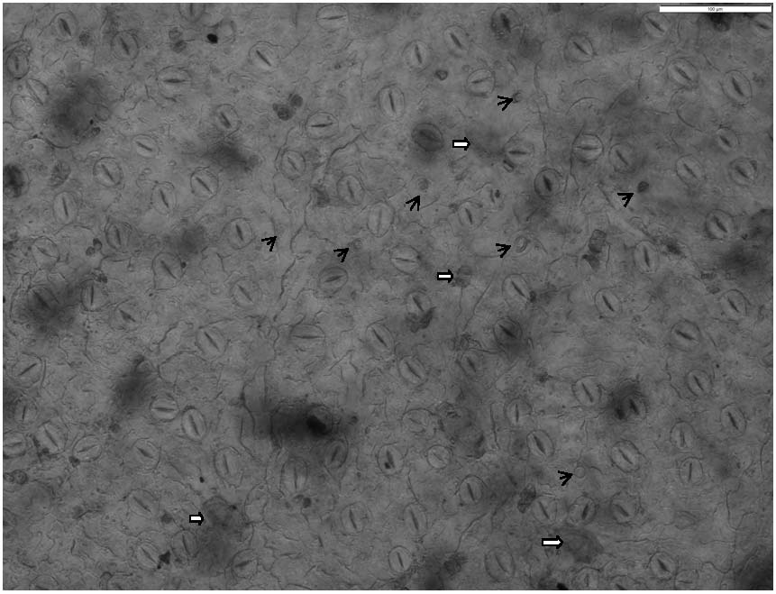
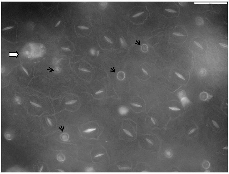
[0021] A kind of microscopic preparation method for observing the characteristics of the lower epidermis of Folium Artemisiae Artifacts of the present invention comprises the following steps:

[0022] (1) Leaf treatment: take fresh mugwort leaves, cut 6-8mm small pieces, and immerse them in methanol at room temperature for 6-10 hours;

[0023] (2) Soaking in hydrochloric acid: Use tweezers to transfer small pieces of leaves from methanol to hydrochloric acid with a mass concentration of 10% to 15%, and soak at room temperature for 8 to 12 hours;

[0024] (3) Separation of mesophyll and bleaching: Take out the small pieces of leaves, rinse them with distilled water for 1 to 3 minutes, and put the small pieces of leaves in a mixture of 5% to 10% sodium hydroxide and 2% to 3% hydrogen peroxide at a volume ratio of 1:1. In the mixed solution, soak at room temperature for 6-12 hours to dissociate the mesophyll tissue and bleach it at the same time;

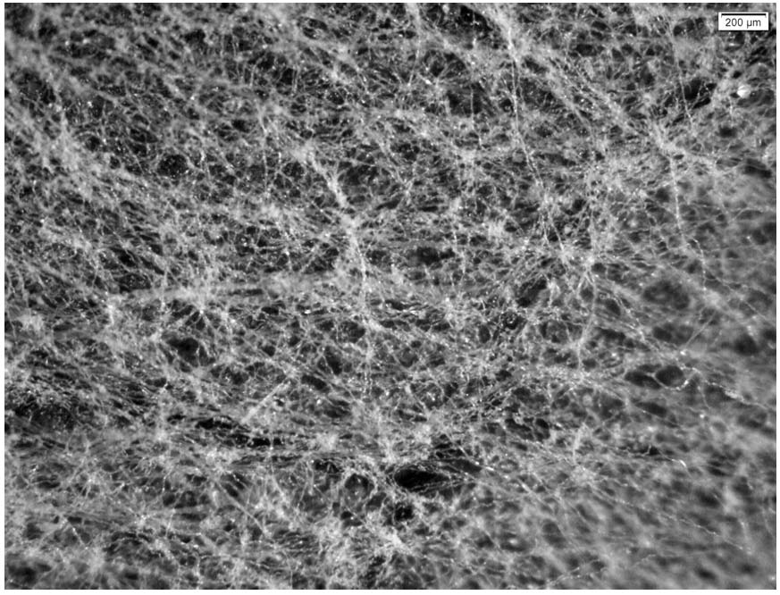
[0025] (4) Remove non-glandula...

Embodiment 2

[0032] A kind of microscopic preparation method for observing the characteristics of the lower epidermis of Folium Artemisiae Artifacts of the present invention comprises the following steps:

[0033] (1) Leaf treatment: take fresh Artemisia argyi leaves, cut 7mm pieces, and immerse them in methanol at room temperature for 8 hours;

[0034] (2) Soaking in hydrochloric acid: Use tweezers to transfer small pieces of leaves from methanol to hydrochloric acid with a mass concentration of 12.5%, and soak at room temperature for 10 hours;

[0035] (3) Separate the mesophyll for bleaching: Take out the small pieces of leaves, rinse them with distilled water for 2 minutes, put the small pieces of leaves in a mixture of 7.5% sodium hydroxide and 2.5% hydrogen peroxide at a volume ratio of 1:1, soak at room temperature for 9 hours, dissociates mesophyll tissue while also bleaching it;

[0036] (4) Remove non-glandular hairs: transfer small pieces of leaves to alcohol with a mass concen...

Embodiment 3

[0042] A kind of microscopic preparation method for observing the characteristics of the lower epidermis of Folium Artemisiae Artifacts of the present invention comprises the following steps:

[0043] (1) Leaf treatment: take fresh Artemisia argyi leaves, cut into 6mm pieces, and immerse them in methanol at room temperature for 6.5 hours;

[0044] (2) Soaking in hydrochloric acid: Use tweezers to transfer small pieces of leaves from methanol to hydrochloric acid with a mass concentration of 14%, and soak at room temperature for 9 hours;

[0045] (3) Separation of mesophyll and bleaching: Take out the small pieces of leaves, rinse them with distilled water for 3 minutes, put the small pieces of leaves in a mixture of 6% sodium hydroxide and 2% hydrogen peroxide at a volume ratio of 1:1, soak at room temperature for 12 hours, dissociates mesophyll tissue while also bleaching it;

[0046] (4) Remove non-glandular hairs: transfer the small pieces of leaves to alcohol with a mass ...


PUM


Abstract

The invention relates to a microscopic slide preparation method for observing the characteristics of the lower epidermis of folium artemisiae argyi, which effectively solves the problem of observing the pore of the lower epidermis of folium artemisiae argyi, the vertical peripheral wall pattern of epidermis cells and the distribution of glandular hair and non-glandular hair by using an optical microscope. Rinsing with distilled water, soaking with a mixed solution of sodium hydroxide and hydrogen peroxide, dissociating mesophyll tissues, bleaching, transferring into alcohol, and removing non-glandular hairs on leaf epidermis; the preparation method comprises the following steps of: adding the raw materials into a mixed solution of chromic acid and nitric acid, standing, dissociating mesophyll tissues, rinsing by using alcohol, separating upper and lower epidermis, removing mesophyll tissues of inner epidermis, dyeing the upper and lower epidermis by using a toluidine blue O solution, and rinsing by using water to remove floating color and water seal sheets, thereby obtaining the high-purity mesophyll tissue. The preparation method is simple to operate, low in cost, small in workload, high in success rate, clear in observation and good in effect; the technical problem of observing the lower epidermis of the folium artemisiae argyi is solved, the technical requirement on operators is not high, and an ideal effect can be achieved.

Description

technical field [0001] The invention relates to the technical field of microscopic identification and observation of plants, in particular to a microscopic film-making method for observing the characteristics of the lower epidermis of the leaves of Artemisia argyi, which can be used to observe and study the characteristics of the leaf epidermis by processing the leaves. Background technique [0002] Artemisia argyi is a commonly used traditional Chinese medicine, and it is produced in most parts of the country. Artemisia argyi leaves are dried and crushed to obtain "Moxa velvet", which is used to make moxa sticks for moxibustion. Glandular hairs are the main source of the volatile oil of Artemisia argyi. Therefore, the quantity and density of the glandular hairs of Artemisia argyi are related to the quality of Artemisia argyi, and the quantity and density of non-glandular hairs in Artemisia argyi are directly related to the yield and quality of Artemisia argyi. The main sour...

Claims


Application Information

Patent Timeline
Patent Type & Authority Applications(China)
IPC IPC(8): G01N1/28G01N1/30
CPCG01N1/2813G01N1/30
Inventor 郭涛冯书营吴易勋位文娜高富陈随清张振强刘孟奇欧阳臻周慧
Owner HENAN UNIV OF CHINESE MEDICINE
Why Eureka
Try Eureka