Method for screening CTL epitopes from autonomously constructed SLA-2-HB01-pCDH/sT2 cell line

A technology of sla-2-hb01-pcdh and sla-2-hb01-flag-pcdh, applied in the field of biomedicine, can solve the problems of low efficiency, difficult folding, harsh experimental conditions and the like

Pending Publication Date: 2022-03-15
DALIAN UNIV
View PDF5 Cites 1 Cited by
  • Summary
  • Abstract
  • Description
  • Claims
  • Application Information

AI Technical Summary

Problems solved by technology

[0011] Although the above methods can simulate the construction of complexes in vitro to screen polypeptide epitopes, the required experimental conditions are harsh and the amount of protein required is very large. Generally, prokaryotic expression is required to meet the requirements. However,

Method used


Image

  • Method for screening CTL epitopes from autonomously constructed SLA-2-HB01-pCDH/sT2 cell line
  • Method for screening CTL epitopes from autonomously constructed SLA-2-HB01-pCDH/sT2 cell line
  • Method for screening CTL epitopes from autonomously constructed SLA-2-HB01-pCDH/sT2 cell line

Examples


Embodiment 1

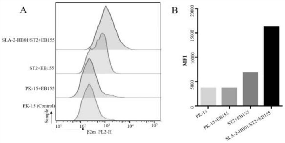
[0091] Example 1 Affinity experiment of PK15, sT2 and SLA-2-HB01-pCDH / sT2 cell lines

[0092] EB155 peptides were used as positive control peptides in PK15, sT2 and SLA-2-HB01-pCDH / sT2 cell lines, and loaded in the three cell lines respectively, because in the previous Western blotting test, the expression of β2m in the cell lines was basically the same , so the porcine β2m monoclonal antibody was selected as the detection of cell surface peptides. Because SLA-I-peptides are presented to the surface of the cell membrane as a whole, the detection of the expression of β2m on the surface of the cell membrane indicates that there are epitope peptides presented to the cell surface by SLA class I molecules. In the experimental group, only PE-labeled IgG was set as the background control group, which was no different from pure PK15 and sT2 cells, indicating that the staining result was the result of flow cytometry detection of the specific combination of PE and β2m monoclonal antibod...

Embodiment 3

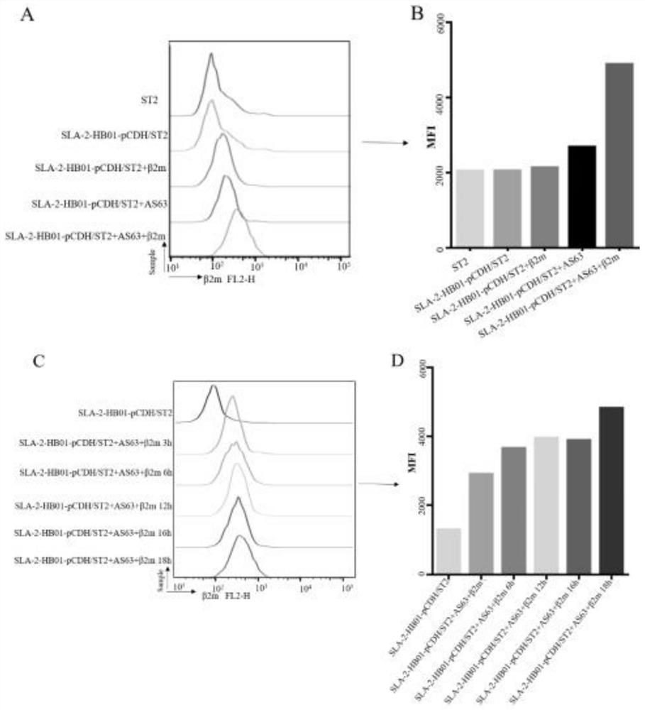
[0095] Example 3 CTL epitope screening function verification

[0096] After determining the peptide screening conditions, use the binding experiment of peptides loaded on SLA-2-HB01-pCDH / sT2 cells to detect whether the peptides can bind to SLA-2-HB01 and whether the affinity is enhanced, 50 μg / mL exogenous Peptides and 3 μg / mL β2m were added to the above cell lines, incubated at 37°C for 18 hours, and then the expression of β2m on the cell membrane surface was measured by β2m monoclonal antibody, so as to determine the expression of SLA-2-peptides and screen out CTL epitope peptides, such as Figure 4 shown. The mean fluorescence intensity (MFI) of β2m on the cell lines was determined by FACs analysis, and the fluorescence index (FI) was calculated. According to the following formula: FI=(MFI[SLA-2-HB01-pCDHsT2 cells+peptide] / MFI[SLA-2-HB01-pCDHsT2 cells without peptide])-1 proves the binding ability of SLA-2 to peptides. The results showed that AS63, Hu62 and EB155 could al...

Embodiment 4

[0099] Example 4 Laser confocal experiment to verify cell line function

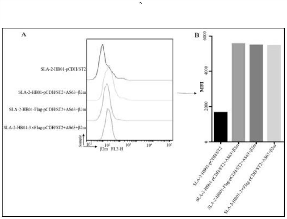
[0100] Add 100 μg / mL Biotin-AS63 peptide and 3 μg / mL β2m to the laser confocal plate of SLA-2-HB01-Flag-pCDH / sT2, SLA-2-HB01-3×Flag-pCDH / sT2 cells and incubate at 37°C for 18 hours Finally, the SLA I-peptide-β2m complex was stained with trimolecular cells, and the specific staining structure was as follows Figure 5 . LSCM detection results show that in the wavelength range of 590-617nm, after loading Biotin-AS63 peptides in SLA-2-HB01-Flag-pCDH / sT2 cells, there is no Alexa594-anti Flag antibody on the surface (1:50 dilution) Fluorescence expressed after binding the Flag tag see Figure 6 B, but in the SLA-2-HB01-3×Flag-pCDH / sT2 cell line under the same conditions, Alexa594-anti Flag antibody (diluted 1:50) was observed to bind to the corresponding 3×Flag fluorescence on the cell membrane surface as Figure 6 d.

[0101] In the wavelength range of 400-420nm, SLA-2-HB01-Flag-pCDH / sT2 and SLA-2-HB01-3×...


PUM


Abstract

The invention belongs to the field of biological medicines, and particularly relates to a method for screening CTL (cytotoxic T lymphocyte) epitopes by an independently constructed SLA-2-HB01-pCDH/sT2 cell line. According to the invention, an exogenous SLA-2-HB01 gene is transfected to an sT2 cell, and a stable SLA-2 gene expression model is established. The method comprises the following steps: loading EB155 and other positive epitope peptides on the surfaces of cells, detecting an SLA-2-peptide-beta2m complex expressed on the surfaces of the cells through FACs, and judging that the SLA-2-HB01-pCDH/sT2 has the function of presenting exogenous polypeptide epitopes. In order to promote the formation of cell surface complexes, the addition of the beta2m is beneficial to the improvement of the presentation efficiency of the antigen polypeptide. In the result of the invention, As63, EB155 and Hu62 peptides can be presented by an SLA-2-HB01-pCDH/sT2 cell line, As63 is presented in the SLA-2-HB01-pCDH/sT2, SLA-2-HB01-Flag-pCDH/sT2 and SLA-2-HB01-3 * Flag-pCDH/in the sT2 cell line, and the presentation efficiency is basically consistent, which indicates that a series of sT2 for expressing the SLA-2-HB01 gene constructed by the invention has the function of presenting the antigen, and the SLA-2-HB01-3 * Flag-pCDH/sT2 can be used for expressing the SLA-2-HB01 gene. Therefore, the SLA-2 heavy chain molecule can be used for screening polypeptide epitopes of the swine viruses on the cellular level, and it is further proved that the antigen presentation function of a cell line is not affected when a Flag tag is added to the C-terminal of the SLA-2 heavy chain molecule.

Description

technical field [0001] The invention belongs to the field of biomedicine, and in particular relates to a method for screening CTL epitopes of a self-constructed SLA-2-HB01-pCDH / sT2 cell line. Background technique [0002] Traditional vaccines are difficult to control pig-derived viruses, such as foot-and-mouth disease virus, and are prone to zoonotic diseases. Therefore, the development of new and safe vaccines is one of the focuses of current research. With the continuous development of immunology and bioinformatics, the use of viral antigens to predict SLA-2-restricted CD8 + CTL epitopes and the construction of peptide epitope vaccines are research hotspots. So how to choose the appropriate viral epitope is the most important. [0003] Epitopes are also called antigenic epitopes, antigenic determinants, chemical groups in antigen molecules that determine antigen specificity, basic units that can specifically bind to TCR or BCR, and finally stimulate the body's immune res...

Claims


Application Information

Patent Timeline
IPC IPC(8): C12N5/077G01N33/68
CPCC12N5/0669G01N33/68
Inventor 高凤山
Owner DALIAN UNIV
Why Eureka
Try Eureka