Detection method of circulating tumor cells and application thereof

Pending Publication Date: 2022-03-18
SOUTH CHINA NORMAL UNIVERSITY
View PDF2 Cites 0 Cited by
  • Summary
  • Abstract
  • Description
  • Claims
  • Application Information

AI Technical Summary

Problems solved by technology

[0006] However, designing precise nanopatterns on the substrate surface often requires cumbersome steps or expensive equipment; in the method of chemically modifying the substrate surface, it is difficult for antibodies or aptamers to modify the substrate surface, and the modification process is generally more complicated; The cell capture efficiency of the above method is not high enough, and there is still a certain gap with the actual application

Method used


Image

  • Detection method of circulating tumor cells and application thereof
  • Detection method of circulating tumor cells and application thereof
  • Detection method of circulating tumor cells and application thereof

Examples


Embodiment 1

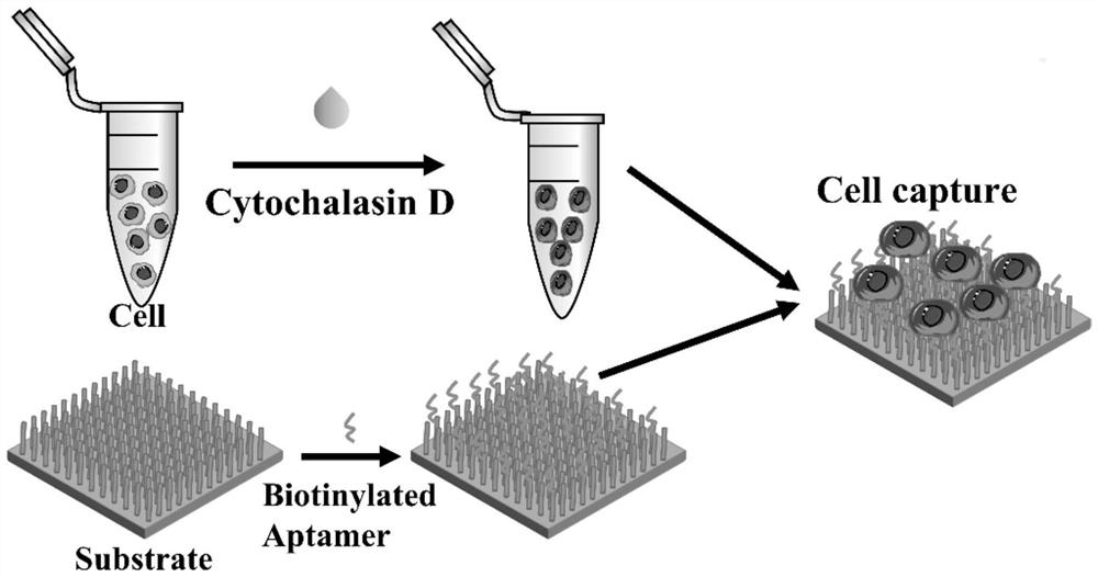
[0063] A method for detecting circulating tumor cells.

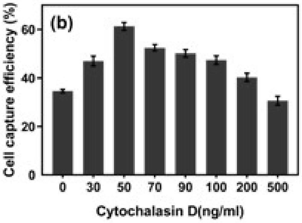
[0064] 1. Softening: Cytochalasin D was used to treat circulating tumor cells, and cytochalasin D was added to the cells at a concentration of 10 5 MCF-7 cells / mL culture medium allows cytochalasin D to enter circulating tumor cells in a subsequent capture step to destroy filamentous actin, thereby softening circulating tumor cells.

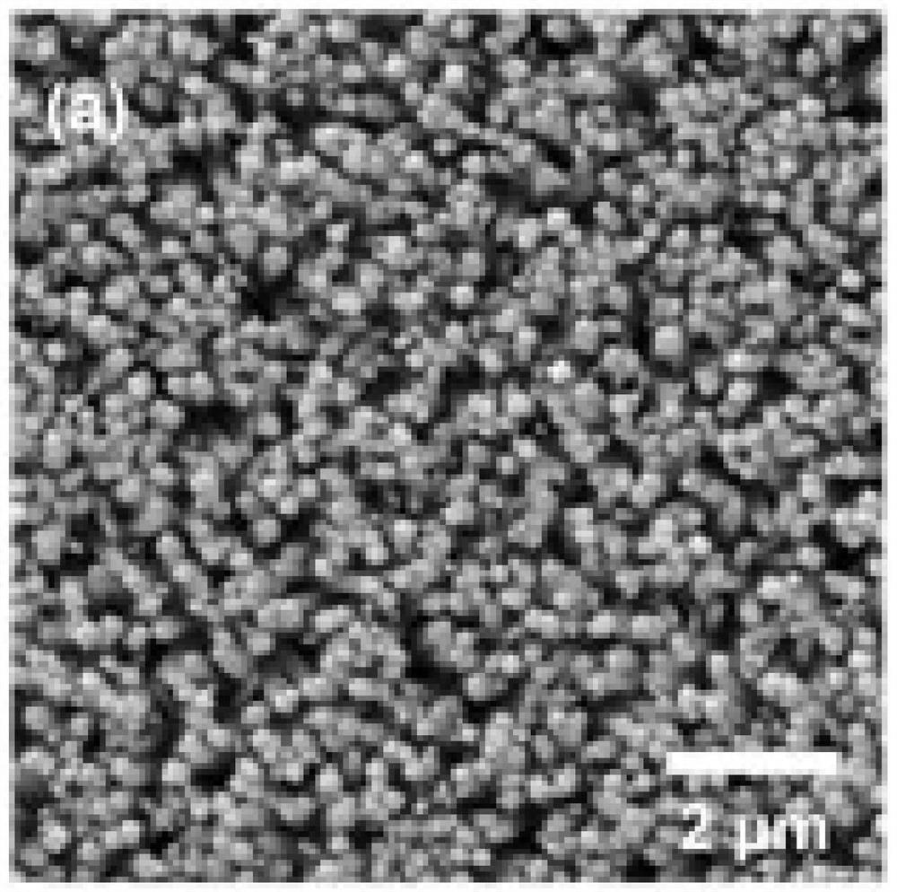
[0065] 2. Capture: Synthesis of TiO by hydrothermal method 2 nanowire array, cell culture medium supplemented with cytochalasin D was added to the TiO 2 nanowire arrays in a petri dish, making circulating tumor cells in TiO 2 The nanowire array was cultured on the substrate surface for 40 minutes at a temperature of 37°C. During this process, the filamentous actin of circulating tumor cells was destroyed by cytochalasin D, and the cells were softened, and the softened circulating tumor cells were treated with TiO 2 Nanowire Array Capture.

[0066] The experimental process of embodiment...

Embodiment 2

[0072] A method for detecting circulating tumor cells.

[0073] Existing techniques usually carry out chemical modification on the surface of nanostructured substrates to further improve cell capture efficiency. Those skilled in the art can modify biomolecules, such as antibodies and aptamers, on the substrate of Example 1 according to requirements. In order to investigate whether cytochalasin D treatment is still effective in improving the cell capture efficiency on the surface of the modified substrate, the inventors set up 4 groups for experimental comparison in this example.

[0074] In the control group, the circulating tumor cells were not treated with cytochalasin D, and the substrate surface of the TiO2 nanowire array was not modified;

[0075] Ap group is circulating tumor cells without cytochalasin D treatment, and biotinylated aptamer modified TiO2 nanowire array substrate surface;

[0076] In the CD group, circulating tumor cells were treated with cytochalasin D,...

Embodiment 3

[0081] A study of the effect of cytochalasin D on cell adhesion.

[0082] 1. Characterize the cell morphology of circulating tumor cells after adhesion.

[0083] The ability to capture cells on the substrate surface is closely related to cell adhesion, strong cell adhesion means high cell capture efficiency. In order to study the effect of cytochalasin D treatment on cell adhesion, this example uses a scanning electron microscope (SEM) to characterize the cell morphology after cell adhesion.

[0084] The result is as Figure 7 As shown, the inventors found that when the cells were not treated with cytochalasin D, the cells captured by the surface of the modified or unmodified substrate showed a distinct protrusion shape. However, cytochalasin D-treated cells exhibited flattening on both modified and unmodified substrate surfaces.

[0085] 2. To study the effect of cytochalasin D on the actin of cells.

[0086] In order to further study the adhesion of cells on the substrat...


PUM

PropertyMeasurementUnit
diameteraaaaaaaaaa

Abstract

The invention relates to a circulating tumor cell detection method and application thereof, and relates to the field of detection and capture of circulating tumor cells. The detection method comprises the following steps: treating circulating tumor cells by adopting cytochalasin D to damage filamentous actin of the circulating tumor cells. According to the detection method, by changing the mechanical property of the cell membrane of the circulating tumor cells, the deformation energy of the treated tumor cells plays a leading role in comparison with the adhesion energy, the tumor cells are promoted to be adhered to the surface of the substrate, and the detection efficiency of the tumor cells is further improved. Compared with the prior art, the detection method is simple and convenient, does not need complex steps, is low in cost, does not need expensive reagents and equipment, and the cytochalasin D used in the detection method does not cause damage to cell activity.

Description

technical field [0001] The invention relates to the field of detection and capture of circulating tumor cells, in particular to a detection method and application of circulating tumor cells. Background technique [0002] Circulating tumor cells (CTCs), a type of cancer cell that spread and survive in the peripheral blood during the development of malignant tumors, play a key role in distant tumor metastasis. The detection and isolation of CTCs is very important for early cancer diagnosis and prognosis. In addition, CTCs harbor a large number of pathogens. Through the analysis of CTCs after detection, personalized treatment can be realized. With the development of nanotechnology, various nanomaterials have been introduced as platforms for the detection of CTCs. Substrate surfaces with nanomorphology facilitate their cell adhesion due to the strong interaction between cells and the nanostructured surface. Utilizing this function, capturing CTCs from blood samples via nanos...

Claims


Application Information

Patent Timeline
Patent Type & Authority Applications(China)
IPC IPC(8): G01N21/64G01N33/569G01N33/574
CPCG01N21/6458G01N33/56966G01N33/574G01N2469/10
Inventor 李心磊明瑞琪陈同生
Owner SOUTH CHINA NORMAL UNIVERSITY
Why Eureka
Try Eureka