Three-dimensional imaging method based on wearable magnetocardiogram three-dimensional measuring device

A three-dimensional imaging and three-dimensional measurement technology, applied in the field of biomedicine, can solve the problems of unable to measure the magnetic signal of the front chest and back vest synchronously, unable to obtain the magnetic characteristic signal of the heart synchronously, and unable to fit the torso at a long distance, so as to improve portability , good promotion, low cost effect

Active Publication Date: 2022-04-08
HANGZHOU INNOVATION RES INST OF BEIJING UNIV OF AERONAUTICS & ASTRONAUTICS
View PDF5 Cites 2 Cited by
  • Summary
  • Abstract
  • Description
  • Claims
  • Application Information

AI Technical Summary

Problems solved by technology

The former is large in size, inconvenient to move, and requires liquid nitrogen cooling, and the maintenance cost is expensive
In addition, the distance between the SQUID sensor and the torso is too far to fit the torso, and the measurement orientation is limited, so it cannot simultaneously measure the magnetic signals of the front chest and back vest
Compared with the SQUID sensor, the OPM cardiomagnetic device has low maintenance cost and is easy to move. Its existing system can only measure the cardiomagnetic signal of the front chest, and cannot acquire all the cardiomagnetic characteristic signals synchronously.

Method used


Image

  • Three-dimensional imaging method based on wearable magnetocardiogram three-dimensional measuring device
  • Three-dimensional imaging method based on wearable magnetocardiogram three-dimensional measuring device
  • Three-dimensional imaging method based on wearable magnetocardiogram three-dimensional measuring device

Examples


Embodiment 1

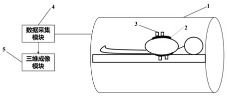
[0074] Such as figure 1 As shown, this embodiment provides a schematic structural diagram of a wearable three-dimensional cardiomagnetic measurement device. The wearable three-dimensional cardiomagnetic measurement device of this embodiment includes: an adjustable wearable vest 2, a data acquisition module 4 and a three-dimensional imaging module 5;

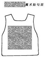
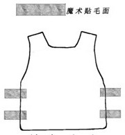
[0075] Each part of the adjustable wearable vest in this embodiment is an independent detachable structure; specifically, Fig. 2(a) is the front of the chest, Fig. 2(b) is the back of the chest, Fig. 2(c ) is the front of the back, and Figure 2(d) is the back of the back. The adjustable wearable vest includes: a front chest part, a back part and a plurality of sticky tapes; the sides of the front chest part and the back part are pasted by sticky tapes; the sticky tapes can be strip-shaped Velcro; that is to say , The front chest part and the back part are completely separated, and the sides can be fixed by a strap or an adhesiv...

Embodiment 2

[0089] Such as Figure 3 to Figure 5 As shown, the embodiment of the present invention provides a three-dimensional imaging method based on a wearable three-dimensional cardiomagnetic measurement device. The method of this embodiment is based on the above-mentioned figure 1 The wearable three-dimensional electrocardiographic measuring device is realized, which belongs to a computer program and is executed in a three-dimensional imaging module. Any electronic device can implement the following three-dimensional imaging method, and the method of this embodiment may include the following steps:

[0090] S10. Screen the transmitted three-dimensional cardiomagnetic signal according to the preset amplitude information, and obtain the filtered cardiomagnetic signal; the three-dimensional cardiomagnetic signal is output by the data acquisition module of the wearable three-dimensional cardiomagnetic measurement device, and the selected amplitude is less than 100PT The channel signal i...


PUM


Abstract

The invention relates to a three-dimensional imaging method based on a wearable magnetocardiography three-dimensional measuring device, and the wearable magnetocardiography three-dimensional measuring device obtains all three-dimensional magnetocardiography signals of a human body through a plurality of measuring sensors fixed by an adjustable vest in a magnetic shielding space. The method comprises the following steps: screening transmitted three-dimensional magnetocardiogram signals according to preset amplitude information, carrying out noise reduction and dimension reduction processing on the screened magnetocardiogram signals by adopting an independent component analysis and empirical mode decomposition method, and establishing a three-dimensional grid model comprising a trunk region, a heart region and a double-lung region based on a CT (Computed Tomography) image of a human body, a lead field matrix of magnetic field conduction from the heart to the position of the in-vitro sensor is obtained in a forward solving mode, and magnetic field distribution, used for real-time display, of the outer membrane surface of the heart is reversely solved based on the lead field matrix and the multi-channel signals. The wearable magnetocardiogram three-dimensional measuring device can synchronously obtain the three-dimensional magnetocardiogram signals, analyzes and displays the three-dimensional magnetocardiogram signals in real time, and is low in cost and convenient to carry.

Description

technical field [0001] The invention relates to the field of biomedical technology, in particular to a three-dimensional imaging method based on a wearable three-dimensional cardiomagnetic measurement device. Background technique [0002] Magnetocardiography is a non-invasive and non-contact heart detection technology. Compared with ECG, it has the advantages of high sensitivity, high signal-to-noise ratio, and sensitivity to eddy current signals. Magnetocardiography has important clinical research value in the detection of fetal heart disease, early diagnosis of coronary artery disease, postoperative evaluation of heart disease and other heart disease diagnosis. Among them, the SERF (Spin Exchange Relaxation Free, SERF) atomic magnetometer that measures the magnetic field of the heart has ultra-high sensitivity. [0003] Existing commercial magnetometry systems mainly include magnetometry based on superconducting quantum interference device (SQUID) and magnetometry based o...

Claims


Application Information

Patent Timeline
Patent Type & Authority Applications(China)
IPC IPC(8): A61B5/243A61B5/256A61B5/279A61B5/00
CPCY02A90/30
Inventor 杨艳菲宁晓琳向岷高阳房建成
Owner HANGZHOU INNOVATION RES INST OF BEIJING UNIV OF AERONAUTICS & ASTRONAUTICS
Why Eureka
Try Eureka