Cell preservation method

A preservation method and cell technology, applied in the field of cell biology, can solve the problems of inability to remove, diagnose cell damage, and not remove red blood cells, and achieve excellent immunohistochemical and molecular pathological test results, complete morphological structure of cells, and huge size. The effect of socioeconomic benefits

Pending Publication Date: 2022-05-13
昆明东环科技有限公司
View PDF0 Cites 0 Cited by
  • Summary
  • Abstract
  • Description
  • Claims
  • Application Information

AI Technical Summary

Problems solved by technology

Similar liquid-based cell preservation solutions are also used in the preservation of fine-needle aspiration cells. There are two main types: one is based on methanol and ethanol, and this type of preservation solution does not have the function of removing red blood cells; the other is based on methanol and ethanol. Add glacial acetic acid, a erythrocyte lysing reagent, to the alcohol fixative solution. Glacial acetic acid is a drastic way to remove erythrocytes, which can quickly remove erythrocytes in the specimen, but it also causes obvious damage to the diagnostic cells and changes their morphology and structure; at the same time , the lysed red blood cell fragments and the released hemoglobin will exist in the form of precipitates in the alcohol fixative, which cannot be removed during the film production process, and the presence of precipitates will block the observation of cell morphology under the microscope
In this way, the cell smear (section) prepared from cells preserved in this type of cell preservation solution makes it difficult to make a cytopathological diagnosis based on the cell morphology and its microstructure, and sometimes even makes a wrong diagnosis.

Method used


Image

  • Cell preservation method
  • Cell preservation method
  • Cell preservation method

Examples


Embodiment approach , specific Embodiment approach 1

[0024] Such as Figure 4 The flow chart for the implementation of the cell preservation method includes two specific embodiments, specific embodiment 1: Unscrew the bottle cap S1, wash the cells into the A liquid S2, let stand for 5 minutes S3, pour the A liquid cell suspension into S4 in liquid B, centrifuge S5, and make slices S6 Specific embodiment two: unscrew the bottle cap S1, take off the isolation pad S2, wash the cells into liquid A S3, let stand for 5 minutes S4, invert the cell culture bottle S5, centrifuge S6, production S7.

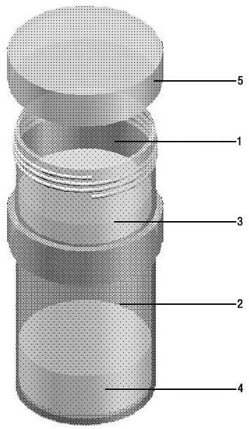
[0025] Specific embodiment one, the upper and lower structure preservation bottle embodiment.

[0026] (1) Unscrew the cap 5 of the cell preservation bottle.

[0027] (2) Inject the fine-needle aspiration cells into the cell preservation solution A solution 3, aspirate the preservation solution to wash the syringe, and then inject the washing solution into the A solution 3.

[0028] (3) Let it stand for about 5 minutes to lyse the red bloo...

specific Embodiment approach 2

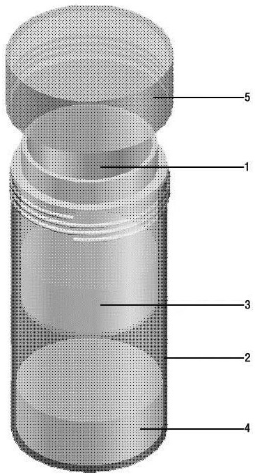
[0031] Specific embodiment two, left and right structure preservation bottle embodiment.

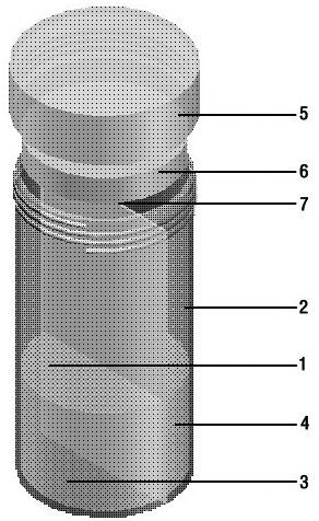
[0032] (1) Unscrew the bottle cap 5 of the cell preservation bottle, and remove the sealing gasket 6.

[0033] (2) Inject the fine-needle aspiration cells into the cell preservation solution A solution 3, aspirate the preservation solution to wash the syringe, and then inject the washing solution into the A solution 3.

[0034] (3) Cover the bottle cap 5 and let it stand for about 5 minutes to lyse the red blood cells until the color of solution A 3 changes from bright red to dark red (the color of red blood cells after lysing).

[0035](4) Turn the cell preservation bottle upside down and mix several times, and mix the cell suspension after lysing red blood cells in chamber A 1 with liquid B 4 in chamber B 2 through the gap left by the airtight (isolation) pad 6, for diagnosis The cells were fixed, and the red blood cell fragments and hemoglobin were dispersed and dissolved in formalde...


PUM


Abstract

The invention discloses a cell preservation method, which comprises a cell preservation bottle, a cell preservation solution and a cell preservation method. The cell preservation bottle is provided with a chamber A and a chamber B which are independent and closed, and the cell preservation liquid is composed of a liquid A and a liquid B which are respectively stored in the chamber A and the chamber B of the cell culture bottle. After a cytological specimen is sequentially treated by the solution A and the solution B, red blood cells in the specimen can be completely removed, meanwhile, the cells are fixed, and cell debris is dissolved in a buffer system. The cell smear or slice prepared by the cell preservation method is clear in cell morphological structure, has no red blood cell and protein precipitation background, and is easy to make pathological diagnosis.

Description

technical field [0001] The patent relates to the field of cell biology, and specifically relates to a cell preservation method, especially a fine needle aspiration cell preservation method. Background technique [0002] Fine needle aspiration (aspiration) cytology (FNA) is an important development direction of cytopathology. Through the method of fine needle aspiration of human cells, clinical diagnostic specimens are obtained, and cell smears, cell wax blocks and cell slices are prepared, and further Carry out immunohistochemical and molecular pathological tests to diagnose, type, detect therapeutic targets and prognostic indicators for diseases. [0003] FNA pathological diagnosis technology has many advantages such as minimally invasive, safe, and simple. Theoretically, the fine needle can reach any part of the human body to take samples for disease diagnosis, reducing the difficulty of sampling for disease diagnosis. However, this technology also has obvious limitations,...

Claims


Application Information

Patent Timeline
Patent Type & Authority Applications(China)
IPC IPC(8): A01N1/02
CPCA01N1/0263A01N1/0221A01N1/021
Inventor 赵稳兴张鹏博赵子浩
Owner 昆明东环科技有限公司
Why Eureka
Try Eureka