
Infectious keratopathy living pathogen detection method combining deep learning and cornea living confocal microscopy

A technology of confocal microscopy and deep learning, applied in the field of detection of live pathogenic bacteria in infectious corneal diseases, can solve the problem that it is difficult to distinguish different fungal genera, and achieve the effect of rapid inspection and strong practicability

Pending Publication Date: 2022-06-03
THE PEOPLES HOSPITAL OF GUANGXI ZHUANG AUTONOMOUS REGION
View PDF0 Cites 0 Cited by
  • Summary
  • Abstract
  • Description
  • Claims
  • Application Information

AI Technical Summary

Problems solved by technology

However, the interpretation of corneal live confocal microscopy still relies on the empirical judgment of the examiner, and the hyphae of different fungi often have similar characteristics in morphology, so it is difficult to distinguish different fungal fungi by current corneal live confocal microscopy belongs to

Method used


Image

  • Infectious keratopathy living pathogen detection method combining deep learning and cornea living confocal microscopy
  • Infectious keratopathy living pathogen detection method combining deep learning and cornea living confocal microscopy
  • Infectious keratopathy living pathogen detection method combining deep learning and cornea living confocal microscopy

Examples


Embodiment

[0046] 1. Combined deep learning and corneal in vivo confocal microscopy for the detection of infectious keratopathy in vivo pathogens:

[0047] 1) Convolutional neural network model construction:

[0048] (1) Data collection: collect the images of the patient's living corneal confocal microscopy, the detection results of pathogenic bacteria, and the patient's medical record. The detection results of pathogenic bacteria include but are not limited to smear microscopy, culture, metagenomic detection and other pathogen species identification As a result, patient medical record data includes, but is not limited to, general patient information, case records, laboratory tests and auxiliary tests, etc.;

[0049] (2) Image screening and labeling: The images are screened by experienced ophthalmologists to ensure that the images are clearly displayed, that the content of the images include disease lesions and the surrounding conditions, that the images can reflect the characteristics o...

experiment example

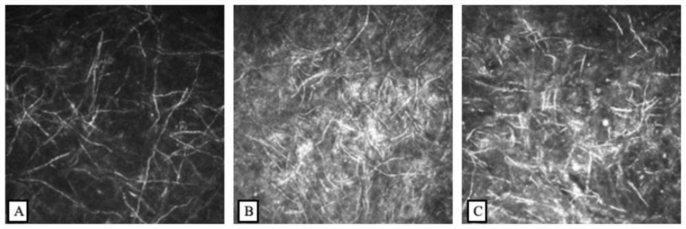
[0066] 1. Image collection: 76 patients who underwent IVCM examination (Heidelberg HRT III / RCM, Germany) for corneal disease in Guangxi Zhuang Autonomous Region People's Hospital from 2017 to 2020 were collected, and 9380 IVCM images were collected.

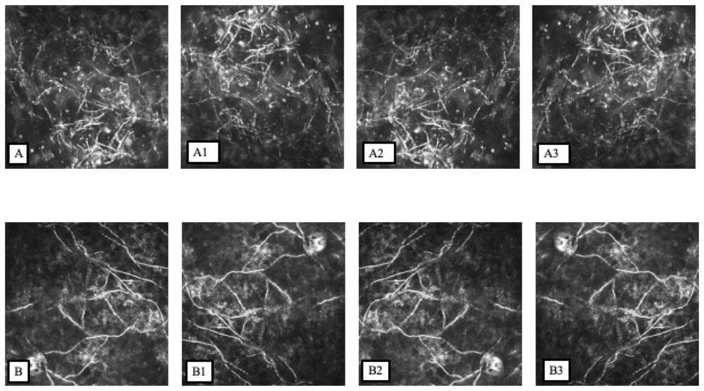
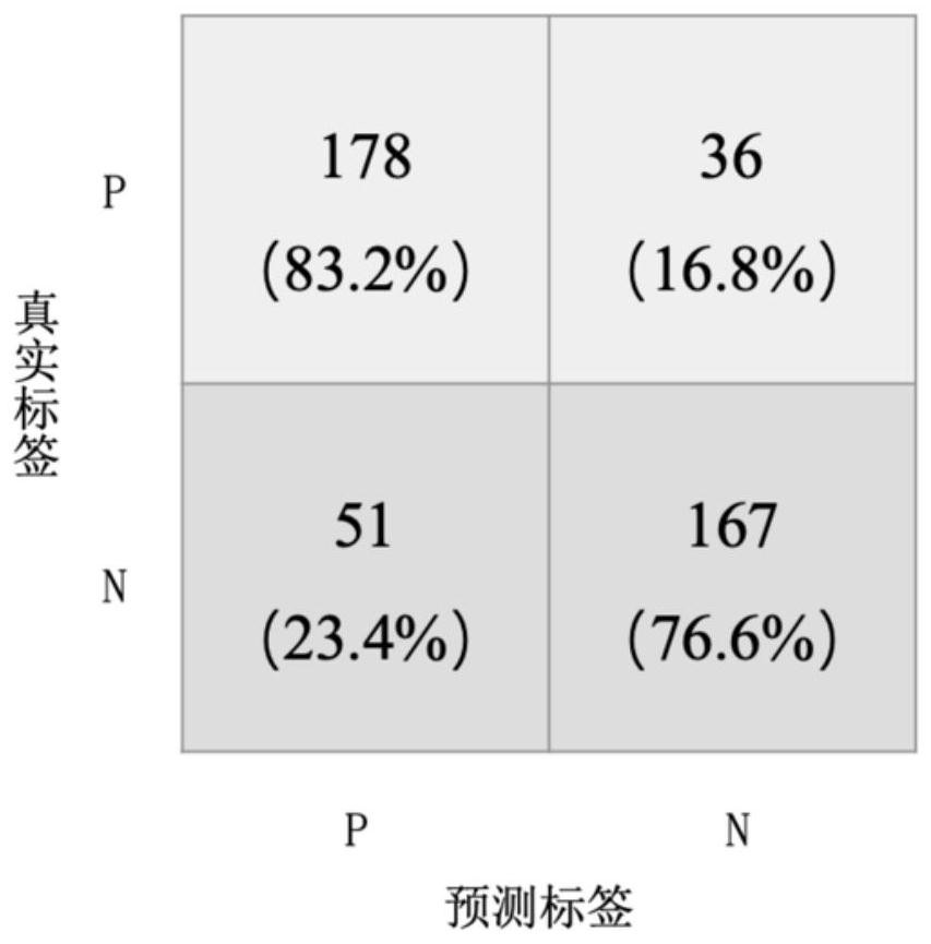
[0067] 2. Image screening and classification: Three experienced ophthalmologists from Guangxi Zhuang Autonomous Region People's Hospital screened the collected images, and screened out the images containing fungal hyphae. When the screening results of the three ophthalmologists were consistent, it was considered that The image contains fungal hyphae; when the screening results of the three ophthalmologists are inconsistent, another chief ophthalmologist with more than 15 years of experience will review the image to determine whether the image contains fungal hyphae. A total of 2157 images contained fungal hyphae; the screened images were classified according to the results of the fungal culture of the patient's corneal scraping, a...


PUM


Abstract

The invention discloses an infectious keratopathy living pathogen detection method combining deep learning and cornea living confocal microscopy, which can realize intelligent detection of the infectious keratopathy living pathogen, and compared with the traditional morphological, biological and pathological examination methods, the method provided by the invention has the advantages that the detection efficiency is greatly improved; the cornea living body confocal microscopy can directly observe the cell image of the cornea structure in a multi-layer three-dimensional manner, so that the steps of traditional tissue slicing, fixing, dyeing and the like are omitted, and the cornea living body confocal microscopy is non-invasive examination and is more beneficial to wide development of examination. According to the method, the image is intelligently detected by combining the neural network model constructed by deep learning, the examination result can be provided in real time, compared with traditional examinations such as corneal blade microscopic examination and corneal blade culture, the method is faster, more convenient and more economical, the result of the method provides a reliable basis for clinical doctors to judge the illness state, and the practicability is high.

Description

【Technical field】 [0001] The invention belongs to the technical field of medical image processing, in particular to a method for detecting live pathogenic bacteria of infectious keratopathy by combining deep learning and corneal live confocal microscopy. 【Background technique】 [0002] Infectious keratitis (infectious keratitis) is a keratitis caused by various pathogenic microorganisms such as bacteria, fungi, viruses, chlamydia, etc. It is a common blinding eye disease in China. Disability of the eye. [0003] Infectious keratitis, traditional early detection methods include corneal scraping microscopy, corneal scraping culture, corneal biopsy, intravital confocal microscopy, etc. Corneal scraping microscopy may have a low positive rate due to the method of scraping and the growth of pathogenic bacteria; corneal scraping takes a long time to culture, and the positive rate of culture is also not high; corneal biopsy due to its required There are many tissues, there is the...

Claims


Application Information

Patent Timeline
Patent Type & Authority Applications(China)
IPC IPC(8): G06V20/69G06T7/00G06N3/08G06N3/04G06K9/62G06V10/774G06V10/82G06V10/764
CPCG06N3/08G06T7/0012G06T2207/30041G06T2207/20081G06T2207/20084G06N3/045G06F18/24G06F18/214
Inventor 徐帆唐宁宁陈琦黄光怡蓝倩倩蒋莉洪祎祎李敏曾思明吕健廖靖林芸茹
Owner THE PEOPLES HOSPITAL OF GUANGXI ZHUANG AUTONOMOUS REGION
Learn More