Modeling method of tumor orthotopic tumor animal model and tumor orthotopic tumor animal model

An animal model and tumor technology, applied in the field of medicine, can solve the problems of many complications, complicated operation, slow postoperative recovery, etc., achieve good application prospects, high success rate of tumor formation, and improve stability

Pending Publication Date: 2022-07-05
ZHEJIANG UNIV
View PDF3 Cites 0 Cited by
  • Summary
  • Abstract
  • Description
  • Claims
  • Application Information

AI Technical Summary

Problems solved by technology

However, the method of establishing tumor models by dissecting animals for orthotopic transplantation has many complications (suc

Method used


Image

  • Modeling method of tumor orthotopic tumor animal model and tumor orthotopic tumor animal model
  • Modeling method of tumor orthotopic tumor animal model and tumor orthotopic tumor animal model
  • Modeling method of tumor orthotopic tumor animal model and tumor orthotopic tumor animal model

Examples


Embodiment 1

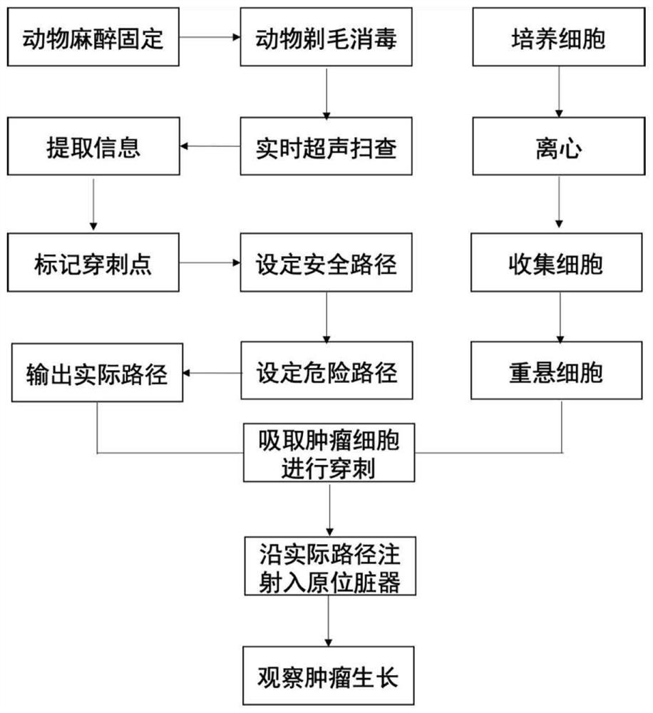
[0054] Example 1 Modeling steps ( figure 1 ):

[0055] A modeling method for a tumor orthotopic tumor animal model, comprising the following steps, such as figure 1 shown:

[0056] 1. C57 mice were anesthetized with 1-1.5% pentobarbital (45g / kg), and the animals were fixed with a mouse fixation plate;

[0057] 2. Shave and sterilize the abdominal area, lay a hole towel, and fully expose the operation area; wherein, the abdominal area refers to the upper to the mouse xiphoid process, the lower to the line connecting the anterior superior iliac spine, and the left to the left mid-axillary line. , right to right mid-axillary line;

[0058] 3. The ultrasound probe is placed in the liver area, and parameters of the liver tissue and surrounding tissue are acquired under real-time ultrasound guidance, the parameters including the parameters of blood vessels, the parameters of the bones and the parameters of the body surface. Ultrasound here includes two-dimensional ultrasound and...

Embodiment 2

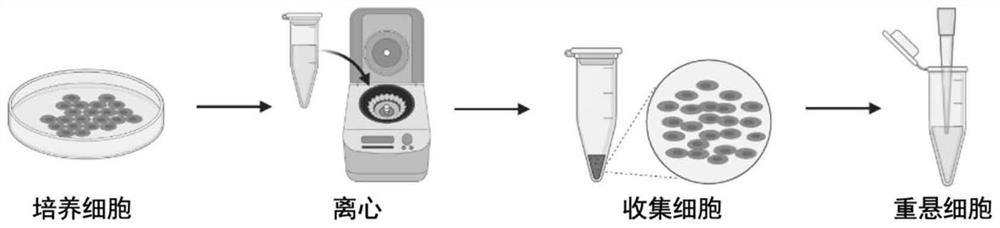
[0066] Example 2 Preparation of tumor cells ( figure 2 ):

[0067] The preparation of tumor cells is a key step in the modeling of orthotopic transplanted tumors under ultrasound guidance. The specific steps are as follows:

[0068] 1. Cultivate tumor cells in a 37°C incubator to observe cell growth rate and cell density; tumor cells here include brain tumors, neck tumors, chest tumors, abdominal tumors, pelvic tumors, skin tumors, limbs, and bone joints tumor etc.

[0069] 2. Then use trypsin to digest the tumor cells, and add medium with serum to stop the digestion; the amount of trypsin depends on the size of the cell culture dish, for example, 3.0-4.0ml of trypsin needs to be added to a 10.0cm diameter cell culture dish Digest. The tumor cells were stopped digested by adding 2 times the volume of trypsin and serum-containing cell culture medium.

[0070] 3. Use a pipette to blow the cells, and collect the digested tumor cells into a centrifuge tube; the tumor cell dig...

Embodiment 3

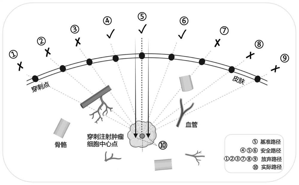
[0074] The selection of the safe path in embodiment 3 ( image 3 ):

[0075] (1) Obtain the surrounding parameters of the target organ under the guidance of ultrasound. The parameters surrounding the target organ include blood vessel parameters, bone parameters, and body surface parameters.

[0076] (2) Marking the body surface puncture needle entry point and the target organ puncture point of interest based on the parameters.

[0077] (3) Connect the body surface puncture needle point and the target organ puncture point to generate multiple reference paths. The reference path does not intersect the great vessels, and the reference path does not intersect the bone.

[0078] (4) N safe paths satisfying the given conditions are then translated from at least one reference path, and the abandoned paths are deleted. A puncture path that does not intersect a large vessel and also does not intersect a bone is output as a safe path. Puncture paths that intersect a bone or a blood...


PUM

PropertyMeasurementUnit
Volumeaaaaaaaaaa

Abstract

The invention discloses a modeling method of a tumor orthotopic tumor animal model and the tumor orthotopic tumor animal model. The modeling method comprises the following steps: performing general anesthesia on an animal; shaving and disinfecting the target visceral organ area of the animal; placing an ultrasonic probe in a target organ area to obtain parameters of the target organ and peripheral tissues; based on the parameters, marking interested body surface puncture needle insertion points and target organ puncture points; connecting the body surface puncture needle insertion point with the target organ puncture point to generate a puncture path; puncturing by using a micro-injection needle according to the puncture path to reach the target visceral organ position; injecting the tumor cells in the injector into a target visceral organ position through a micro-injection needle; the tumor cell injection process is observed in real time through ultrasound, and after all tumor cells are injected, the needle is rotated out to obtain the tumor in-situ tumor animal model. The method has the advantages of small trauma, high tumor formation rate, fast recovery and stable operation, avoids large trauma such as laparotomy, pelvic opening and thoracotomy, has no influence on surrounding organs, and has great application potential.

Description

technical field [0001] The present application relates to the field of medicine, and in particular, to a modeling method for an orthotopic tumor animal model and an orthotopic tumor animal model. Background technique [0002] Effective treatment of tumors is a major medical problem. According to the latest World Cancer Report released by the World Health Organization, the global cancer burden is increasing. The treatment of tumors has thus become the focus of exploration and research in the medical field. [0003] An important part of the field of oncology research is recognized as being able to provide model systems for the study of carcinogenesis and therapeutic treatments. To evaluate the effects of cancer therapy in preclinical and clinical studies, animal models of tumors in which human tumor cell lines or patient-derived tumors are transplanted into immunodeficient mice are commonly used. Tumor animal models provide a good alternative for clinical trials, and are a ...

Claims


Application Information

Patent Timeline
IPC IPC(8): A01K67/027
CPCA01K67/027A61K9/5123A01K2207/12A01K2227/105A01K2267/03
Inventor 谢丽婷蒋天安赵齐羽许敏
Owner ZHEJIANG UNIV
Why Eureka
Try Eureka