Human exfoliated cell immunofluorescent staining kit and method

A technology of immunofluorescence staining and exfoliated cells, which is applied in the field of fluorescent staining reagents, can solve the problems of inability to distinguish nucleoli and nucleoplasm, the troubles of malignant tumor cell morphology detection, and the inability to clearly mark nucleoli.

Pending Publication Date: 2022-08-02
青岛言鼎生物医疗科技有限公司
View PDF0 Cites 0 Cited by
  • Summary
  • Abstract
  • Description
  • Claims
  • Application Information

AI Technical Summary

Problems solved by technology

Traditional histopathological examination, exfoliated cytology examination and blood cell examination are mainly chemical staining that has been used for many years. The basic structure of malignant tumor cells such as the morphological structure of cell membrane, cell paddle and nucleus can be seen, and the nuclear enlargement of malignant cells can be displayed. , nuclear abnormalities and some cytopathological characteristics of nuclear abnormalities, the main technical disadvantages are that the shape and structure of the nucleoli cannot be clearly displayed, the nucleoli cannot be clearly marked, and the nucleoli and nuclear plasma cannot be distinguished. Morphological detection brings some troubles

Method used


Image

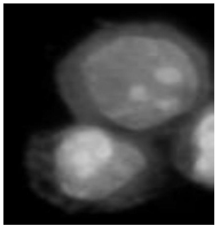
  • Human exfoliated cell immunofluorescent staining kit and method
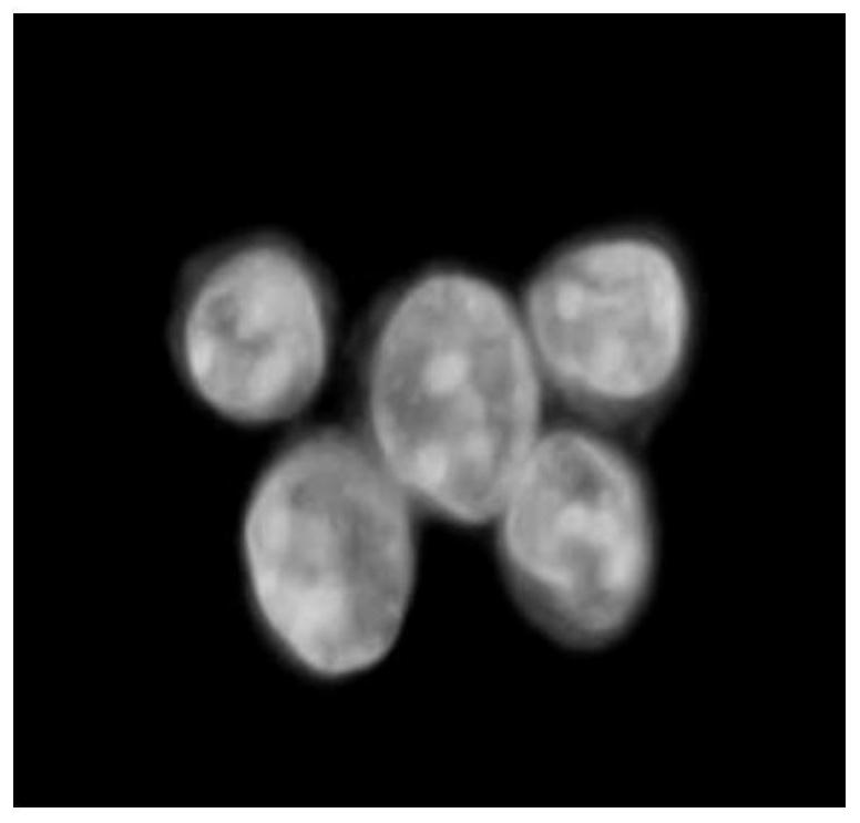
  • Human exfoliated cell immunofluorescent staining kit and method
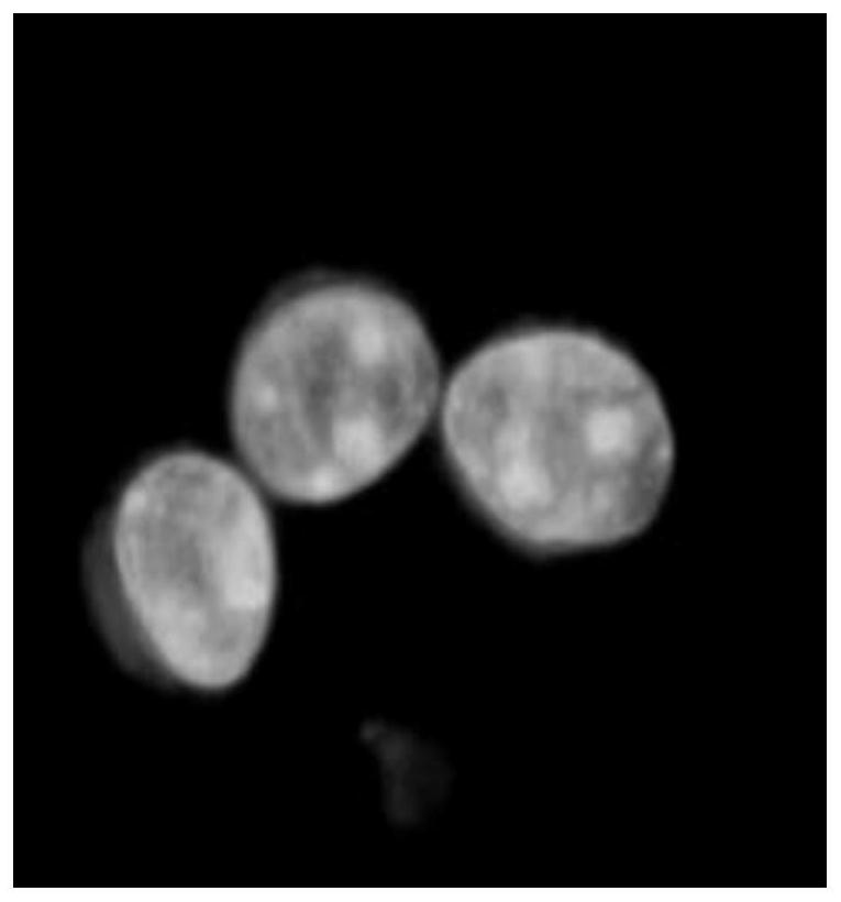
  • Human exfoliated cell immunofluorescent staining kit and method

Examples


Embodiment 1

[0072] Example 1 Fluorescent staining of CTC cells

[0073] (1) The reagents of the kit include:

[0074] Cell fixative: methanol, acetone, paraformaldehyde.

[0075] Cell blocking solution includes: pH 7.2-7.4 PBS (containing 1% BSA or calf serum).

[0076] The rinse solution includes: pH 7.2-7.4 PBS (with Tween-20).

[0077] Diluents include: pH 7.2-7.4 PBS (containing 1% BSA or calf serum).

[0078] Cell membrane and nuclear membrane permeabilizers include: 0.2-0.5% Triton X-100.

[0079] Nucleolar marker fluorescent dye Syto RNA select green.

[0080] Monoclonal antibody red fluorescent marker for CK, EpCAM, and vimentin.

[0081] Other reagents: phosphate buffered saline (PBS), bovine serum albumin (BSA), donkey plasma (clear), Tween-20, Tris-HCl, etc.

[0082] (2) Fluorescence staining method:

[0083] ① Prepare CTC cell detection sheet.

[0084] ②Fix the cells with methanol or 4% paraformaldehyde for 10-20 minutes.

[0085] ③ Wash with rinsing solution (pH7.2-7...

Embodiment 2

[0091] Example 2 Fluorescent staining of exfoliated cells in pleural and ascites

[0092] (1) Kit reagents:

[0093] Cell fixative: methanol, acetone, paraformaldehyde.

[0094] Cell blocking solution includes: pH 7.2-7.4 PBS (containing 1% BSA or calf serum).

[0095] The rinse solution includes: pH 7.2-7.4 PBS (with Tween-20).

[0096] Diluents include: pH 7.2-7.4 PBS (containing 1% BSA or calf serum).

[0097] Cell membrane and nuclear membrane permeabilizers include: 0.2-0.5% Triton X-100.

[0098] Nucleolar marker fluorescent dye Syto RNA select green.

[0099] EpCAM, CK monoclonal antibody red fluorescent marker.

[0100] Other reagents: phosphate buffered saline (PBS), bovine serum albumin (BSA), donkey plasma (clear), Tween-20, Tris-HCl, etc.

[0101] (2) Fluorescence staining method:

[0102] ①Preparation of pleural and ascites exfoliated cell detection sheet.

[0103] ②Fix the cells with methanol or 4% paraformaldehyde for 10-20 minutes.

[0104] ③ Wash with...

Embodiment 3

[0110] Example 3 Fluorescent staining of blood disease cells

[0111] (1) Kit reagents

[0112] Cell fixative: methanol, acetone, paraformaldehyde.

[0113] Cell blocking solution includes: pH 7.2-7.4 PBS (containing 1% BSA or calf serum).

[0114] The rinse solution includes: pH 7.2-7.4 PBS (with Tween-20).

[0115] Diluents include: pH 7.2-7.4 PBS (containing 1% BSA or calf serum).

[0116] Cell membrane and nuclear membrane permeabilizers include: 0.2-0.5% Triton X-100.

[0117] Nucleolar marker fluorescent dye Syto RNA select green.

[0118] CD45 monoclonal antibody red fluorescent marker.

[0119] Other reagents: phosphate buffered saline (PBS), bovine serum albumin (BSA), donkey plasma (clear), Tween-20, Tris-HCl, etc.

[0120] (2) Fluorescent staining method

[0121] ①Prepare blood disease cell test pieces.

[0122] ②Fix the cells with methanol or 4% paraformaldehyde for 10-20 minutes.

[0123] ③ Wash with rinsing solution (pH7.2-7.4PBS (containing Tween-20)) f...


PUM


Abstract

The invention discloses a fluorescent staining kit and method for human exfoliated cells, which comprises the following steps: staining cell nuclei and cell nuclei into green by using a fluorescent dye SYTO RNA Select with special affinity to the cell nuclei, especially RNA of the cell nuclei, and marking a red fluorescent dye by combining monoclonal antibodies of epithelial cell antigens and the like (EpCAM, CK, waveform protein and the like). And dyeing the cytoplasm and the cell surface into red. According to the staining method and the staining kit, the cell nucleus can be clearly and completely displayed, especially the nucleolus of the cell nucleus can be calibrated, and the number, the shape, the volume and the distribution of the nucleolus can be clearly seen. The method is a key characteristic for analyzing human body cell pathology, and an immunofluorescence staining method is used for characteristic display of antigens of cytoplasm and cell membranes, so that the human body exfoliated cells can be subjected to all-form observation and analysis.

Description

technical field [0001] The present invention relates to the field of medical examination and malignant cell analysis, in particular to human exfoliated cells, including circulating tumor cells (CTCs) exfoliated from solid tumor cells into the blood circulation, exfoliated cells of pleural effusion, exfoliated cells of cerebrospinal fluid, exfoliated cells of vagina and cervix, Fluorescent staining reagents and staining methods for urethral exfoliated cells, gastrointestinal exfoliated cells and blood disease cells. Background technique [0002] Human cells are divided into four parts: cell membrane, cytoplasm (cytoplasm), nucleus and nucleolus. Nucleolus is the key to cell growth, development, differentiation and proliferation. Malignant tumor lesions are precisely the problem of cell differentiation and development. The nucleolus is the first pathological change in the process, so the pathological changes of the nucleus and nucleolus should be paid attention to in the patho...

Claims


Application Information

Patent Timeline
Patent Type & Authority Applications(China)
IPC IPC(8): G01N33/52G01N33/533G01N33/536G01N33/569G01N33/574G01N21/64
CPCG01N33/52G01N33/533G01N33/536G01N33/56966G01N33/574G01N21/6428G01N21/6458G01N2021/6439
Inventor 肖乐义刘元柱肖晨亮米明仁孙丽君杨勤英李娟张腾业
Owner 青岛言鼎生物医疗科技有限公司
Why Eureka
Try Eureka