Treatment of inner ear hair cells
A technology of inner ear hair cells and supporting cells, which can be applied to or improve inner ear tissue, promotion, and induction, and can solve problems such as irreversible and permanent hearing damage
- Summary
- Abstract
- Description
- Claims
- Application Information
AI Technical Summary
Problems solved by technology
Method used
Image
Examples
Embodiment 1
[0107] Research on the characteristics of hair cells
[0108] It was determined that cultured utricular epithelial cells were determined to exhibit characteristics of epithelial cells, but not fibroblasts, glial cells, or neuronal cells.
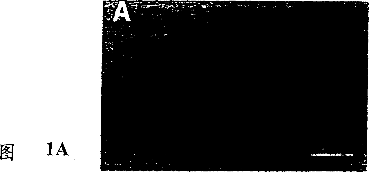
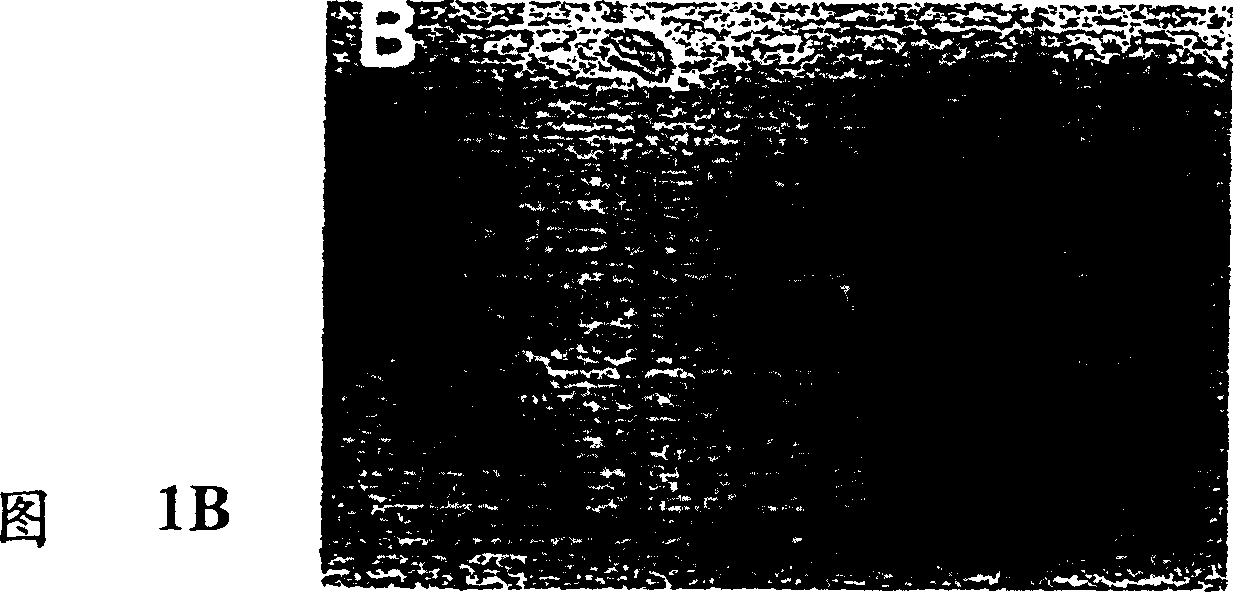
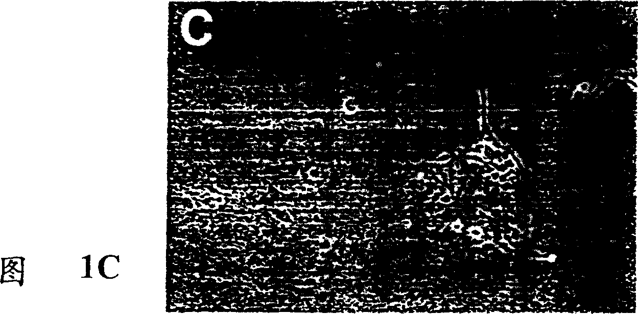
[0109] Using 0.5 mg / ml thermolysin (Sigma; in Hank's calcium-magnesium-free balanced salt solution) at 37°C for 30 min, according to a previously reported method (Corwin et al., 1995), from postpartum days 4-5 ( Utricle epithelial sheets were isolated from P4-5) Wistar rats. Epithelial sheets were then incubated at 37°C for 8 minutes in a mixture of 0.125% trypsin and 0.125% collagenase (Fig. 1A). Enzyme activity was inactivated with a mixture of 0.005% soybean trypsin inhibitor (Sigma) and 0.005% DNase (Worthington) and then pipetted up and down in 0.05% DNase BME solution 10 times with a 1 ml pipette tip. In this way, epithelial sheets were partially dissociated into small pieces containing approximately 10-80 cells (Fig. 1B). Since we ...
Embodiment II
[0129] Stimulates the regeneration of hair cells
[0130] In order to test whether the currently known growth factors can stimulate the proliferation of Utricle Sertoli cells, we used tritiated thymidine incorporation test to measure the DNA synthesis. To measure DNA synthesis, after 24 hours of incubation, add 3 H-thymidine (2 μCi / well) was used for 24 hours, and then the cells were harvested with a Tomtec cell harvester. Because epithelial cells were grown on a polylysine matrix, trypsin (1 mg / ml) was added to the culture wells for 25 minutes at 37°C to detach the cells and then harvested. Counts (Cpm / well) were performed using a Matrix 9600 gas phase counter (Packard Instrument Company, IL) as previously described (Gao et al., 1995). The data of 5-10 wells for each test group were collected and presented in the form of mean ± mean square error (s.e.m.). Statistical analysis was performed using a two-tailed, unpaired t-test. Under control culture conditions, moderate lev...
Embodiment III
[0146] Comparison of Mitogens
[0147]To compare the potency of FGF-2 with IGF-1 and TGF-α, a dose-dependent study was performed in utricular epithelial cell cultures in the range of 0.1-100 ng / ml (Figure 4). At a concentration of 0.1 ng / ml, none of the three growth zones showed a detectable effect (p > 0.05). At a concentration of 1 ng / ml, FGF-2 exhibited a significant mitogenic effect (p<0.01) whereas IGF-1 and TGF-α had no detectable effect. At higher doses (10-100 ng / ml), all three growth factors showed significant mitogenic effects (p<0.05) compared to control cultures. However, FGF-2 was more effective than IGF-1 or TGF-α (p<0.01; Figure 4). Higher potency of FGF-2 than IGF-1 or TGF-α was also observed by BrdU immunocytochemical analysis (Table 2).
[0148] To determine whether FGF-2 acts synergistically with IGFF-1 or TGF-α, FGF-2 was added to the cultures together with IGF-1, or with TGF-α. Both tritiated thymidine incorporation and BrdU immunocytochemical analysis...
PUM
 Abstract
Description
Claims
Application Information
 - R&D
- Intellectual Property
- Life Sciences
- Materials
- Tech Scout
- Unparalleled Data Quality
- Higher Quality Content
- 60% Fewer Hallucinations
Browse by: Latest US Patents, China's latest patents, Technical Efficacy Thesaurus, Application Domain, Technology Topic, Popular Technical Reports.
© 2025 PatSnap. All rights reserved.Legal|Privacy policy|Modern Slavery Act Transparency Statement|Sitemap|About US| Contact US: help@patsnap.com


