Wagnieres George

A technology of imaging diagnosis and equipment, applied in the direction of diagnosis, diagnosis recording/measurement, instruments used for radiological diagnosis, etc., can solve the problems of distinguishing, non-existing distinguishability, insufficient distinguishing characteristics, etc.

Inactive Publication Date: 2004-08-18
RICHARD WOLF GMBH
View PDF3 Cites 4 Cited by
  • Summary
  • Abstract
  • Description
  • Claims
  • Application Information

AI Technical Summary

Problems solved by technology

[0005] However, the discriminative properties of these lesions represented by the above systems are problematic: in known autofluorescence images, inflamed and deformed tissue cannot be practically distinguished from dysplasia and carcinoma in situ, i.e. initial and early malignant lesions However, the two tissue forms of inflamed tissue and deformed tissue have not or have not yet developed into initial or early malignant transformation, which are collectively referred to as "non-malignant tissue changes" in the following description, so this distinction should be made in the diagnosis of patients In order to be able to draw reliable and final conclusions and to rule out (incipient) malignancy, it is necessary to carry out all the obvious places that appear in the autofluorescence map, both for initial and early malignant lesions and for inflamed and deformed tissues. Histologically examined and provided to the pa

Method used


Image

  • Wagnieres George
  • Wagnieres George
  • Wagnieres George

Examples


Embodiment Construction

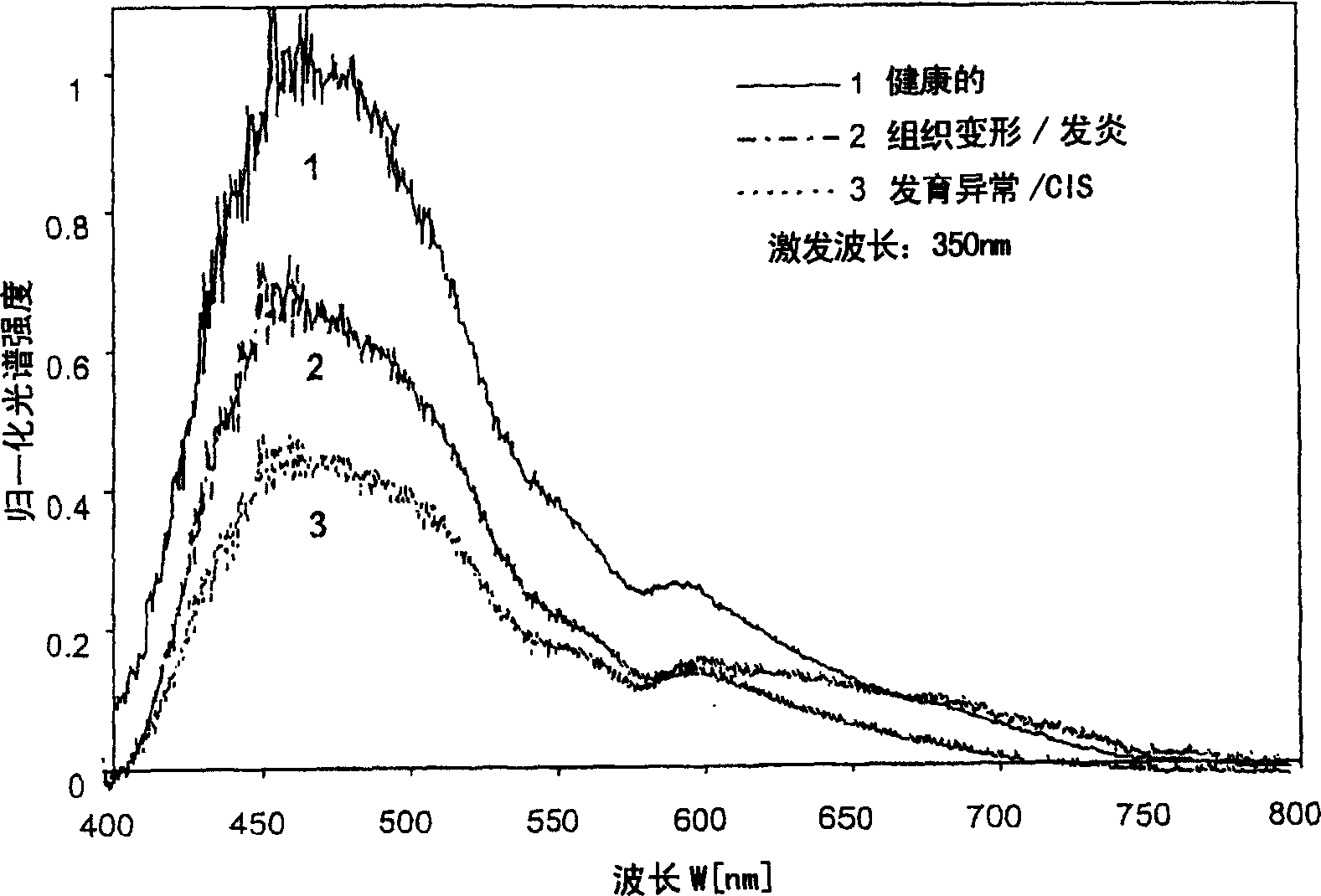
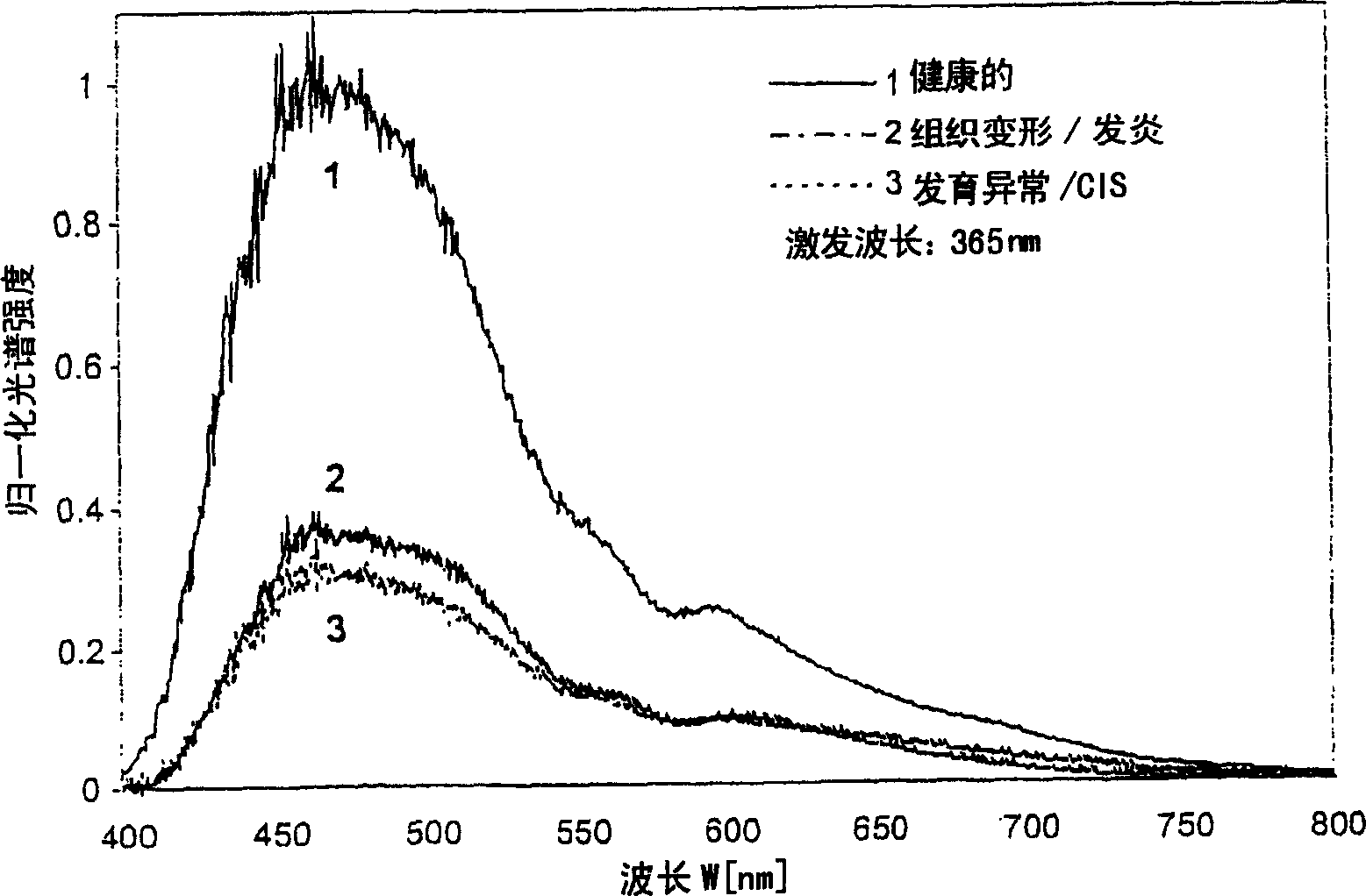
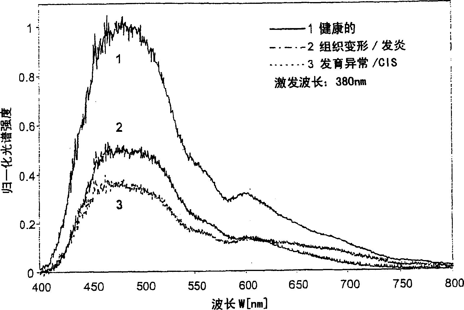
[0064] Figure 1 to Figure 11 Typical fluorescence spectral intensities of human bronchial tissue for different tissue states when using different excitation wavelengths are shown. A detailed explanation of the graphs has been given above.

[0065] Figure 12 Shown schematically, i.e. in block diagram form, for complementary diagnostic white light endoscopy DWLE, diagnostic autofluorescent endoscopy DAFE or DAFEI and optionally diagnostic autofluorescent endoscopy The device structure of DAFEII.

[0066] The diagnostic imaging device for tissue 1 comprises a light source 2 that provides white light for illumination in the DWLE operating mode and for illuminating the tissue 1 through the light guide 3 in the DAFEI and DAFEII operating modes Different light beams for each fluorescent excitation form. The light guide 3 can consist of a single optical fiber, a fiber bundle or a liquid light guide or a combination of these elements.

[0067] Not only the images formed by the r...


PUM

PropertyMeasurementUnit
Bandwidthaaaaaaaaaa
Widthaaaaaaaaaa

Abstract

The apparatus for imaging diagnosis of tissue (1) has selectivity to use at least two methods of diagnosis. One working mode uses diagnostic white light endoscopy (DWLE) and another mode uses diagnostic auto fluorescence endoscopy (DAFE). Optionally, a further DAFE diagnosis mode can be used. A lamp (2) delivers light to the tissue through an optic fiber (3). A lens (4) is used either by the human eye, or in combination with a camera (5) linked to an image processing unit (6) and a monitor (7). Most of the light used for both DAFE modes is generated from a narrow spectral band not more than30 nm, with a wavelength of 405 nm.

Description

technical field [0001] The present invention relates to a device for imaging diagnosis of tissue for the optional use of two or three diagnosis methods, namely a working mode DWLE for white light endoscopy for diagnosis, and a working mode for autofluorescence endoscopy for diagnosis The first mode of operation DAFE or DAFEI and, if necessary, the second mode of operation DAFEII for autofluorescent endoscopy for diagnosis, the imaging diagnostic equipment of the tissue has a light source, the light emitted by the light source is detected by the endoscope Guided to the tissue, it also has an image transmission unit and an image receiving unit, and an image processing unit is connected with the image receiving unit, and the image signal is provided for a monitor through the image processing unit, and the image transmission unit can also be connected with people Eyes are directly connected, in this case, the image receiving unit, image processing unit and monitor can be omitted. ...

Claims


Application Information

Patent Timeline
IPC IPC(8): G01N21/64A61B1/00A61B1/04A61B1/06A61B5/00
CPCA61B1/043A61B5/0071A61B1/0669A61B1/00186A61B1/0638A61B5/0084A61B1/0646
Inventor 贝恩德·克劳斯·韦伯托马斯·戈尔马丁·多尔特斯特凡·米勒菲利普·艾德纳尼古拉斯·佩雷拉-德尔加多胡贝特·范登贝格乔治·瓦格尼尔斯托马斯·格兰茨曼塔尼娅·加布雷希特
Owner RICHARD WOLF GMBH
Why Eureka
Try Eureka