3D controlled stacking formation method of cell-material units

A cell and three-dimensional technology, applied to biochemical equipment and methods, fixed on/in organic carriers, microorganisms, etc., can solve the problems of different cell positioning and fixed-point placement, blood vessel growth restriction, cell migration and Reproduction is not particularly ideal and other issues, to achieve the effect of facilitating proliferation and differentiation

Inactive Publication Date: 2005-04-27
TSINGHUA UNIV
View PDF1 Cites 15 Cited by
  • Summary
  • Abstract
  • Description
  • Claims
  • Application Information

AI Technical Summary

Problems solved by technology

These methods have the following defects when constructing tissues and organs with complex functional gradient structures: first, for relatively thick tissue engineering scaffolds, the migration and propagation of cells are not particularly ideal, and the growth of blood vessels is limited; Different cells cannot be accurately positioned and placed at fixed points in space to meet the needs of constructing functionally graded structures of tissues and organs. However, in the process of artificial manufacturing of complex tissues and organs, different cells and materials must be accumulated in a specified position in space and for them. Create conditions for interaction
This method is also based on the forming mechanism of material curing, and the environment after curing is also not suitable for the survival of cells.

Method used


Image

  • 3D controlled stacking formation method of cell-material units
  • 3D controlled stacking formation method of cell-material units

Examples


Embodiment 1

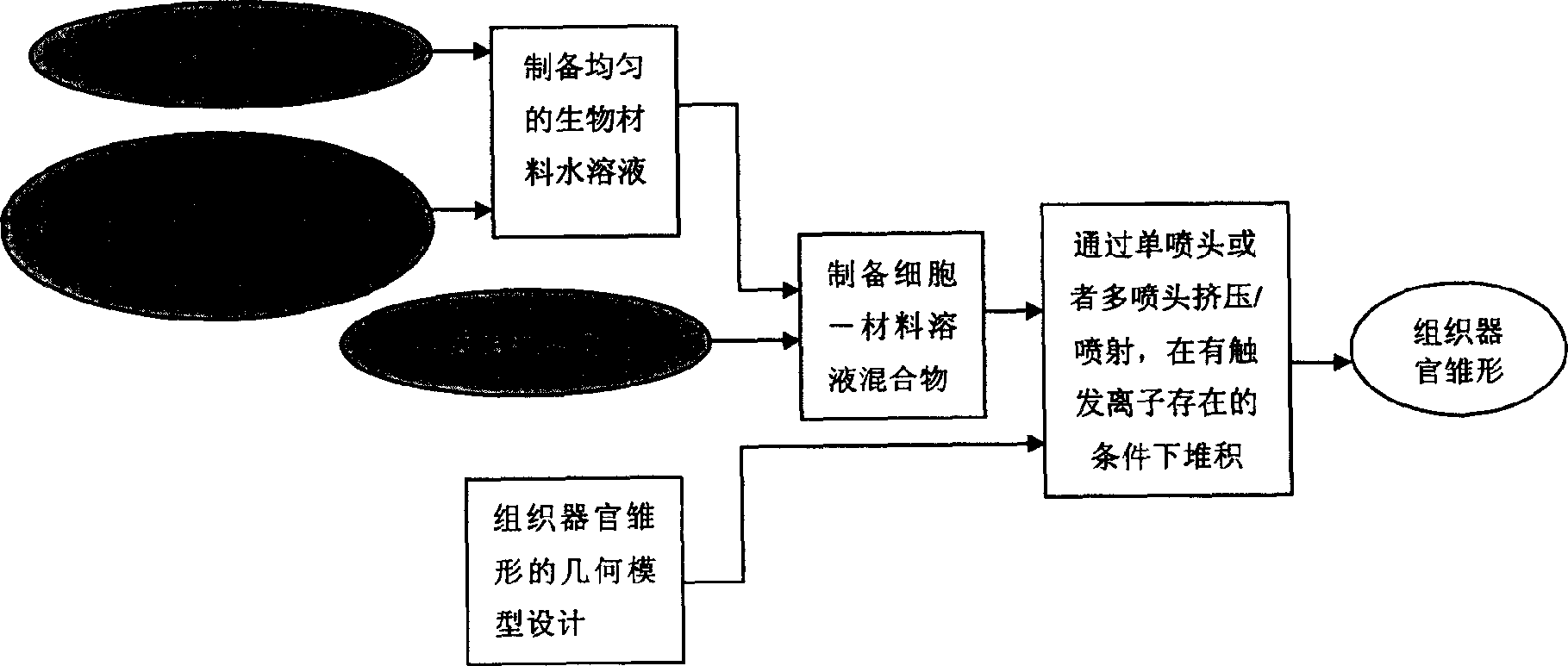
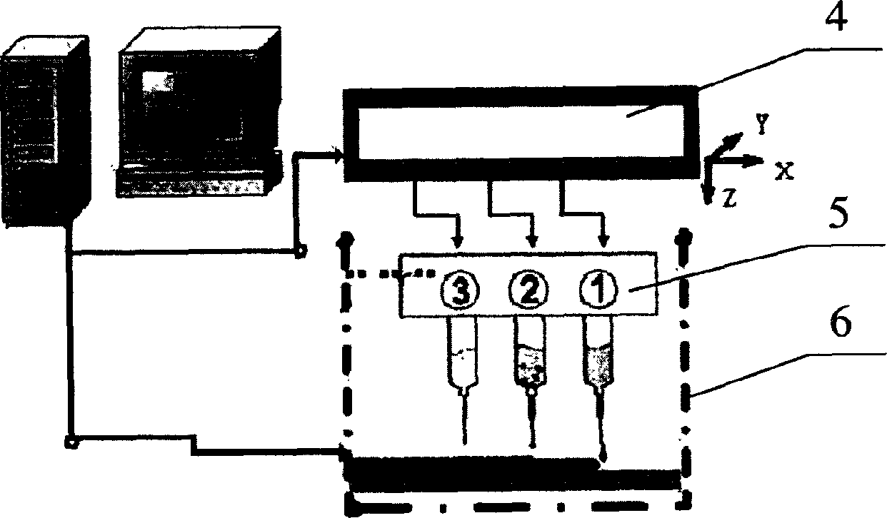
[0022] Example 1: The three-dimensional structure of chondrocyte-gelatin-calcium alginate manufactured by this process. The mixture of sodium alginate and gelatin that promotes cell adhesion and growth is prepared into an aqueous solution with a concentration of 100g / L, wherein the mass ratio of sodium alginate and gelatin is 1:10, and it is sterilized for use; the chondrocytes are mixed with the material solution evenly , to obtain a mixture of chondrocyte-gelatin-sodium alginate solution; prepare 10% calcium chloride solution as a trigger and make it atomized in the forming space; according to the pre-designed structure and defined planning path, the above mixture It is sprayed into a room-temperature sterile forming chamber filled with an atomization trigger by means of droplet spraying; after the solution is sprayed to a designated position, it undergoes a sol-gel transition rapidly to form a chondrocyte-gelatin-calcium alginate gel; gradually Layer stacking results in a t...

Embodiment 2

[0023] Example 2: The three-dimensional structure of hepatocyte-gelatin-hepatocyte growth factor-calcium alginate produced by this process. The mixture of sodium alginate, gelatin and hepatocyte growth factor that promotes cell adhesion growth is prepared into an aqueous solution with a concentration of 30g / L, wherein sodium alginate: (gelatin+hepatocyte growth factor) is 1: 0.67 (mass ratio) , sterilized for later use; the hepatocytes and the material solution were evenly mixed to obtain a mixture of hepatocytes-gelatin-hepatocyte growth factor-sodium alginate solution; 5% calcium chloride solution was prepared as a trigger and input into the trigger nozzle; according to With the pre-designed structure and the defined and planned path, the above-mentioned mixture is sprayed to the designated position in the forming chamber by the method of droplet spraying, and the trigger agent sprays the calcium chloride solution at the same position, so that the cell-material unit rapidly u...

Embodiment 3

[0024] Example 3: The three-dimensional structure of hepatocyte-endothelial cell-gelatin-calcium alginate produced by this process. The mixture of sodium alginate and gelatin that promotes cell adhesion and growth is prepared into an aqueous solution with a concentration of 10g / L, wherein the mass ratio of sodium alginate and gelatin is 1:0.5, and it is sterilized for use; liver cells and endothelial cells are mixed with Mix evenly with the material solution to obtain a mixture of two cell-gelatin-sodium alginate solutions; input the two mixtures into different nozzles respectively; prepare 20% calcium chloride solution as a trigger and input it into a special trigger nozzle; Design the structure and define the planned path. After the above mixture is sprayed to the specified position by the method of droplet injection, the trigger nozzle sprays the calcium chloride solution at the same position, and the mixture of cell-material solution undergoes a sol-gel transition rapidly. ...


PUM


Abstract

The present invention is 3D controlled stacking formation method of cell-material units. Ion concentration sensitive material and cell tactophily promoting biodegradable material(s) are first prepared into aqua and sterilized; one or several kinds of cell are then mixed with the solution to form cell-material solution mixture; and various kinds of cell-material solution mixture are extruded or jetted in preset structure and path to form cell-material units stacked in different spatial positions, to form cell-material gel with certain configuration and strength in corresponding triggering condition, and to obtain tissue organ rudiment via stacking. The present invention is based on the basic dispersion / stacking principle for fast formation to realize the accurate locating and formation of one or several kinds of cell in 3D space without damaging cell and other live biological body. The present invention is favorable to cell amplification and differentiation.

Description

technical field [0001] The invention relates to a process method for manufacturing tissue and organ prototypes by three-dimensional controlled stacking of cell-material units, which belongs to the technical field of tissue engineering. Background technique [0002] The repair and replacement of human diseased tissues and organs is an eye-catching scientific frontier in the 21st century. In order to solve this scientific problem, tissue engineering technology was born in the mid-1980s. Its essence is to combine living cells with matrix materials or scaffolds in a certain way to create new tissues. After about two decades of development, tissue engineering has made great progress in scaffolds, cells, growth factors, construction techniques, biological evaluation, detection techniques and standards, clinical trials, and industrialization. However, in terms of construction technology, the composite method of cells and matrix materials or scaffolds usually adopts methods such as...

Claims


Application Information

Patent Timeline
Patent Type & Authority Applications(China)
IPC IPC(8): C12N5/08C12N11/04
Inventor 颜永年熊卓王小红林峰吴任东张人佶卢清萍
Owner TSINGHUA UNIV
Why Eureka
Try Eureka