
Nuclear medical diagnosing apparatus and data collecting method for nuclear medical diagnosis

A diagnostic device and a technology of nuclear medicine, applied in the field of data collection of nuclear medicine diagnosis, can solve problems such as positional resolution deterioration, sensitivity deterioration, inability to reliably detect breathing state, etc., and achieve the effect of good positional resolution and sensitivity

Active Publication Date: 2005-07-13
TOSHIBA MEDICAL SYST CORP
View PDF1 Cites 3 Cited by
  • Summary
  • Abstract
  • Description
  • Claims
  • Application Information

AI Technical Summary

Problems solved by technology

[0007] However, in the case of the above-mentioned breath-synchronized data collection, basically, since the collection statistics corresponding to one phase are reduced, there is a problem that statistical noise and other disturbances are large, and there is a problem that image degradation is caused by this.
On the contrary, when the respiratory phase is roughly divided, although the image quality degradation caused by the decrease of the collection statistics can be reduced, the effect of suppressing the body movement caused by the breathing is poor, and the opposite situation is caused.
[0008] Furthermore, the above-mentioned breath-synchronized data collection is performed with a breath-synchronized monitor, but it is also pointed out that the breath-synchronized monitor cannot reliably detect the breathing state of the subject.
If such a reliable respiratory state detection cannot be performed, the data collection time will be extended and the collection statistics will be reduced, and the number of patients that can be detected will decrease and the above-mentioned image quality will deteriorate significantly.
[0009] Furthermore, in the case of data collection synchronized with respiration, since it is imaging under the average body motion of the subject over the entire time of one respiration phase for data collection, the difference between the subject and the subject stopping breathing When comparing the data collected in the state, the image quality is unsatisfactory due to the influence of a large amount of body movement caused by breathing.
[0010] On the other hand, as another problematic point, if it is possible to collect data only during the breath stop of the subject and generate an image using only the data collected during the stop of breath, it is possible to suppress the noise caused by the body movement of the subject. Deterioration of positional resolution and contrast, on the contrary, also because no data is collected during breathing, so the number of samples decreases and the smoothness of the image, that is, the problem of sensitivity degradation can be cited

Method used


Image

  • Nuclear medical diagnosing apparatus and data collecting method for nuclear medical diagnosis
  • Nuclear medical diagnosing apparatus and data collecting method for nuclear medical diagnosis
  • Nuclear medical diagnosing apparatus and data collecting method for nuclear medical diagnosis

Examples


Embodiment 1

[0039] refer to Figure 1 to Figure 4 The nuclear medicine diagnostic apparatus and the data collection method for nuclear medicine diagnosis according to Embodiment 1 of the present invention will be described.

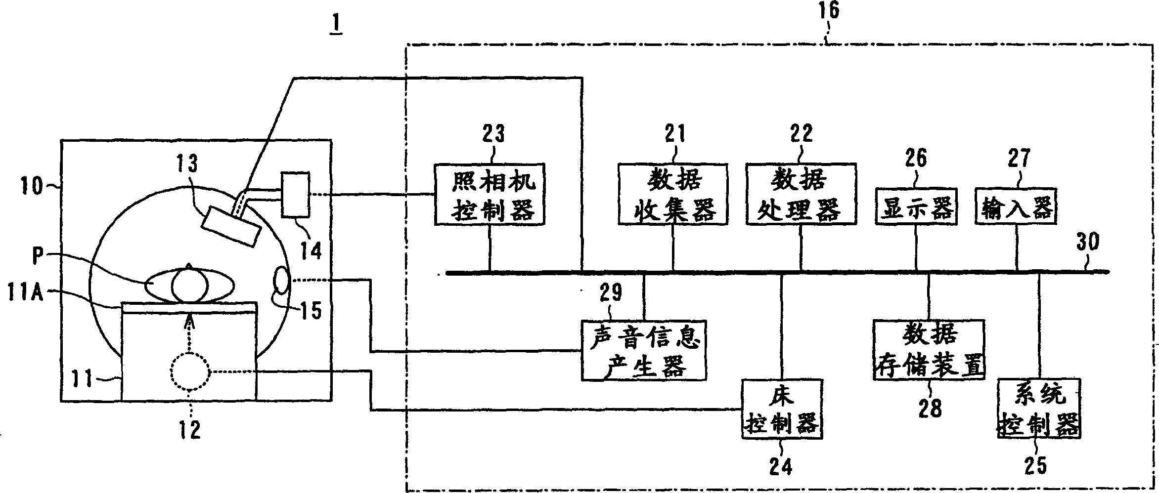
[0040] figure 1 A schematic configuration of the nuclear medicine diagnostic apparatus 1 of this embodiment is shown. This nuclear medicine diagnostic apparatus 1 includes: a bed 11 equipped with a top plate 11A on which a subject P such as a patient usually lies on its back; a bed driver 12 built in the bed 11; a stand 10 arranged adjacent to the bed 11; A gamma camera 13 held on the gantry 10 ; a camera driver 14 arranged in the gantry 10 and capable of driving the gamma camera 13 to move; a speaker 15 and a control processing device 16 .

[0041] The control processing device 16 includes a data collector 21, a data processor 22, a camera controller 23, a bed controller 24, a system controller 25, a display 26, an input device 27 operated by an operator, and a da...

Embodiment 2

[0072] Below, refer to Figure 5-7 Example 2 of the present invention will be described.

[0073] The nuclear medicine diagnostic apparatus of the second embodiment adopts a structure in which collected data corresponding to signals from an operation switch as an example of a signal generating unit is divided into different areas and recorded.

[0074] That is, including the operating switch, the system controller 25, the data collector 21, and the data recording device 28 form a breathing identification unit for identifying whether the breathing stops or the breathing state, and the data recording device 28 for collecting data is recorded. The area serves as identification information for identifying whether the breathing is stopped or breathing.

[0075] Furthermore, other configurations and functions are the same as those of the nuclear medicine diagnostic apparatus of Embodiment 1, so explanations are omitted.

[0076] The operation switch of the nuclear medicine diagnos...

Embodiment 3

[0091] Below, refer to Figures 9 to 11 Embodiment 3 of the present invention will be described.

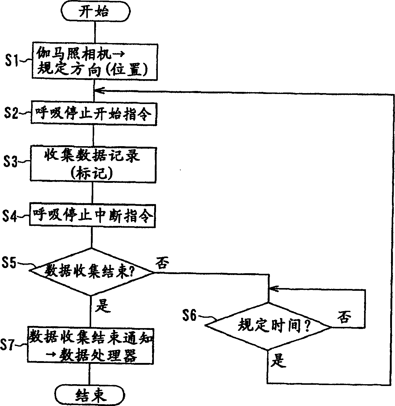
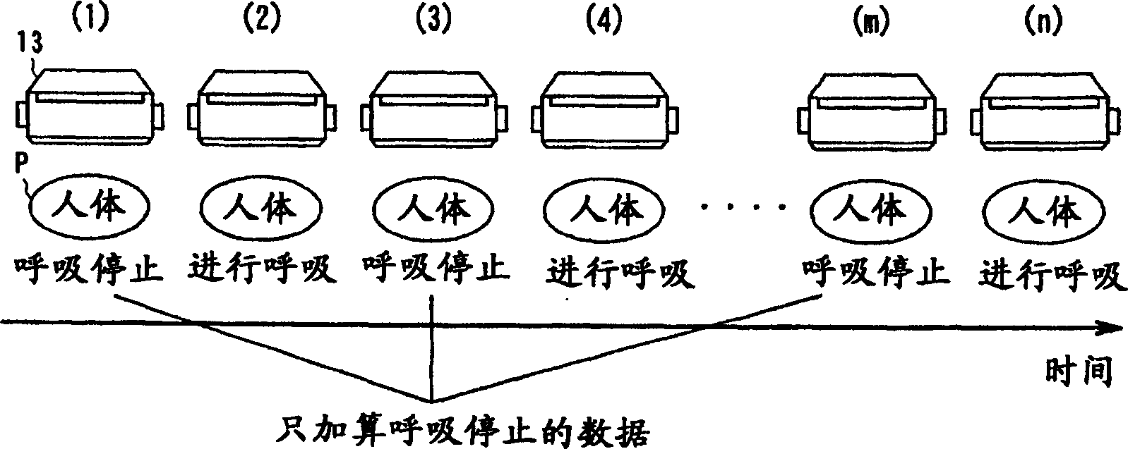
[0092] The nuclear medicine diagnostic apparatus of this Example 3 is related to the configuration of the SPECT method using the "intermittent data collection method". In particular, this embodiment is characterized in that the gamma camera 13 rotates around the subject P in a stepwise manner, and when imaging is performed in each of a plurality of imaging directions (positions), the temporary apnea and the apnea of ​​the apnea operation are alternately repeated. Take a breath. At this time, a plurality of imaging directions (positions) are set twice as fine as the usual SPECT method (for example, each step length is 3 degrees, and the collection time is 10 to 15 seconds), and the collection is repeated (for example, the number of repetitions is 60 times). Figure 9An outline of this processing is shown. Furthermore, the hardware configuration and figure 1 as shown.

[0093...


PUM


Abstract

The nuclear medicine diagnostic apparatus of the present invention detects radiation emitted by an atomic nuclide injected into a subject as projection data with a gamma camera, and obtains an image showing the distribution of atomic nuclides in the subject based on the projection data. The device includes: a rotation unit that rotates the radiation detector around the subject; a breath recognition unit that recognizes breathing stoppage and respiration caused by the stop of breathing of the subject; a data storage unit for storing radiation detection data collected by the radiation detector; and an image generation unit for generating an image based on the radiation detection data stored in the data storage unit based on the identification result of the breathing identification unit.

Description

technical field [0001] The present invention relates to a nuclear medicine diagnostic device that uses a gamma (γ) camera to detect radiation emitted from an atomic nuclide (radioisotope: RI) administered to a subject, and images the distribution of RI from the detected information, and a nuclear medicine diagnostic device. Data Collection Methods. Background technique [0002] With recent technological advancements in hardware and software, nuclear medicine diagnostic devices, which are medical equipment, have also made great progress. [0003] This nuclear medicine diagnostic apparatus includes a measurement unit called a gamma camera, and is used for nuclear medicine examination of a subject. As described above, a drug labeled with a radioactive isotope (hereinafter referred to as RI) is administered into the subject, and the RI distribution in the body is imaged with a gamma camera to perform a nuclear medicine examination. Among the main methods of nuclear medicine in...

Claims


Application Information

Patent Timeline
Patent Type & Authority Applications(China)
IPC IPC(8): G01T1/161A61B5/05A61B6/00G01T1/164G01T1/166G03B42/02
CPCA61B6/037A61B6/541
Inventor 本村信笃林万寿夫堀内承治二沢佳史
Owner TOSHIBA MEDICAL SYST CORP
Learn More