Multi-aptamer-based, cell-specific, one-step tumor cell detection assays

a tumor cell and multi-aptamer technology, applied in the field of clinical chemistry, oncology, and diagnostic medical assays, can solve the problems of multiple step protocols, limiting the sensitivity of current assays, and no such assays available, and achieves high sensitiveness, high accuracy and reliability, and easy implementation and execution.

Active Publication Date: 2018-07-10
THE METHODIST HOSPITAL
View PDF7 Cites 0 Cited by
  • Summary
  • Abstract
  • Description
  • Claims
  • Application Information

AI Technical Summary

Benefits of technology

[0014]The present invention overcomes these obstacles, as well as numerous additional limitations inherent in the prior art, by providing novel and non-obvious molecular biomarker detection assays that employ a cell-specific, intracellularly-activated, one-step detection system. This system, which is both highly sensitive and capable of detecting single tumor cells in populations of millions of normal cells, is highly accurate and reliable, and is facile in implementation and execution. Significantly, the assay system described herein can be accomplished rapidly—using a very small quantity of biological sample and a one-step, single-reagent protocol. In illustrative embodiments, the present assay system has been employed to rapidly and specifically detect single or multiple distinct biomarkers from different types of human tumor cells using only a single drop of the patient's blood as the specimen sample.
[0015]The present assay system provides a highly-desirable method for rapid tumor cell detection. Importantly, the disclosed assay may be readily performed as a one-step procedure—either in a traditional clinical laboratory environment, or even at the point-of-care, including remote, and distant locales outside the conventional clinical environs. In its simplest form, the disclosed assay system involves simply mixing a small volume of a biological sample (e.g., a single drop of a patient's blood will suffice) with a minimal number of reagents (in particular embodiments, a single reagent solution), incubating the test mixture briefly (in certain embodiments for as short a time as a few minutes), and then rapidly obtaining the results in a cost-effective, reproducible, and highly-sensitive manner. Particularly advantageous with the present invention, the results can be readily achieved, with little to no “off-target,”“false-positive,” or extraneous “background” signals. In particular commercially-relevant formats, the present assay can be scaled in a suitable form to permit adaptation for use in conventional, rapid, high-volume, high-throughput assay.
[0016]The assays of the present invention provide such desirable features, and affords a wholly-new approach to rapid, low-cost, screening of clinical samples, even at the point-of-care. FIG. 1A presents a schematic overview of the process. To achieve this goal, an assay system was developed that is both tumor cell-specific, and “active” (i.e., detectable) only within particular cells of interest that express the target molecule(s) for which the aptamer is specific (see, e.g., FIG. 1B). The new method is in sharp contrast to existing antibody-based assay systems found in the prior art (see e.g., FIG. 1C) that are constantly active, and consistently produce extraneous, off-target, and / or background signals.

Problems solved by technology

To date, there are no such assays available.
The presence of the constant, “off-target” background signals limits the sensitivity of current assays, and the complex nature of the existing assay systems require tedious, multiple-step protocols to achieve sufficient results.
Although the final imaging and detection can be completed rapidly in this methodology, the multiple steps involved in sample preparation are both time- and labor-intensive.
Moreover, the cost of this system and reagents presents a barrier to entry for many laboratories and clinical settings.
However, all of these technologies use fluorescent antibodies for cell staining, and require multiple, labor-intensive steps to prepare and assay the sample.
Importantly, none of the existing commercial methodologies represents a suitable platform for “point-of-care” screening, or facile, rapid sample analysis.
Moreover, these systems are limited in their applicability to high-throughput platforms, multi-sample scale-up, and / or robotics-based analytical systems.

Method used


Image

  • Multi-aptamer-based, cell-specific, one-step tumor cell detection assays
  • Multi-aptamer-based, cell-specific, one-step tumor cell detection assays
  • Multi-aptamer-based, cell-specific, one-step tumor cell detection assays

Examples


example 1

Tumor Cell Detection Assay System

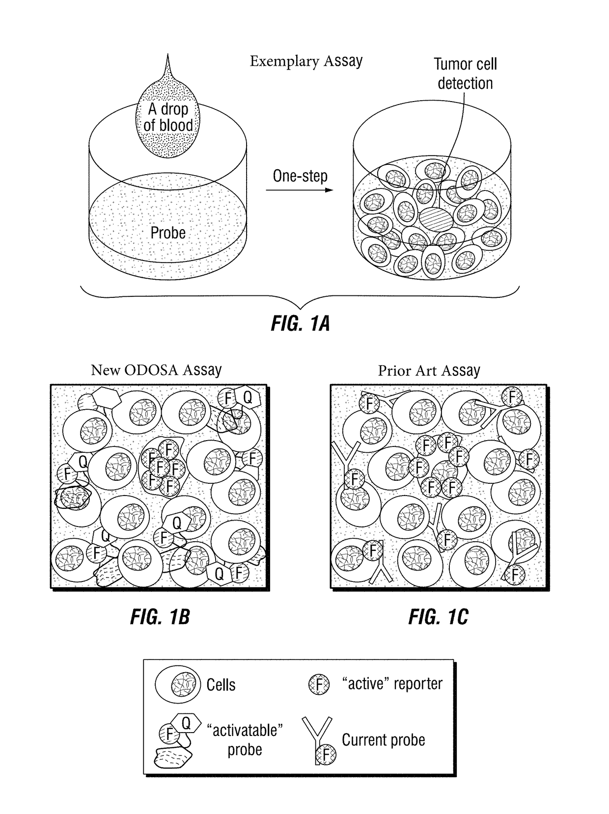
[0159]To develop an exemplary assay system with significant potential for clinical application, RNA-based aptamers that are specific for one or more cellular biomarkers (e.g., CD30 protein) were synthesized (Zhang et al., 2009) and each were conjugated to a specific fluorochrome reporter molecule (such as Cy 3, Cy5.5, etc.) operably linked to a quencher molecule (e.g., BHQ2).

[0160]Freshly cultured CD30-positive lymphoma cells (ATCC, Manassas, Va., USA) were stained with the aptamer probes or anti-CD30 antibody (BD Biosciences) as a standard control. Flow cytometry analysis showed that the aptamer probe is able to selectively bind to and detect lymphoma cells that express CD30 (Zhang et al., 2009). Specific cell binding of aptamer probes was also confirmed by fluorescence microscopy at a final concentration as low as 0.3 nM. Multicolor flow cytometry demonstrated that comprehensive immunophenotyping of lymphoma cells could be performed by using oligon...

example 2

Aptamer Probes as Replacements for Conventional Antibody-Based Diagnostics

[0178]In this example, the use of a synthetic aptamer probe for cell immunophenotyping, tissue immunohistochemical (IHC) stain, and blood circulating tumor cell detection was demonstrated.

[0179]Experimental Methods

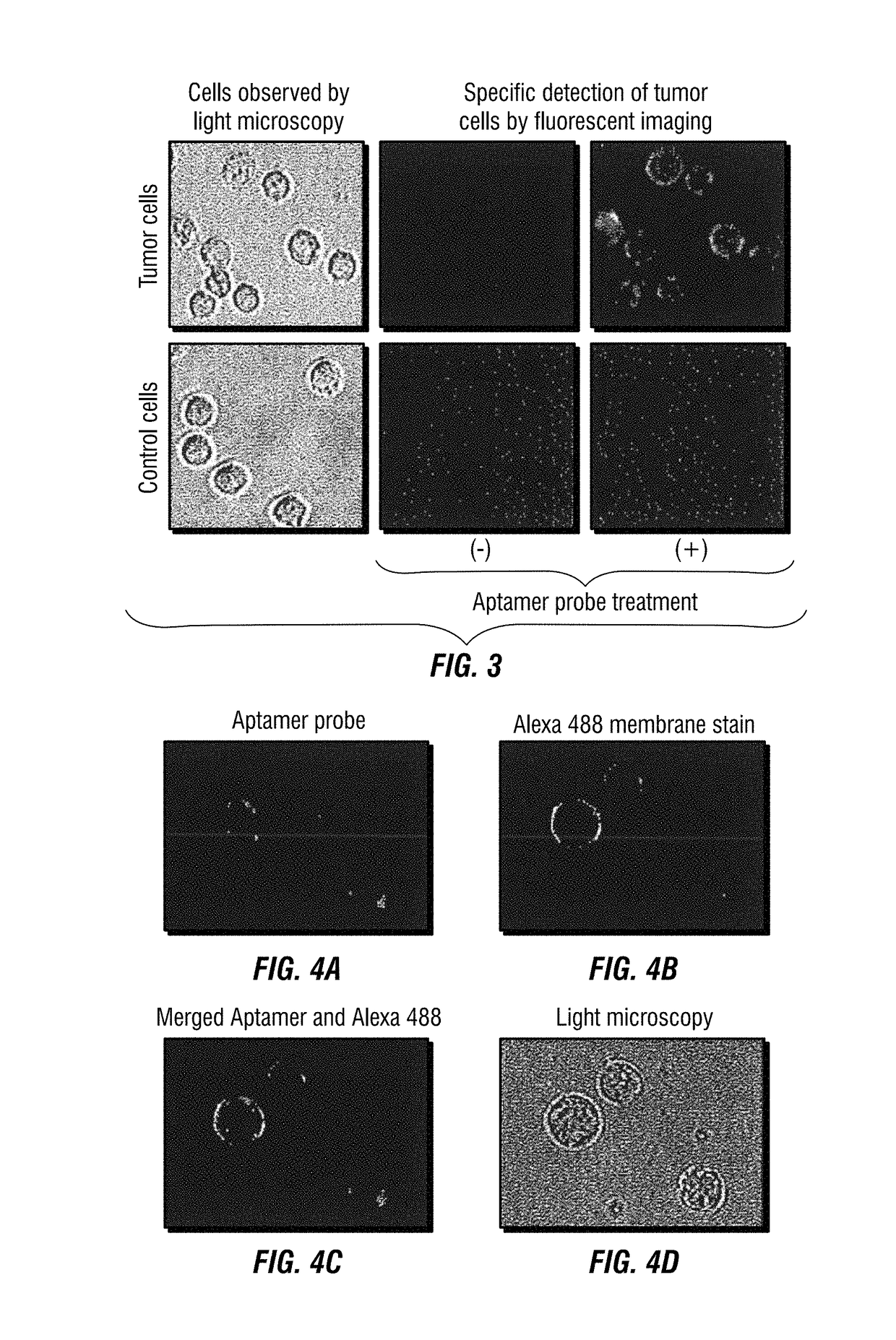
[0180]For immunophenotyping a CD30-specific aptamer probe (FIG. 4A) was synthesized and conjugated with a fluorochrome Cy5.5 reporter. Cultured lymphoma cells as showed in FIG. 16 were stained with the aptamer probe in combination with antibodies and analyzed by multicolor flow cytometry as published previously (Zhang et al., 2009). For tissue IHC study, biotinylated aptamer probes were generated (Integrated DNA Technologies, Coralville, Iowa, USA). Formalin-fixed and paraffin-embedded lymphoma tissues were stained with the aptamer probes (FIG. 17) and visualized by a HRP-conjugated streptavidin color development kit (Dako).

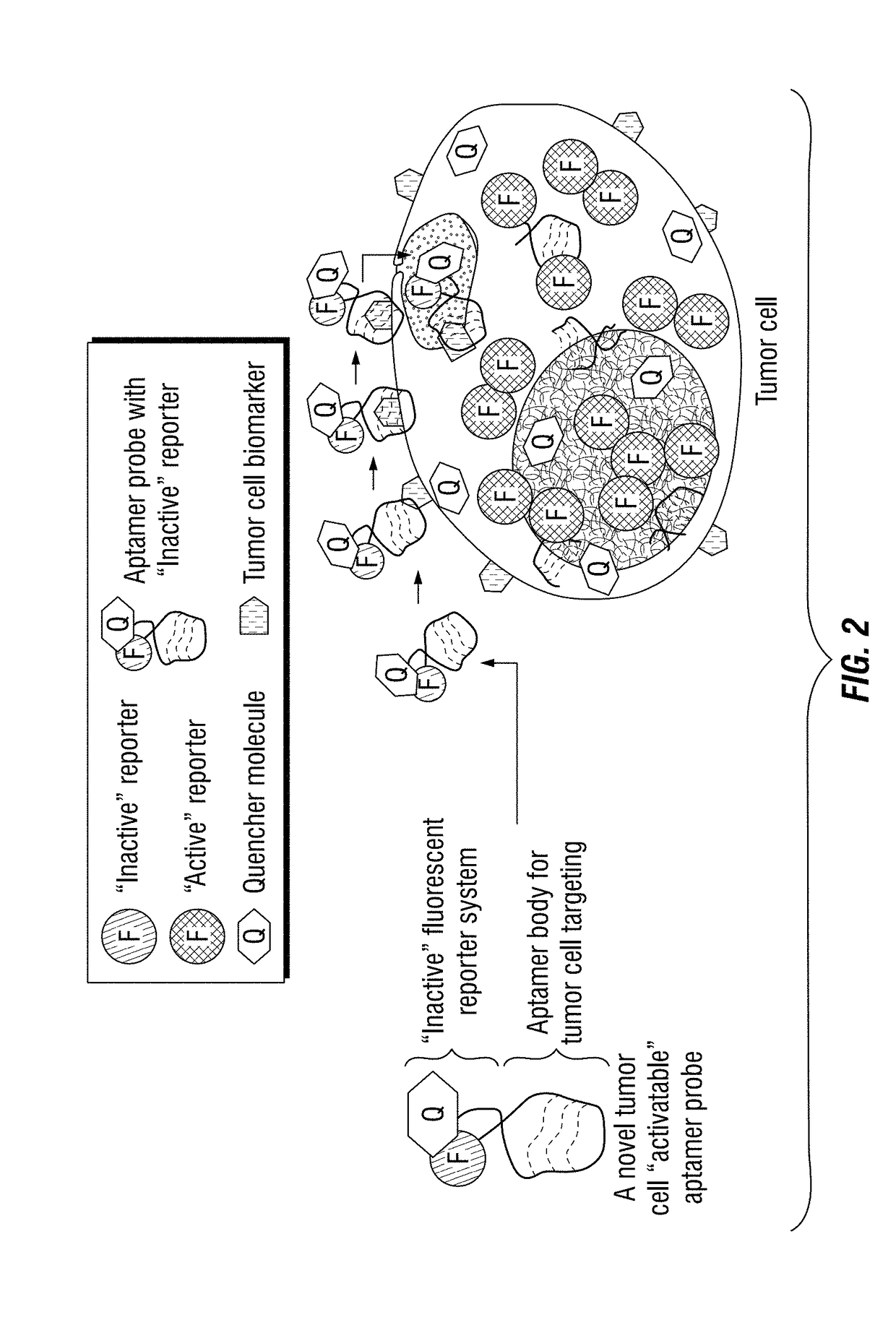
[0181]For circulating tumor cell detection a novel “intracellular-activatable...

example 3

Design of “Intra-Tumor Cell-Activatable” Aptamer Probes

[0184]To develop the one-step assay technology for circulating tumor cell detection, the inventors chose to use aptamers instead of traditional antibody probes for target recognition and signal reporting. Aptamer sequences were synthesized, and conjugated to the fluorochrome Cy3 (Integrated DNA Technologies) and quencher BHQ2 molecules (Integrated DNA Technologies) at the 5′- and 3′ ends, respectively (FIG. 4B). Notably, in the absence of cells of interest, the aptamer probes are optically silent (i.e., “inactive”), because the fluorochrome is completely quenched / inactivated by the paired quencher molecule that is present on the same aptamer molecule (FIG. 4C). After specifically binding to targeted tumor cells and being internalized, however, subsequent lysosomal degradation of the aptamer sequence activates the previously-optically-silent reporter system by freeing the fluorochrome from the paired quencher molecule, resulting ...


PUM

PropertyMeasurementUnit
volumeaaaaaaaaaa
volumeaaaaaaaaaa
volumeaaaaaaaaaa

Abstract

Disclosed are methods and compositions for the detection of one or more different types of cellular biomarkers in a biological sample, and in particular, methods and compositions for the rapid, one-step, highly-cell specific detection of circulating tumor cells from minute quantities of mammalian biological fluids, including, for example, from a single drop of human blood. In certain embodiments, distinctly-labeled, multi-aptamer detection reagents are provided for detecting and quantitating selected cancer cells in clinical samples such as patient specimens and / or tissues. Aptamer-based imaging methodologies are also provided for use in a variety of diagnostic assay protocols.

Description

CROSS-REFERENCE TO RELATED APPLICATIONS[0001]The present application is a continuation of PCT International Patent Application No. PCT / US2013 / 74797, filed Dec. 12, 2013, which claims priority to U.S. Provisional Patent Application No. 61 / 736,452, filed Dec. 12, 2012, and U.S. Provisional Patent Application No. 61 / 801,523, filed Mar. 15, 2013; the contents of each is specifically incorporated herein in its entirety by express reference thereto.BACKGROUND OF THE INVENTIONStatement Regarding Federally Sponsored Research or Development[0002]Not Applicable.NAMES OF THE PARTIES TO A JOINT RESEARCH AGREEMENT[0003]Not Applicable.FIELD OF THE INVENTION[0004]The present invention relates to the fields of clinical chemistry, oncology, and diagnostic medical assays. The invention provides multi-aptamer-based detection reagents, and corresponding diagnostic assays and methods for detection of target biomolecules. In exemplary embodiments, fluorescently-labeled detection reagents are provided for...

Claims


Application Information

Patent Timeline
Patent Type & Authority Patents(United States)
IPC IPC(8): G01N33/574C12Q1/6816C12Q1/6886G01N33/542G01N33/53
CPCG01N33/57492C12Q1/6816C12Q1/6886G01N33/5308G01N33/542G01N2333/70578C12Q2525/205C12Q2565/101
Inventor ZU, YOULI
Owner THE METHODIST HOSPITAL
Why Eureka
Try Eureka