
System for detecting protease

Inactive Publication Date: 2006-09-19
AHRAM BIOSYST
View PDF17 Cites 51 Cited by
  • Summary
  • Abstract
  • Description
  • Claims
  • Application Information

AI Technical Summary

Benefits of technology

[0021]Practice of the present invention provides a number of important advantages.
[0022]For example, the invention provides chimeric proteins that are cleaved specifically by the subject protease to produce labeled (and unlabeled) component proteins. Preferred practice of the invention links the subcellular location and, preferably, the signal intensity of the labeled proteins to the presence (or absence) of the subject protease. This “two-factor” detection strategy provides for highly sensitive and reliable protease detection. That is, both the location of labeled protein and its signal intensity within the cells or tissues can be taken to be indicative of the presence of the active protease. The invention is also highly selective ie., it can readily discriminate between presence of different proteases or isozymes as well as presence of inactive and active versions of the same protease inside cells. Preferred chimeric proteins of the invention can be made with available reagents and standard recombinant manipulations making the invention easy to use.
[0023]Additionally, the invention is flexible and can be used to detect active protease in a wide range of cells, typically eukaryotes, including those derived from plants, yeast, fungi, animals, and insects. Preferred chimeric proteins have minimal impact on the gene expression in the cells or tissue, thereby avoiding potentially complicating genetic effects. The invention is also compatible with a variety of suitable protease cleavage sites and detectable amino acid sequences.
[0038]By addressing the technical problems involved, the present invention provides more realistic in vivo methods for analyzing the activity of a specific protease and screening inhibitors for the protease, in which the protease and its specific substrate co-exist in a living cell so that the cleavage of the substrate by the protease can take place in a living cell and the result can be directly observed from the living cell.
[0052]As discussed, the invention is well-suited to detect protease inhibitors. Known protease inhibitors in accord with the invention are recognized viral disease inhibitors. Because the viral protease is essential for replication and reassembly of viral capsids, the protease inhibitor can be used for treatment of viral diseases by inhibiting the protease to suppress the viral proliferation. Animal viruses can cause diseases such as AIDS and hepatitis etc., and plant viruses reduce crop yield by causing wilting leaves and mottling.
[0053]In one aspect, the present invention provides a framework for effectively developing inhibitors specific to various proteases by supplying the substrates and the method for in vivo screening of inhibitors specific to various proteases. This method can be also used to determine the efficiency of the drug candidates screened by the conventional in vitro method.

Problems solved by technology

However, several side effects were reported during such treatments (Miller, T. L. et al., 2001), and mutants having mutated protease genes were reported in the cases of prolonged treatments (Jacobsen, H. et al., 1996; Cote, H. et al., 2001).
However, in the in vitro screening method, it is not possible to examine various complicated effects such as the transport efficiency of drug candidates into the cell, the stability and cytotoxicity of drug candidates in the cell, etc.
Many additional time-consuming experiments are thus necessary before examining the drug candidates selected by the in vitro screening method in a living body.
Accordingly, such methods do not always reflect intracellular environments that may substantially impact protease function.
Moreover, many of the prior methods are believed to be limited in terms of sensitivity, selectivity, and convenience.
These and other drawbacks are believed to have lowered the efficiency and reliability of past screening attempts.

Method used


Image

  • System for detecting protease
  • System for detecting protease
  • System for detecting protease

Examples


example 1

Detection of Chimeric Proteins and Trafficking to the Subcellular Organelles

[0180](a) Construction of Recombinant Plasmids for Expression of the Chimeric Proteins

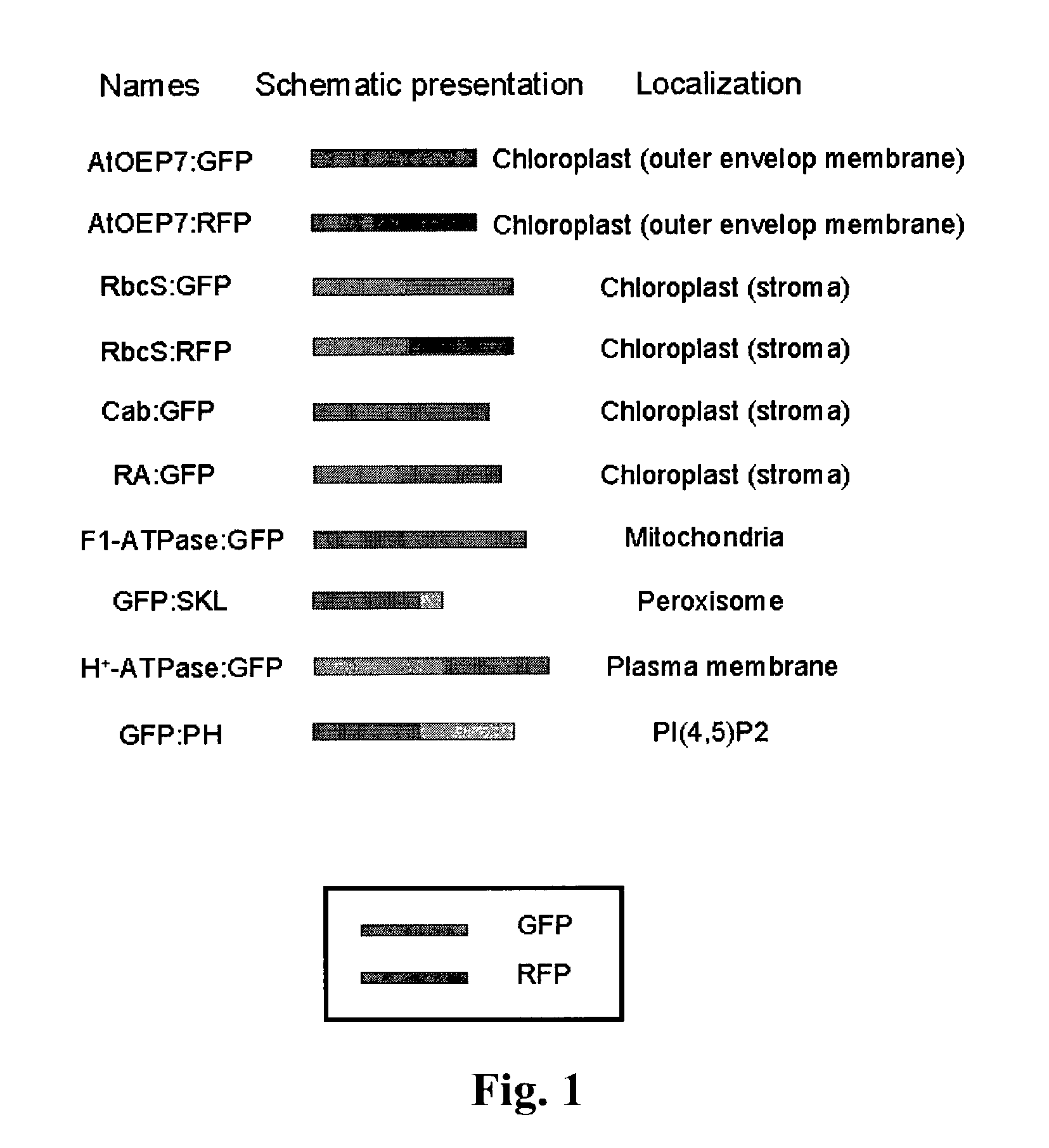
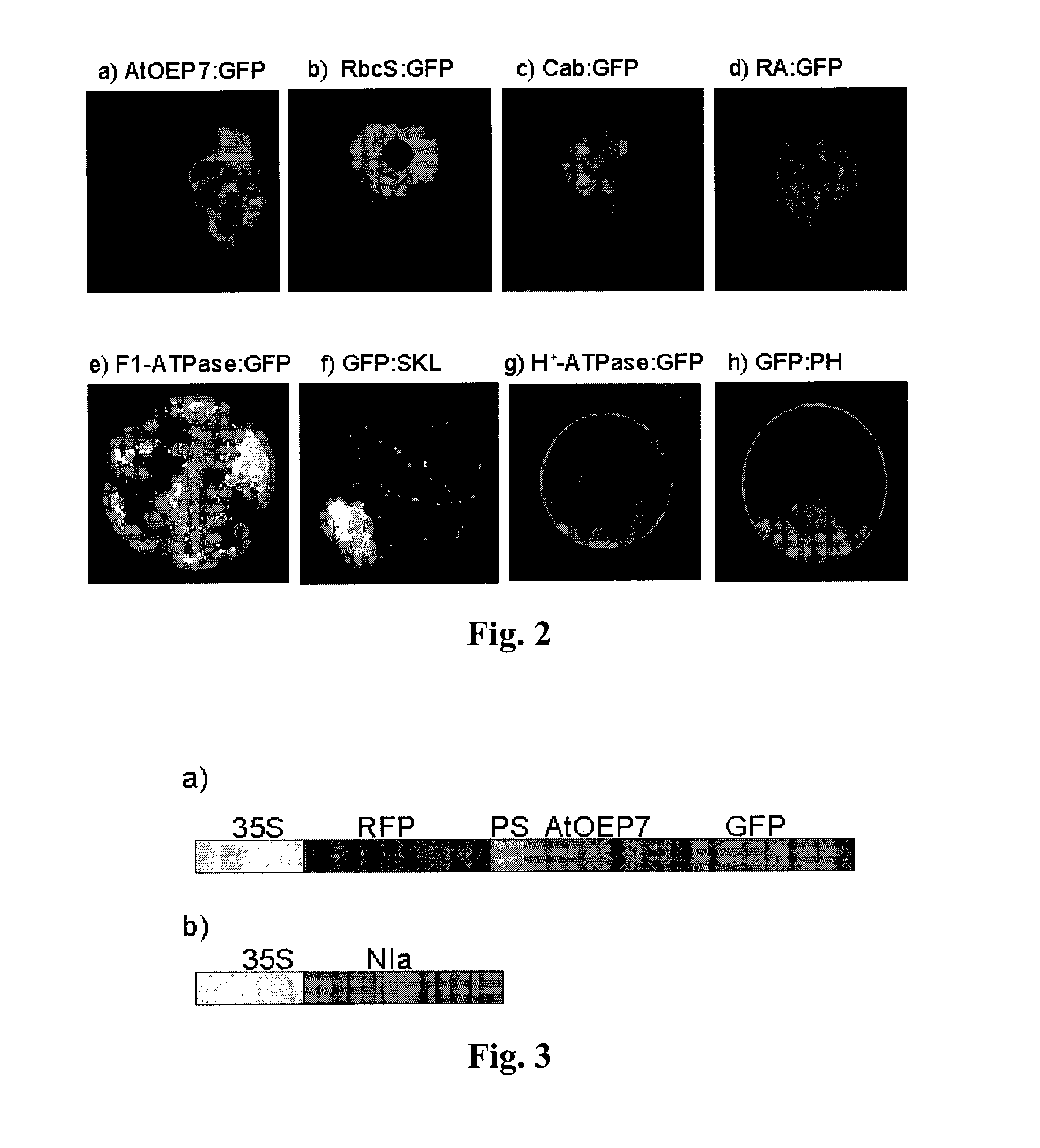
[0181]The coding region of the outer envelope membrane protein of Arabidopsis, AtOEP7, that is a homolog of OEP14 of pea, was amplified by polymerase chain reaction (PCR) from Arabidopsis genomic DNA using two specific primers (5′-GACGACGACGCAGCGATG and 5′-GGATCCCCAAACCCTCTTTGGATGT) designed to remove the natural termination codon. Then, it was ligated in frame to the 5′ end of the coding region of the green or red fluorescent protein to construct recombinant plasmids for AtOEP7:GFP and AtOEP7:RFP, respectively. The ligated genes were regulated by the 35S promoter in the recombinant plasmids. The same method was used for construction of other recombinant plasmids described hereafter.

[0182]For expression of the chimeric protein of Rubisco (ribulose bisphospate carboxylase) complex protein, the coding region for the transit p...

example 2

Detection of Cleavage of the Chimeric Substrate Protein by Protease

[0202](a) Construction of the Recombinant Plasmids

[0203]The recombinant plasmid for NIa protease was constructed by placing the coding region of NIa protease under the control of the 35S promoter in a pUC vector.

[0204]The recombinant plasmid for Arabidopsis outer envelope membrane protein:green fluorescent protein (AtOEP7:GFP) was constructed by ligating the AtOEP7 coding region without the termination codon to the 5′ end of the coding region of the green fluorescent protein in the 326GFP vector (obtained from Arabidopsis Biological Resource Center, Ohio University, USA). The cleavage site of the protease, VRFQ, was ligated to the N-terminus of AtOEP7:GFP by PCR amplification of this plasmid with two primers (5′ primer, 5′-CCCGGGGTGTGCGCTTCCAGGGAAAAACTTCGGGAGCG and 3′ primer, 5′-GAGCTCTTATTTGTATAGTTCATC). The PCR product (SmaI and XhoI fragment) was then ligated to HindIII (filled in) and XhoI sites of the 326RFP-nt ...

example 3

In vivo Screening System for HIV-1 Protease Inhibitors

[0214]A convenient in vivo screening system for detecting inhibitors of the human immunodeficiency virus (HIV-1) protease was performed as follows.

[0215](a) Construction of Recombinant Plasmids for Expression of HIV-1 Protease

[0216]To construct a recombinant plasmid for HIV-1 protease, the coding region of HIV-1 protease was PCR amplified with two primers: (5′-TCTAGAATGCCTCAGGTCACTCTTTGG-3′ and 5′-CTCGAGTCAAAAATTTAAAGTGCAACC-3′) using pHX2BΔRT as a template. The pHX-2BΔRT is a plasmid clone containing HX2B (GenBank accession number K03455) without the reverse transcriptase coding region. The amplified product was subcloned into pBluescript-T vector and subsequently cloned into XbaI and XhoI sites of a pUC vector under the control of the 35S promotor.

[0217]The plasmids maps for HIV-1 protease is shown in FIG. 9(a) and the nucleic acid and protein sequences are given in SEQ ID NOs: 55 and 56, respectively.

[0218](b) Construction of ...


PUM


Abstract

Disclosed is a system for detecting a protease inside a cell. In one embodiment, the system includes a chimeric protein that comprises as covalently linked components: 1) at least one optionally masked signal protein; 2) at least one protease-specific cleavage site; and 3) at least one detectable amino acid sequence. The invention has a wide spectrum of applications including use in the detection of novel protease inhibitors inside cells and tissue.

Description

CROSS-REFERENCE TO RELATED APPLICATION[0001]The present application is a continuation of Korean patent application No.10-2001-0048123 as filed on Aug. 10, 2001 and entitled A System For In Vivo Screening Of Protease Inhibitors, the disclosure of which is hereby incorporated by reference.FIELD OF THE INVENTION[0002]The present invention generally relates to system for detecting protease inside a cell. A preferred system includes a chimeric substrate protein that includes as covalently linked components: 1) at least one optionally masked signal protein; 2) at least one protease-specific cleavage site; and 3) at least one detectable amino acid sequence. The invention has a broad spectrum of important applications including use in screens to detect compounds that block proteases produced by one or more human pathogens.BACKGROUND[0003]Protease is an enzyme that cleaves a specific peptide bond of proteins. In biological organisms, proteases having specific proteolytic activities, and thei...

Claims


Application Information

Patent Timeline
IPC IPC(8): C07K14/00G01N33/50C07K19/00C12N5/10C12N15/09C12Q1/02C12Q1/37G01N21/76G01N33/15
CPCC07K19/00C12Q1/37G01N2500/04
Inventor HWANG, INHWANKIM, DAE HEONLEE, YONG JIK
Owner AHRAM BIOSYST
Learn More