
Intraocular drug delivery device and associated methods

a drug delivery and intraocular technology, applied in the field of ophthalmology, can solve the problems of retinal detachment, endophthalmitis, and other complications, and achieve the effects of reducing the risk of side effects, and improving the safety of patients

Active Publication Date: 2018-09-04
UNIV OF UTAH RES FOUND
View PDF211 Cites 1 Cited by
  • Summary
  • Abstract
  • Description
  • Claims
  • Application Information

AI Technical Summary

Benefits of technology

The present invention relates to a system, method, and device for delivering pharmaceutical active agents to the eye for the treatment of glaucoma, age-related macular degeneration, and other ophthalmic diseases. The invention provides a sustained and targeted delivery of the active agents to the eye, overcoming the limitations of current therapies that require multiple applications per day and often result in poor patient compliance. The invention also addresses the need for a more effective and efficient way to manage inflammation after surgery and reduce the risk of postoperative complications. The invention provides a device that can be implanted in the eye to deliver the active agents for an extended period of time, minimizing the need for frequent systemic adverse events and improving patient outcomes.

Problems solved by technology

These dosing regimens are often difficult for subjects to consistently follow, and many individuals progress to needing surgical treatments such as intraocular shunts or trabeculectomies, which have significant attendant complications.
Such injections are painful and may lead to retinal detachment, endophthalmitis, and other complications.
Furthermore, these injections are generally performed only by retinal surgeons, a small fraction of the ophthalmic community, producing a bottleneck in eye care delivery and increased expense.
Poor compliance compromises the efficacy of topical drugs, which are further limited by corneal absorption and have highly variable intraocular concentrations during the therapeutic course.

Method used


Image

  • Intraocular drug delivery device and associated methods
  • Intraocular drug delivery device and associated methods
  • Intraocular drug delivery device and associated methods

Examples


example 1

[0068]A standard clear-corneal phacoemulsification with intraocular lens (Acrysof SA60AT; Alcon) implantation was performed on 35 rabbits. At the time of each surgery, an intraocular device containing an active agent was inserted into a lens capsule of each rabbit. The rabbits were divided into 4 groups, depending on the active agent in the intraocular device. Devices were loaded with 5-15 mg of either Avastin, Timolol, Brimonidine, or Latanoprost. Each group was evaluated to determine the intraocular device and lens stability, capsular fibrosis, and healing of cataract wounds and anterior segment. A subgroup of eyes was evaluated weekly for 4 weeks for inflammation and harvested at 1 month for histopathologic evaluation of capsular and CDR integrity.

example 2

[0069]The surgery and setup as described in Example 1 was repeated, with the exception that aqueous and vitreous taps were performed biweekly and assayed for drug concentrations with HPLC and / or ELISA. In each drug group, half of the eyes were harvested at one month and the other half at two months. This was accomplished as follows: immediately after sacrificing the rabbit and enucleating the eye, the eye was frozen in liquid nitrogen to prevent perturbation and redistribution of drug in eye tissues. The eye was then dissected into 3 parts (aqueous humor, vitreous and retina / choroid layer) to evaluate anatomic toxicity and tissue drug concentration. The intraocular device was retrieved and assessed for remaining drug amounts. The distribution profile of the intraocular device was compared with the conventional intravitreal injection of 2.5 mg / 0.1 cc Avastin® for direct comparison of the different delivery methods.

[0070]At 2 and 4 months, eyes from the remaining subgroups of rabbits ...

example 3

[0071]Three intraocular devices were implanted into eyes of New Zealand white rabbits under general anesthesia after lens extraction (phacoemulsification technique). Two of the devices were loaded with Avastin and one was loaded with the contrast agent Galbumin as a control. Proper intraocular device position was verified by MRI as well as clinical examination.

[0072]The rabbits were sacrificed and the eyes are removed and assayed after 1 week post implantation. Avastin was detected by ELISA in the retina and vitreous at concentrations of 24-48 mcg / mL, and was not present in the control rabbit eye. FIG. 5 shows the amount of Avastin assayed per ocular region at 1 week post implantation.


PUM

PropertyMeasurementUnit
Timeaaaaaaaaaa
Timeaaaaaaaaaa
Thicknessaaaaaaaaaa

Abstract

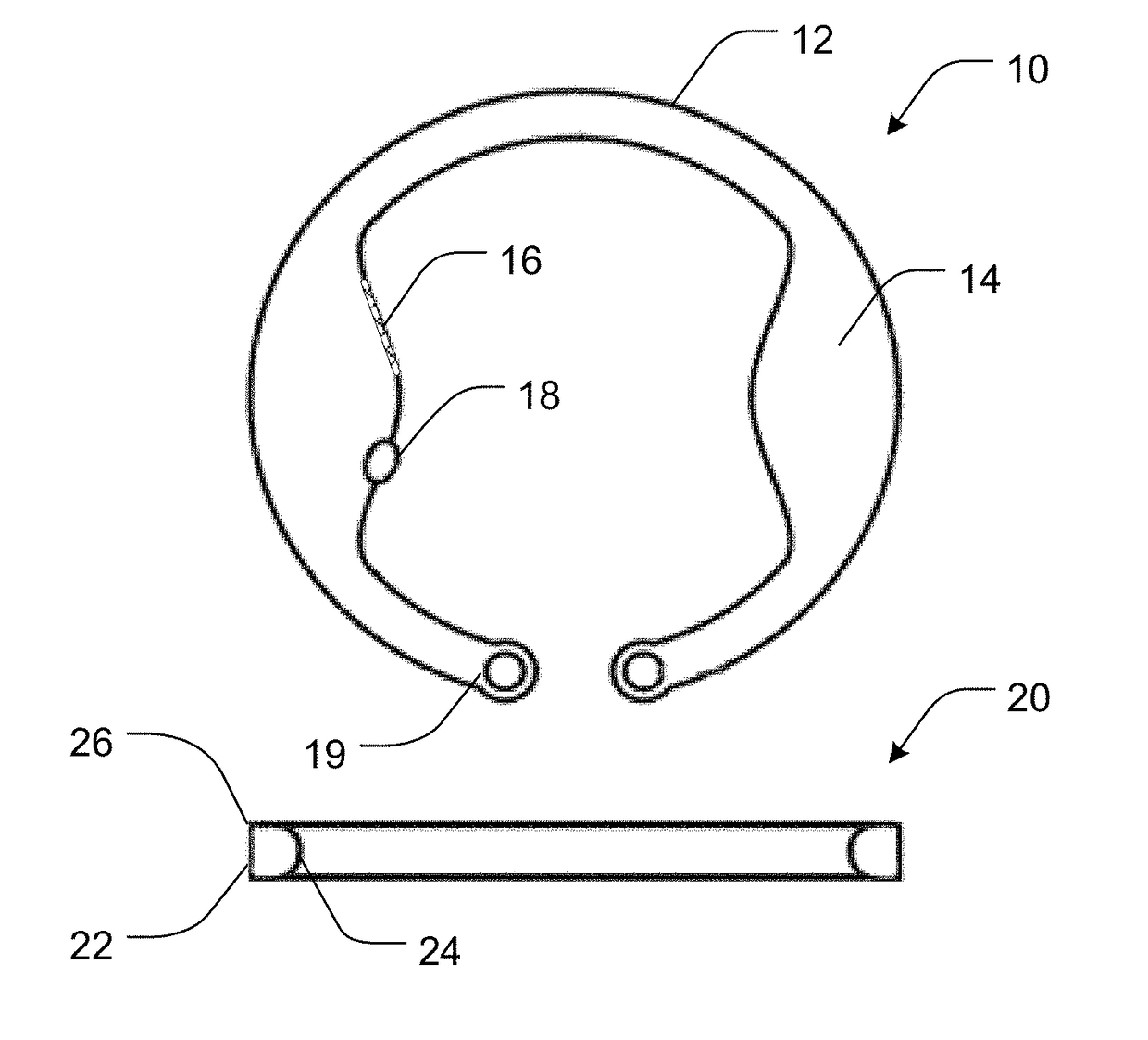
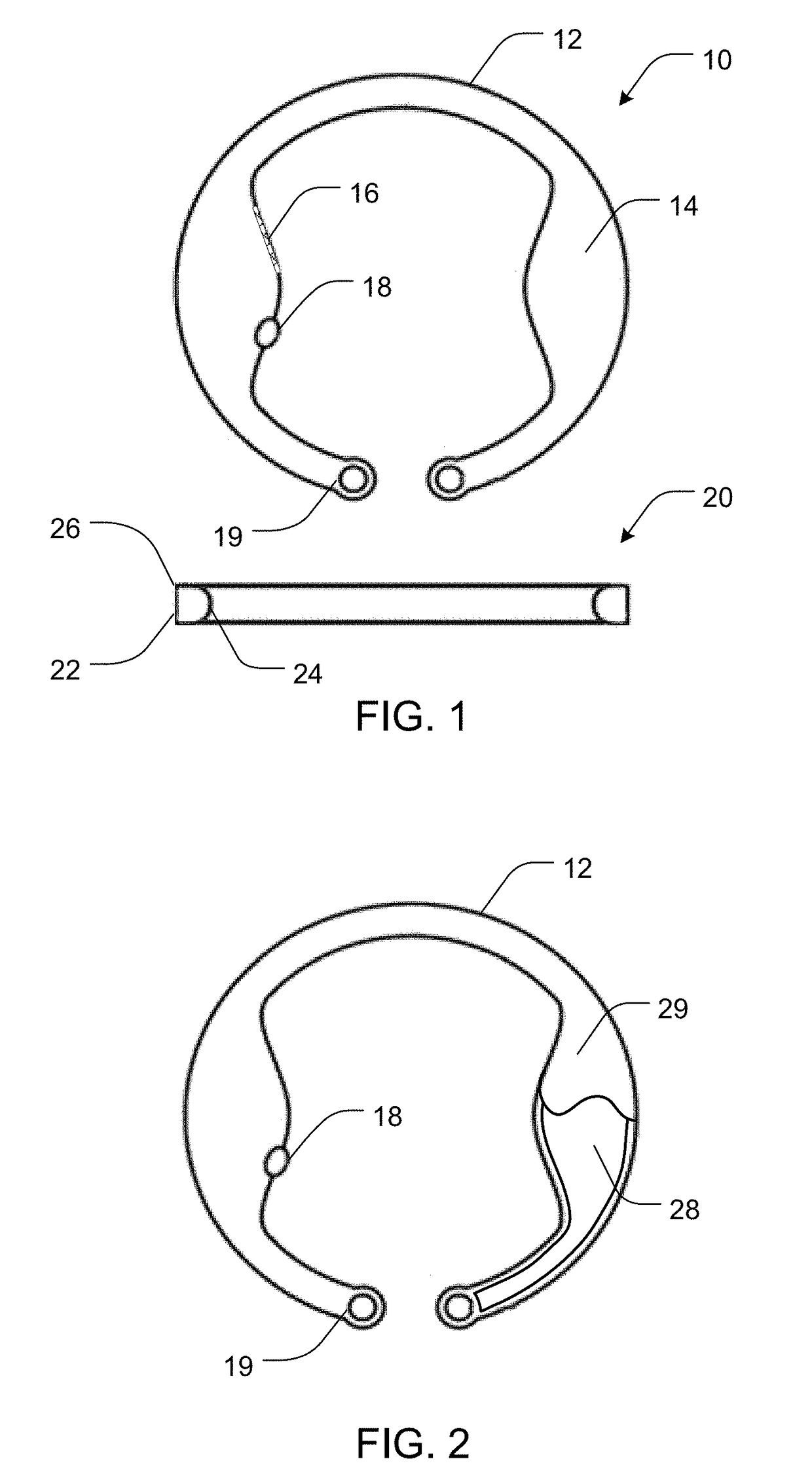
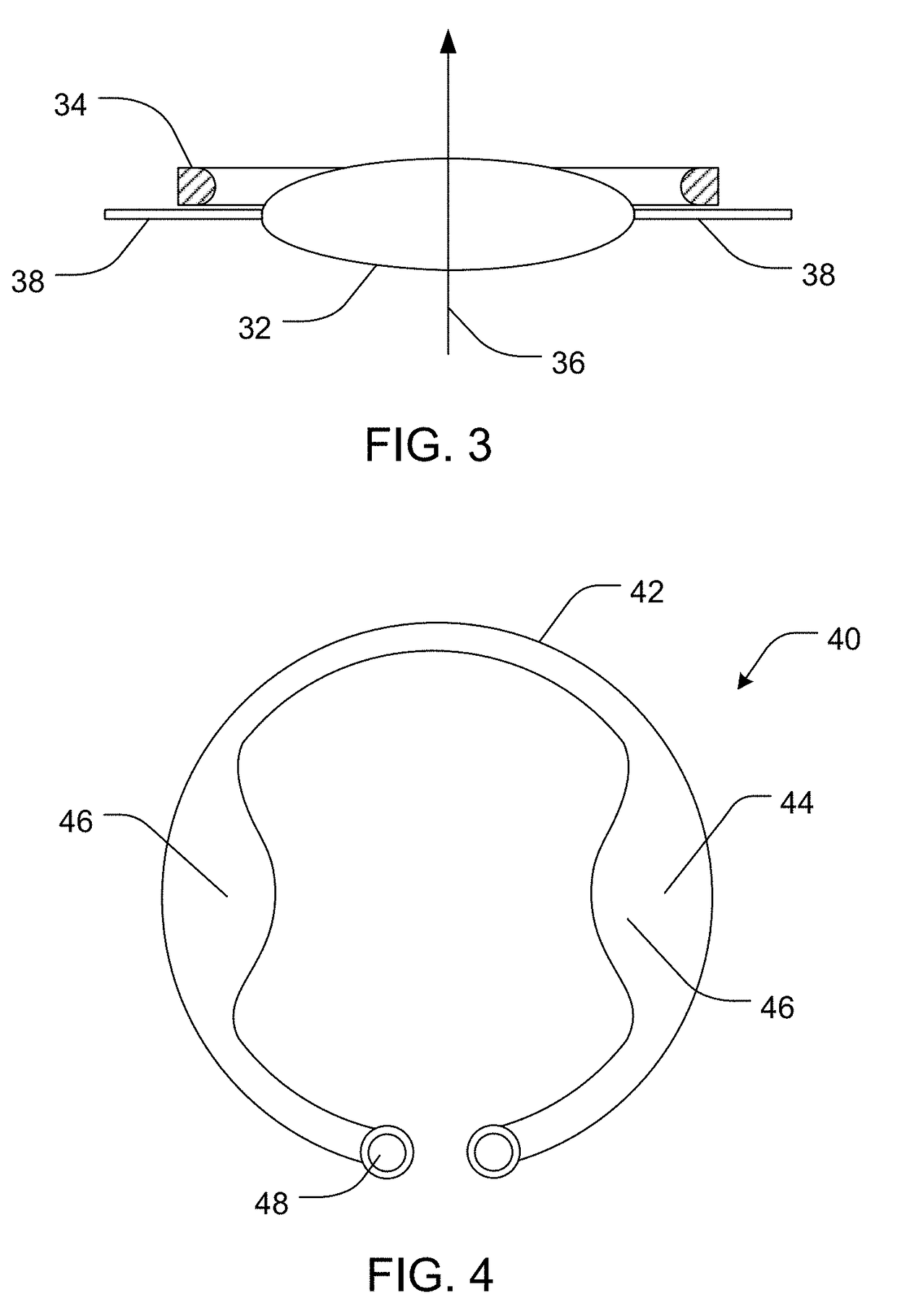
Devices, systems, and methods for delivery of an active agent into the eye of a subject can include an intraocular active agent delivery device including an active agent dispersed within a biodegradable active agent matrix. The active agent includes dexamethasone and the delivery device is adapted to fit within a lens capsule or ciliary sulcus of an eye. The delivery device can be inserted into the lens capsule or ciliary sulcus of an eye during cataract surgery or for treatment of uveitis.

Description

RELATED APPLICATIONS[0001]This application is a continuation-in-part of U.S. application Ser. No. 13 / 211,169, filed Aug. 16, 2011, which is a continuation-in-part of U.S. application Ser. No. 12 / 945,428, filed Nov. 12, 2010, which is a continuation-in-part of International Application No. PCT / US2009 / 043566, filed May 12, 2009, which claims priority to U.S. Provisional Application No. 61 / 052,507, filed May 12, 2008, which are each incorporated herein by reference.FIELD OF THE INVENTION[0002]The present invention relates to systems, methods, and devices for the sustained and targeted (local) delivery of a pharmaceutical active agent into a subject's eye. Accordingly, the present invention involves the fields of polymer chemistry, material science, polymer science, drug delivery, formulation science, pharmaceutical sciences, and medicine, particularly ophthalmology.BACKGROUND[0003]Age-related macular degeneration (AMD) and glaucoma are two of the leading causes of blindness in the Unit...

Claims


Application Information

Patent Timeline
IPC IPC(8): A61M31/00A61K9/20A61K9/00A61K31/573
CPCA61K9/0051A61K31/573A61K9/2031A61K9/0017
Inventor AMBATI, BALAMURALI K.GALE, BRUCE K.CHENNAMANENI, SRINIVAS RAO
Owner UNIV OF UTAH RES FOUND
Learn More