
Patient-specific temporary implants for accurately guiding local means of tumor control along patient-specific internal channels to treat cancer

a technology for cancer and local control, applied in the direction of catheters, ultrasonic/sonic/infrasonic image/data processing, therapy, etc., can solve the problems of limited radiation intensity, large amount of energy delivered, adverse side effects of use, etc., and achieve the effect of convenient design and printing

Active Publication Date: 2019-05-14
RGT UNIV OF CALIFORNIA
View PDF15 Cites 4 Cited by
  • Summary
  • Abstract
  • Description
  • Claims
  • Application Information

AI Technical Summary

Benefits of technology

[0104]An advantage of the present invention is the ability to register the delivery of the effects of the tissue ablation means with the region of diseased tissue. Moreover, the device of the invention can be designed with reference to data acquired from the subject through one or more diagnostic imaging modality (FIG. 9). One or more of the designs of the device, proper placement of the device and calculation of the dosage of the tissue ablation means delivered to both diseased and normal tissues by the device is readily correlatable with diagnostic images acquired from the subject. Moreover, the width, length arc and position of the at least one curvature constrained channel can be determined using such imaging data (FIG. 7, FIG. 8). These images can be taken either or both before and after installation of the device. The invention provides a device allowing the ready registration tissue ablation means with the region of diseased tissue using one or more imagable fiducial marker incorporated into the device. In an exemplary embodiment, the device and its incorporated fiducial marker are configured for registration between at least one region of anatomy of the subject and at least one diagnostic image of the region of anatomy. In an exemplary embodiment, the one or more region of anatomy is selected from a region of diseased tissue and a region of normal tissue proximate to the area of normal tissue.
[0105]As will be appreciated by those of skill in the art, the imaging modality of use in the present invention is any modality capable of providing useful data regarding the fiducial marker and the region of anatomy of the subject. Exemplary modalities include, without limitation, MRI, CT, gamma camera scintigraphy, PET, ultrasonography and a combination thereof. Thus, the invention provides a device in which the fiducial marker is imageable by MRI, CT, gamma camera scintigraphy, PET, ultrasonography or a combination thereof.
[0106]In an exemplary embodiment, the device of the invention is substantially solid with the exception of the curvature constrained channels, and other channels or compartments that contain various materials, e.g., shielding materials. The invention, however also encompasses light weight devices that are substantially hollow in which the curvature constrained channels and other channels and compartments are luminal structures disposed within a device body that, with the exception of these channels and compartments, is substantially hollow.
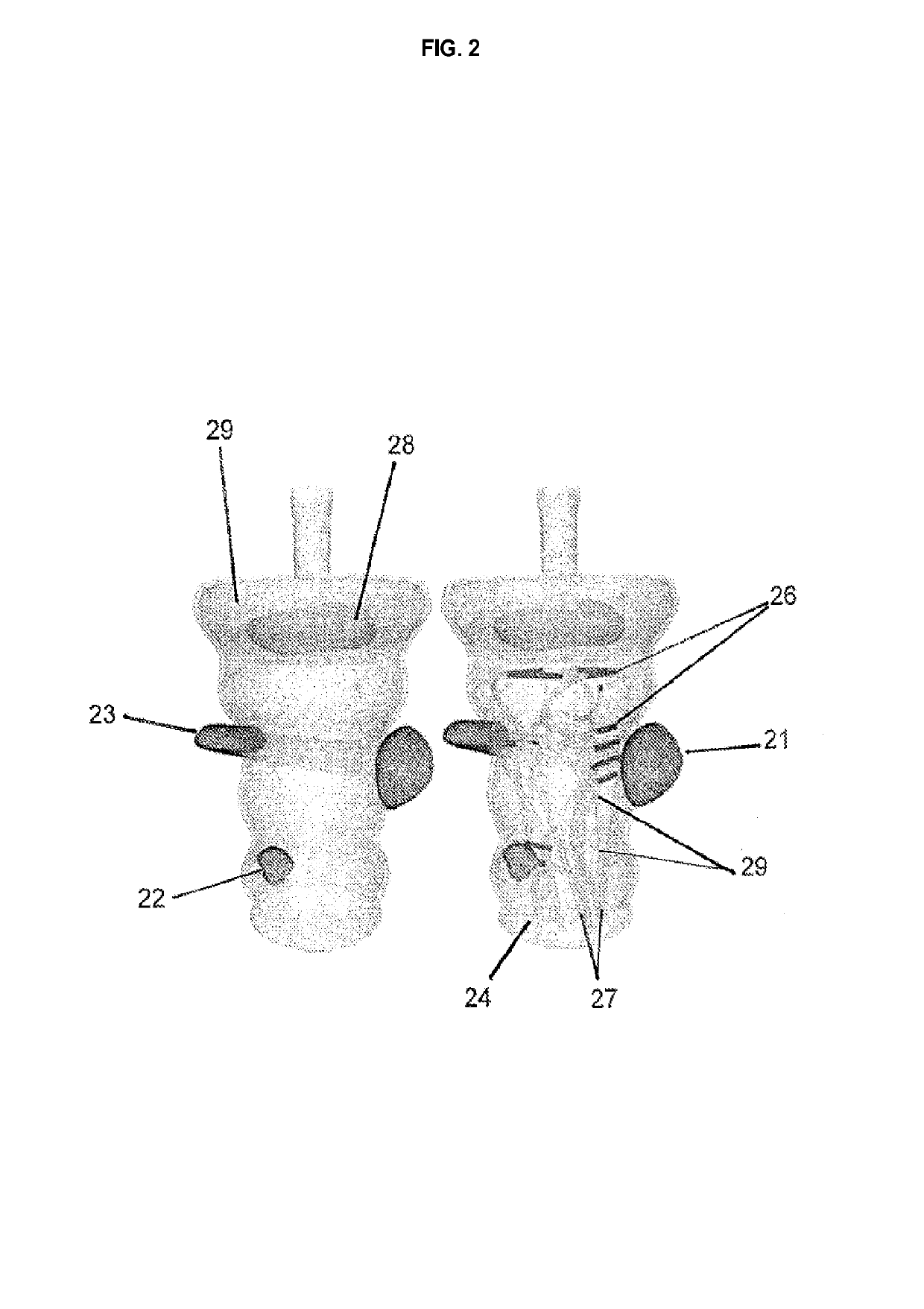
[0107]The at least one local means of tumor control is located at any useful point or region within the device. In an exemplary embodiment, the at least one local means of tumor control is disposed at a position within at least one curvature constrained channel, forming at least one dwell point. Various devices of the invention include two or more dwell points in registration with one or more regions of diseased tissue within said patient upon implantation of the customized device of the invention (FIG. 2, 15B). The device optionally includes one or more shielding regions adjacent to a dwell point. The device further optionally includes one or more fiducial markers providing an imageable means for determining the position of the means of tissue ablation.
[0108]In various embodiments, the device of the invention is not intended for implantion but rather to be overlaid on a region of a subject's body for the treatment of tumors on or below the skin. In various embodiments, the curved channels discussed in the context of the impantable device are also a component of the overlaid device. In various embodiments, the curved channels are designed such that they place a local means of tumor control sufficiently proximal to a tumor to ablate the diseased tissue and, in a preferred embodiment, provide therapeutic advantage. The other design and structural elements discussed in the context of the implantable device, e.g., local means of tumor control source, shielding and the like can be components of the overlaid device. As will be apparent to one of skill in the art, in some embodiments, it may be efficacious for the device to have a combination of implantable and overlaid regions and it can be readily designed and printed to have any desirable combination of such regions.The Methods
[0109]In addition to the devices disclosed herein, the invention provides a method of treating a region of diseased tissue using one or more devices of the invention. For example, the invention provides a method of treating a disease in a patient by implanting a device of the invention in a body cavity of the subject such that a therapeutically effective dose of a local means of tumor control contained within the device is delivered to a locus of the disease from at least one dwell point in the device. In an exemplary embodiment, the disease is a neoplastic disease. In various embodiments, local means of tumor control is a source of ionizing radiation.

Problems solved by technology

The higher temperatures required by thermotherapy require delivery of large amounts of energy to the target tissues.
Chemotherapy involves the administration of various anti-cancer drugs to a patient but due to the requirement that it be administered systemically, its use is accompanied by adverse side effects.
Methods employing beams of photons or other sub-atomic or atomic particles generated outside the body and penetrating into the body and tumor location (external beam) can accumulate radiation at specific internal points, but radiation intensity is limited by the dose delivered to intervening non-cancerous tissue.
A problem with needles is that they are difficult to place precisely, their paths are limited to primarily linear forms, and that radiation is emitted uniformly in all directions when the seed is stationary. FIG. 10 displays representative conventional brachytherapy devices.

Method used


Image

  • Patient-specific temporary implants for accurately guiding local means of tumor control along patient-specific internal channels to treat cancer
  • Patient-specific temporary implants for accurately guiding local means of tumor control along patient-specific internal channels to treat cancer
  • Patient-specific temporary implants for accurately guiding local means of tumor control along patient-specific internal channels to treat cancer

Examples


example 1

[0137]Automation science has been applied to a number of healthcare applications to improve quality of treatment by improving repeatability and reliability. Huang et al. (Y. Y. Huang and K. H. Low, “Comprehensive planning of robotic therapy and assessment of task-oriented functions via improved {QFD} applicable to hand rehabilitation,” in Automation Science and Engineering (CASE), 2010 IEEE Conference on, pp. 252-257, IEEE, 2010) studied planning of robotic therapy and assessment of task-oriented functions for hand rehabilitation. Tervo et al. (K. Tervo, L. Palmroth, and H. Koivo, “Skill Evaluation of Human Operators in Partly Automated Mobile Working Machines,”IEEE Transactions on Automation Science and Engineering, 7:133-142, January 2010) and Solis et al. (J. Solis and A. Takanishi, “Towards enhancing the understanding of human motor learning,” in 2009 IEEE International Conference on Automation Science and Engineering, pp. 591-596, IEEE, August 2009) explored the use of automati...

example 2

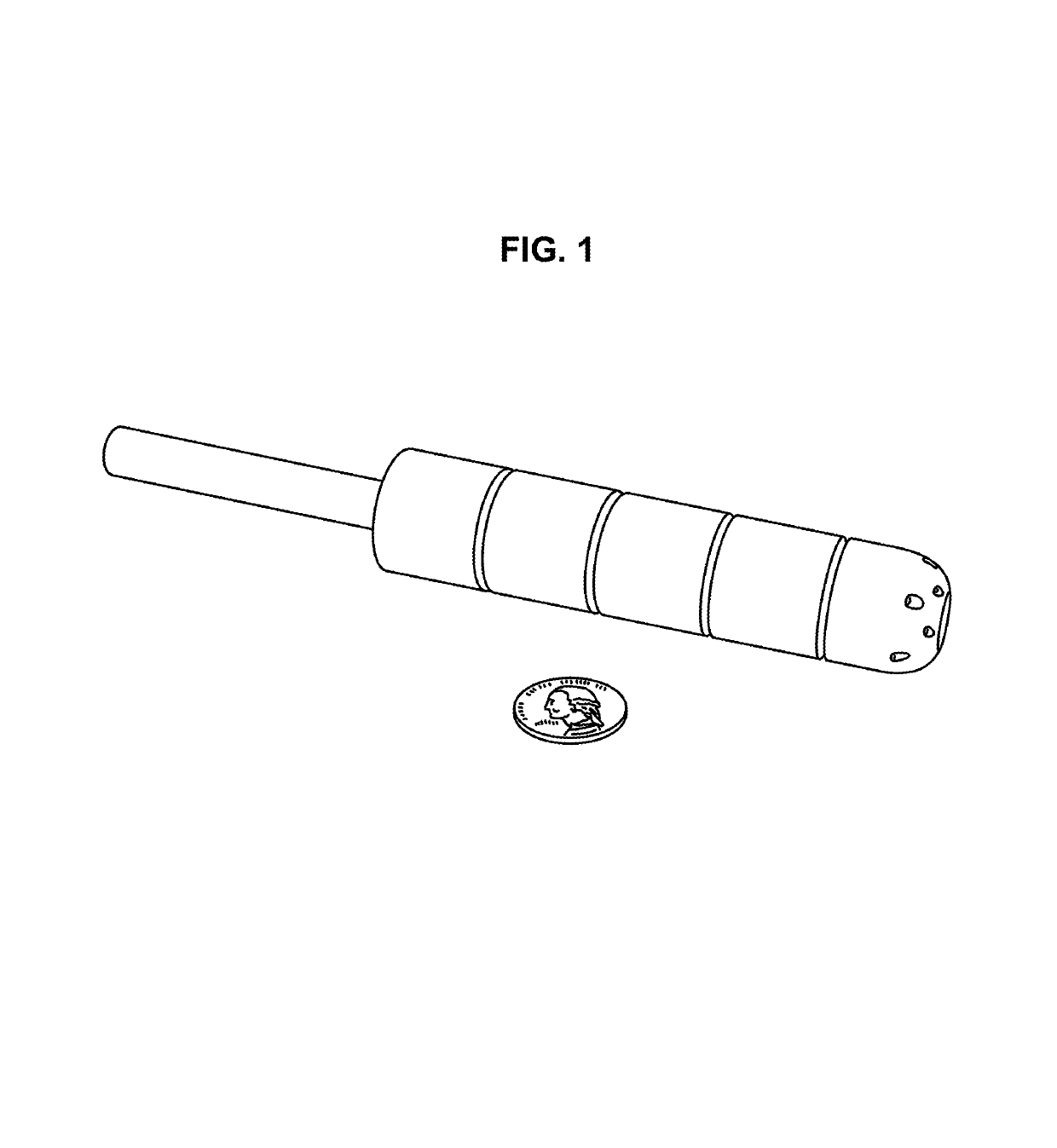
This Example illustrates the 3D printing of an exemplary device of the invention . . . . The patient had cancerous tumors on cervix and vaginal wall.

I. Patient Scan and Physician Contouring

[0184]Anonymized patient data from the UCSF patient database was used for this study. The patient, Anon1, was scanned with CT-Scan and found to have cervical cancer. In the clinic, Anon1 was treated using a commercially available, non-customized (standard shaped) Interstitial Ring. In this case study an alternative approach is developed which replaces the Interstitial Ring with a custom implant that is contoured to patient anatomy and includes curved interior channels that can guide radioactive sources to deliver radiation closer to tumors and with more accuracy than with standard Interstitial Ring methods.

[0185]Anon1 had been scanned using a CT-scanner and anatomical images were generated as slices in axial, coronal, or sagittal planes. (The CT-Scanner usually takes one helical scan which is then...

example 3

Clinical Applications of Custom-Made Vaginal Cylinders Constructed Using 3D Printer

[0234]Purpose:

[0235]3D printing technology allows physicians to rapidly create highly customized devices for patients. This technology has already been adapted by various fields in medicine. We report a proof of concept and first clinical use of this technology for vaginal brachytherapy.

[0236]Introduction:

[0237]There are currently many medical applications of 3D printing in development, for example, medical modeling for maxillofacial surgical management (Chow, et al., Journal of Oral and Maxillofacial Surgery, 65(11):2260-2268, 2007; Anchieta, et al., Advanced Applications of Rapid Prototyping Technology in Modern Engineering. Rijeka, Croatia: InTech, pp. 153-72, 2011), bone reconstructions (Cohen, et al., Oral Surgery, Oral Medicine, Oral Pathology, Oral Radiology, and Endodontology, 108)(5):661-666, 2009; Schrank, et al., Journal of Biomechanical Engineering, 135(1):101011, 2013), and oral surgeries...


PUM

PropertyMeasurementUnit
temperatureaaaaaaaaaa
δaaaaaaaaaa
diameteraaaaaaaaaa

Abstract

The present invention offers an alternative for cancer treatment where radiation, thermotherapy, or another therapeutic modality must be delivered to an internal cavity of a subject, for example to treat mouth, anal, cervical, and vaginal cancers. The invention is a new approach that builds on recent results in 3D printing and steerable needle motion planning to create customized implants containing customized curvature-constrained internal channels that fit securely, minimize air gaps, and precisely guide treatment sources through internal printed channels to accurately reach tumors and minimize damage to healthy tissue.

Description

CROSS-REFERENCE TO RELATED APPLICATIONS[0001]This application claims under 35 USC 119(e) the benefit of U.S. Provisional Application No. 61 / 859,096, filed Jul. 26, 2013, and U.S. Provisional Application No. 61 / 859,687 filed Jul. 29, 2013, each of which is incorporated herein by reference in its entirety for all purposes.STATEMENT AS TO RIGHTS TO INVENTIONS MADE UNDER FEDERALLY SPONSORED RESEARCH OR DEVELOPMENT[0002]This invention was made with Government support under Grant No. 0905344 and Grant No. IIS-1227536 awarded by the NSF. The Government has certain rights in the invention.FIELD OF THE INVENTION[0003]The technology relates generally to the focused delivery of therapy such as phytotherapy, cryosurgery, thermotherapy, radiation therapy, and chemotherapy to regions of diseased tissue as exemplified by devices for brachytherapy and methods of using and making such devices.[0004]The present invention offers an alternative for cases where radiation, thermotherapy, or another thera...

Claims


Application Information

Patent Timeline
Patent Type & Authority Patents(United States)
IPC IPC(8): A61M31/00A61B6/03A61N5/06A61N5/10A61B8/08A61B18/18A61N7/02B29C64/20B29C64/112A61F7/12A61B90/00B33Y80/00A61B18/02A61B18/00A61B18/14B33Y10/00B29K101/12B29L31/00
CPCA61M31/002A61B6/037A61B8/085A61B18/1815A61N5/0603A61N5/1014A61N7/022A61B6/032B33Y80/00A61B18/14A61B2018/00559A61B2018/00577A61B2018/00791A61B2018/0212A61B2018/1861A61B2090/3983A61F2007/126A61M2207/00A61N5/062A61N5/1016A61N5/1049A61N2005/0606A61N2005/0608A61N2005/0611A61N2005/1018A61N2005/1094B29C64/112B29C64/20B29K2101/12B29K2995/0056B29L2031/753B33Y10/00
Inventor POULIOT, JEANGOLDBERG, KENHSU, I-CHOWCUNHA, J. ADAM M.GARG, ANIMESHPATIL, SACHINABBEEL, PIETERSIAUW, TIMMY
Owner RGT UNIV OF CALIFORNIA
Learn More