
Method, learning apparatus, and medical imaging apparatus for registration of images

a learning apparatus and image registration technology, applied in the field of learning apparatus and medical imaging apparatus for image registration, can solve the problems of difficult registration between images acquired using different imaging modes/apparatus, soft tissue structures that are observable in magnetic resonance images may not be visible (or not easily observable), and the ability of domain classifier to reduce the ability

Active Publication Date: 2020-11-10
SIEMENS HEALTHCARE GMBH +1
View PDF16 Cites 2 Cited by
  • Summary
  • Abstract
  • Description
  • Claims
  • Application Information

AI Technical Summary

Benefits of technology

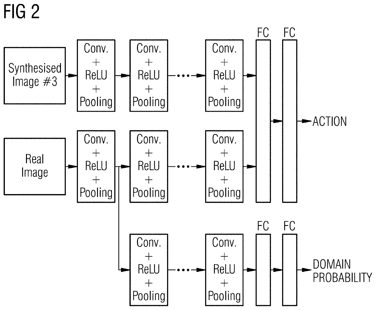
[0016]A second training operation is executed using the second and third source image data without using labels associated therewith, in which the computer system is trained to determine a transformation between respective coordinate frames of the view of the imaged structure and the view of the synthesized structure, such that the ability of the domain classifier to discriminate between the synthesized structure and the imaged structure is reduced.
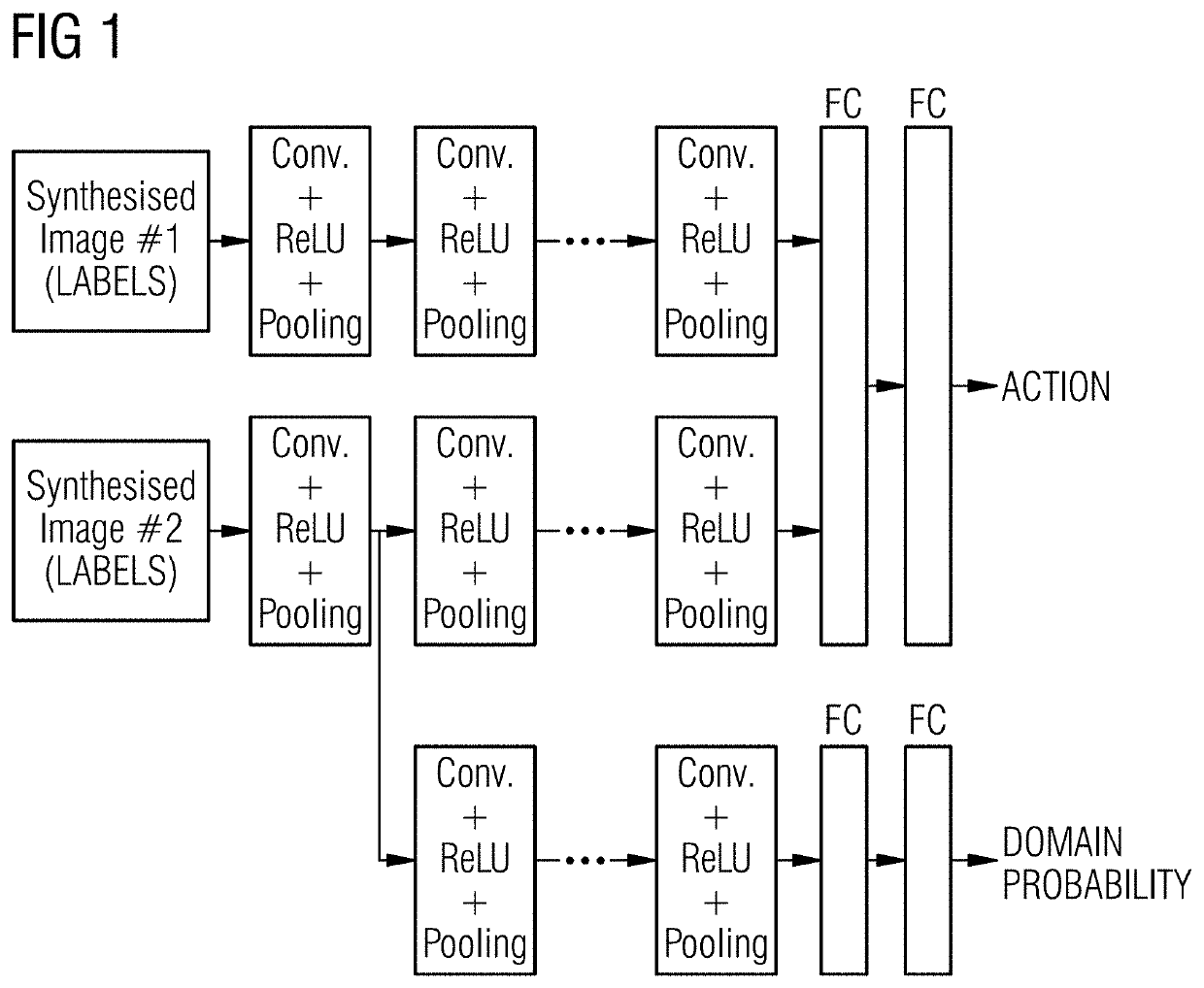
[0020]In this way, the invention provides a method of training a learning agent to identify suitable transformations between different synthesized views of a model of a structure, such as an anatomical structure (e.g. an organ, muscle, bones, vasculature etc.) of a human body. These synthesized views are labeled image data. It is very easy to rapidly produce and correctly label large quantities of images of the labeled structure in many different views. This labeled, synthesized image data are used to train the learning agent in the first training operation, so that the learning agent is increasingly able to identify transformations between labeled, synthesized image data. Importantly, in the process of this first training operation, the feature map (or maps) of the learning agent is (are) trained appropriately to identify the relevant image features useful for determining transformations.
[0022]During the second training operation the learning agent is trained in such a way that the agent is deemed more optimal when the domain classifier comes less able to discriminate between the synthesized second source data and the “real” third source data. The aim and goal of this is to train the learning agent to employ domain-agnostic features of the second and third image data for the purposes of determining the transformation between corn frames of image data. This means that the image features from within images that are employed for that purpose, will be less likely to contain synthetic artefacts of the model used to synthesize the second image data, and will be more likely to be an anatomical feature common to both the synthetic second images and the “real” third images. This means that fully labeled synthetic image data (the first image data) can be used to train the learning agent without the need to use “real” training images that have been pre-labeled manually.
[0023]The domain classifier may be configured to generate from said feature map a probability estimate that received image data is one of the third source image data (a first domain) or the second source image data (a second domain). Preferably, the second training operation includes training the computer system to determine the transformation between respective coordinate frames of the view of the imaged structure and the view of the synthesized structure, such that the probability estimate approaches a value of 0.5 thereby reducing the ability of the domain classifier to discriminate between the synthesized structure and the imaged structure.

Problems solved by technology

The registration between images acquired using different imaging modes / apparatus is challenging because structures observable in an image acquired using one imaging mode / apparatus of imaging may appear to be different (or not visible at all) in an image acquired using a different imaging mode / apparatus.
As an example, soft tissue structures that are observable in a magnetic resonance image may not be visible (or not easily observable) in an X-ray fluoroscopy image.
Finding a transformation between two image datasets that were acquired using fundamentally different imaging modes / apparatus can therefore be challenging.
In this procedure, differences between preoperative and intraoperative image data can be significant.
In contrast, X-ray fluoroscopy performed intraoperatively may have a wider field of view and show dense structure, such as bones or instruments, but may not show the soft tissue anatomy clearly.
This may require a change in clinical procedures and may also require additional imaging hardware in the operating room.
However, there are a number of challenges in achieving accurate and robust registration between imaging modalities.
Such data is difficult to obtain for certain combinations of imaging modalities.
Obtaining such data sets is a challenge.

Method used


Image

  • Method, learning apparatus, and medical imaging apparatus for registration of images
  • Method, learning apparatus, and medical imaging apparatus for registration of images
  • Method, learning apparatus, and medical imaging apparatus for registration of images

Examples


Embodiment Construction

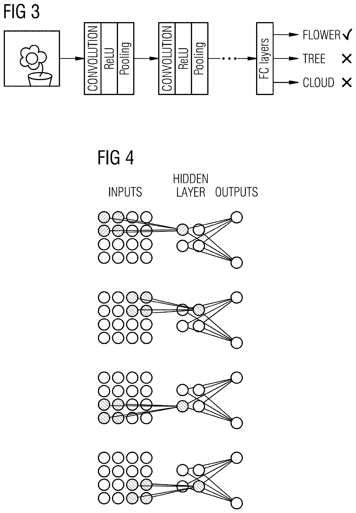
[0063]FIG. 3 schematically illustrates a convolutional neural network (CNN) as a machine learning algorithm used for deep learning. This CNN is specifically arranged for image data as input. CNNs differ from other types of neural networks in that the neurons in a network layer of a CNN are connected to sub-regions of the network layers before that layer instead of being fully-connected as in other types of neural network. The neurons in question are unresponsive to the areas outside of these sub-regions in the image.

[0064]These sub-regions might overlap, hence the neurons of a CNN produce spatially-correlated outcomes, whereas in other types of neural networks, the neurons do not share any connections and produce independent outcomes. In a neural network with fully-connected neurons, the number of parameters (weights) may increase quickly as the size of the input image increases.

[0065]A convolutional neural network reduces the number of parameters by reducing the number of connectio...


PUM


Abstract

A method of training a computer system for use in determining a transformation between coordinate frames of image data representing an imaged subject. The method trains a learning agent according to a machine learning algorithm, to determine a transformation between respective coordinate frames of a number of different views of an anatomical structure simulated using a 3D model. The views are images containing labels. The learning agent includes a domain classifier comprising a feature map generated by the learning agent during the training operation. The classifier is configured to generate a classification output indicating whether image data is synthesized or real images data. Training includes using unlabeled real image data to training the computer system to determine a transformation between a coordinate frame of a synthesized view of the imaged structure and a view of the structure within a real image. This is done whilst deliberately reducing the ability of the domain classifier to discriminate between a synthesized image and a real image of the structure.

Description

BACKGROUND OF THE INVENTIONField of the Invention[0001]The present invention concerns a method for determining a correspondence transformation between coordinate frames of sets of image data. In further aspects, the present invention concerns a medical imaging apparatus and a non-transitory, computer-readable data storage medium that implements such a method. In particular the invention is concerned with a method of training a computer system, according to a machine learning algorithm, for determining a correspondence transformation between coordinate frames of sets of image data.Description of the Prior Art[0002]For medical applications that involve preoperative planning and intraoperative guidance, there is often benefit in being able to determine a transformation between a coordinate frame of an image acquired preoperatively and a coordinate frame of an image take intraoperatively. Such a determination of a transformation is often referred to as registration.[0003]The registratio...

Claims


Application Information

Patent Timeline
Patent Type & Authority Patents(United States)
IPC IPC(8): A61B34/37G06T11/00G06N3/08G06N3/04G06N20/20G06T7/00
CPCG06N3/08G06T7/0002G06N3/0454G06T11/003G06N20/20G06T2207/20084G06T2207/20081G06T2210/41G06T7/30G06T2207/10116G06N3/045G06N3/006G06N3/084G06T7/33G06T2207/10072G06T2207/20221G06T2207/30004G06N3/047G06N3/048
Inventor CECCALDI, PASCALKURZENDORFER, TANJAMANSI, TOMMASOMOUNTNEY, PETERPIAT, SEBASTIENTOTH, DANIEL
Owner SIEMENS HEALTHCARE GMBH
Learn More