Prosthetic mitral valve coaptation enhancement device

a technology of prosthetic mitral valve and enhancement device, which is applied in the field of implantable prosthetic mitral valve coaptation enhancement device, can solve the problems of reducing the heart's pumping efficiency, reducing the efficiency of heart pumping, and causing backward flow of valve blood, so as to minimize the gradient between the left atrium and the ventricle, reduce or even eliminate the effect of mitral regurgitation

Active Publication Date: 2021-01-12
MEDIRA AG
View PDF30 Cites 2 Cited by
  • Summary
  • Abstract
  • Description
  • Claims
  • Application Information

AI Technical Summary

Benefits of technology

[0021]With the prosthetic mitral valve coaptation enhancement device according to the invention, the space left by malcoaptation of the native mitral leaflets can filled out, and, thus, mitral regurgitation can be reduced or even eliminated when placing the device according to the invention inside the annulus of a native mitral valve at the respective coaptation line.
[0022]With the dimensions of the presently described device being smaller than a native mitral annulus, the disclosed prosthetic mitral valve coaptation enhancement device allows filling of the ventricle through the device and alongside the device during diastole, and thus minimizes the gradient between the left atrium and ventricle.
[0027]With the device according to the invention, a unidirectional flow is created, preventing flow from the ventricle to the atrium in systole and permitting flow from the atrium to the ventricle in diastole: The leaflets of the native mitral valve coapt against the implanted device according to the invention, thus sealing the mitral annulus during systole and recreating the valvular function of the structure. With the lower radial rigidity in the sealing section compared to the valve-bearing section, in connection with a circular. D- or crescent shape of the proximal end, it is guaranteed that the space left between the two malcoapting mitral leaflets is filled out without expanding the space and without forcing the not properly closed valve even further open.
[0044]According to a preferred embodiment, the valve-element, upon implantation of the device, is sized and configured such, that the native mitral valve's function is supported without replacing or impairing the native mitral valve's function.
[0054]This embodiment has the advantage that interference or contact with the subvalvular mitral apparatus (chordae tendinae, papillary muscles) and the left ventricle can be avoided in difficult anatomies.
[0068]In this connection, a “bar” element or “ligament” element or means is any lengthy structure of sufficient rigidity and flexibility for being coupled, via its one end, to the device according to the invention, and, via its second end, to a structure of the heart and / or to another anchoring element, e.g. a plug. The lengthy structure / bar / ligament element can be of any material suitable for these purposes, and is preferably of an inert, substantially rigid material, such as, metals, e.g. Nitinol, stainless steel. Titanium, polymers, e.g. polyether ether ketone (PEEK), polyoxymethylene (POM), polyether (PE), polyamide (PA), polytetrafluoroethylene (PTFE), which materials may also be reinforced by fibres to improve stability), ceramics, materials of animal or human origin or generally synthetic materials. The structures may also be substantially hollow structures, which are inflatable or fillable to attain their stability. They may also be combinations of the aforementioned. To promote the biocompatibility, the surface of the structures may be coated or otherwise treated.

Problems solved by technology

Valve regurgitation (backward flow) is also a common problem, and occurs when a heart valve doesn't close tightly, as a consequence of which the valve does not seal and blood leaks backwards across the valve.
This condition—also called valvular insufficiency—reduces the heart's pumping efficiency: When the heart contracts blood is pumped forward in the proper direction but is also forced backwards through the damaged valve.
However, the different percutaneous repair approaches do still not offer the same degree of efficacy as a surgical repair of the mitral valve.
While the technology of percutaneous mitral valve replacement is a possible alternative in a selected group of patients with a low probability of successful repair, the challenges of this technique are very high: the mitral annulus has an asymmetrical saddle shape, and different anchoring designs might be required for different mitral regurgitation etiologies.
Further, left ventricular outflow obstruction might occur due to retained native valve tissue and paravalvular leaks might also pose problems.

Method used


Image

  • Prosthetic mitral valve coaptation enhancement device
  • Prosthetic mitral valve coaptation enhancement device
  • Prosthetic mitral valve coaptation enhancement device

Examples


Embodiment Construction

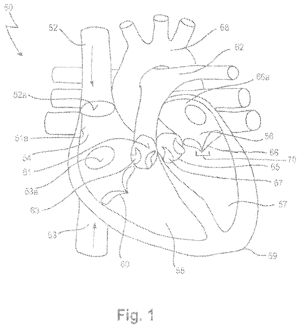
[0083]In FIG. 1, a human heart 50 is depicted, having a right atrium 54, a right ventricle 55, a left atrium 56 and a left ventricle 57. Also depicted in FIG. 1 is a portion of the vena cava superior 52, entering the heart 50 via the right atrium 54, and a portion of the vena cava inferior 53.

[0084]In more detail, the superior vena cava 52 returns the blood from the upper half of the body, and opens into the upper and back part of the right atrium 54, the direction of its orifice 52a being downward and forward. Its orifice 52a has no valve.

[0085]The inferior vena cava 53, which has a larger diameter than the superior vena cava 52, returns the blood from the lower half of the body, and opens into the lowest part of the right atrium 54, its orifice 53a being directed upward and backward, and guarded by a rudimentary valve, the valve of the inferior vena cava (Eustachian valve, not shown).

[0086]The right ventricle 55 has a triangular in form, and extends from the right atrium 54 to nea...


PUM


Abstract

The present invention relates to a prosthetic mitral valve device for implanting at the native mitral valve region of a heart. The prosthetic mitral valve coaptation enhancement device comprises a main body consisting of a stentframe and a valve element attached thereto, wherein the main body has a sealing section and a valve-bearing section, the valve-bearing section; the shape of the sealing section of the main body, in the compressed state, has a form that is adapted to the coaptation zone of the native mitral valve during systole, and the sealing section of the main body, the radial rigidity of the stentframe is lower than the radial rigidity of the valve-bearing section; the prosthetic mitral valve coaptation enhancement device further comprises at least one anchoring element, which anchoring element is coupled to the main body and is capable to anchor the main body within the native mitral valve region of a heart.

Description

CROSS REFERENCES TO RELATED APPLICATIONS[0001]This application is claiming priority from European patent application EP 15202121.8, filed on Dec. 22, 2015. The entire content of this priority application is incorporated herein by reference.BACKGROUND OF THE INVENTION[0002]The present invention concerns an implantable prosthetic mitral valve coaptation enhancement device, in particular for treating mitral valve regurgitation and the use of such a device for treating diseased or otherwise dysfunctional mitral valves.[0003]Nowadays, prosthetic mitral valve devices are used as a possible treatment of mitral valve insufficiency, also referred to as mitral regurgitation. Such prosthetic valves are delivered by traditional surgical implantation methods, or by less invasive percutaneous catheter or minimally invasive transapical methods.[0004]The mammalian heart comprises four chambers, i.e. two atria, which are the filling chambers, and two ventricles, which are the pumping chambers. In a ...

Claims


Application Information

Patent Timeline
Patent Type & Authority Patents(United States)
IPC IPC(8): A61F2/24
CPCA61F2/246A61F2/2418A61F2/2448A61F2/2457A61F2220/0008A61F2220/0016A61F2230/0069A61F2250/006A61F2250/0018A61F2/2409A61F2230/0006A61F2230/0034
Inventor KUETTING, MAXIMILIANKAWA, EMILIACENTOLA, MARCOS
Owner MEDIRA AG
Why Eureka
Try Eureka