Cell-permeable imaging sensors and uses thereof

a cell-permeable, imaging sensor technology, applied in the direction of instruments, material analysis, measurement devices, etc., can solve the problems of limited imaging techniques and limited imaging techniques for measuring large-scale signaling dynamics in intact organisms, and achieve the effect of facilitating imaging of cellular ion signaling

Active Publication Date: 2021-03-02
MASSACHUSETTS INST OF TECH
View PDF6 Cites 0 Cited by
  • Summary
  • Abstract
  • Description
  • Claims
  • Application Information

AI Technical Summary

Benefits of technology

[0005]Aspects of the disclosure relate to sensors and related methods for imaging. In some embodiments, cell-permeable imaging sensors are provided that facilitate imaging of cellular ion signaling (e.g., calcium signaling). In some embodiments, magnetic resonance imaging (MRI) provides a powerful technique for evaluating ion signaling (e.g., calcium ion) in animals and humans (e.g., in wide-field, deep-tissue context) using the sensors provided herein. In some embodiments, MRI achieves a combination of unlimited depth penetration, relatively high 3D spatial resolution (<100 μm in some contexts), and sensitivity to a wide variety of contrast mechanisms. Accordingly, in some aspects, novel magnetic resonance imaging (MRI) sensors are disclosed herein. In some embodiments, these sensors are specific for detecting ions in cells, such as calcium ions. In some embodiments, the sensors provided herein may be applied to noninvasive neuroimaging, muscular (including cardiac) imaging, immunological activation, or imaging of secretory cells in other organs of animals and humans. Furthermore, in some embodiments, sensors provided herein are powerful tools for functional MRI (MRI) and have various applications, including but not limited to: functional visualization of brain activity in healthy and disease / disordered models at preclinical studies, neuroimaging relevant to surgical planning, peripheral neuroimaging relevant to significant neuropathies, tests of cardiac or skeletal muscle function, and monitoring lymphatic function in inflammation and cancer.

Problems solved by technology

Available imaging techniques for measuring large-scale signaling dynamics in intact organisms have been limited.
Thus, imaging techniques are often limited by sharp trade-offs between depth and resolution.

Method used


Image

  • Cell-permeable imaging sensors and uses thereof
  • Cell-permeable imaging sensors and uses thereof
  • Cell-permeable imaging sensors and uses thereof

Examples


example 1

Sensing Intracellular Calcium Ions Using a Manganese-Based MRI Contrast Agent Building Block for Intracellular MRI Calcium Sensors

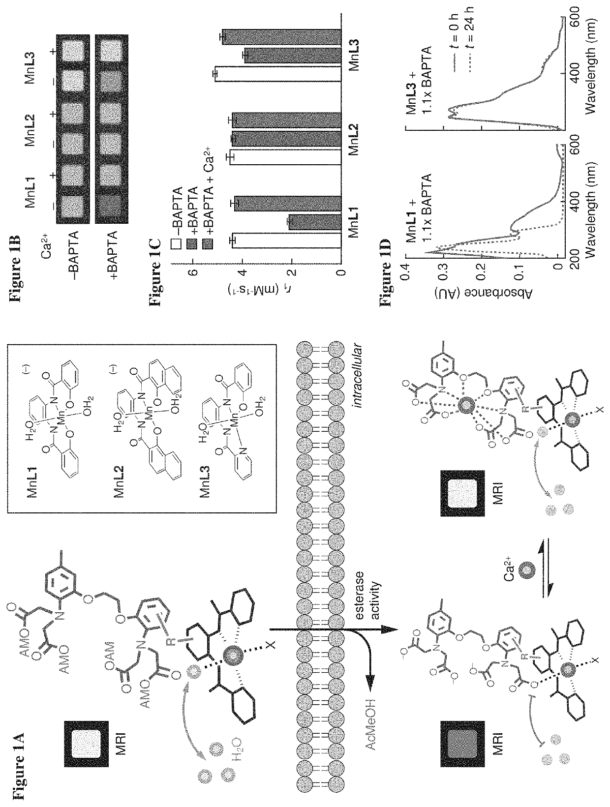
[0071]To examine the potential of Mn-PDA contrast agents to function as building blocks for the sensor design of FIG. 1A, MRI measurements were performed to assess the potential of BAPTA to interact with three paramagnetic chelates (FIG. 1A, inset). Addition of 1.1 equivalents of BAPTA to two of the Mn-PDA complexes, MnL1 and MnL3, produced significant effects on the T1-weighted MRI contrast induced by these compounds in buffer. These BAPTA-dependent contrast changes were reversed by addition of equimolar Ca2+ (FIG. 1B). This was consistent with Mn-PDA moieties competing with Ca2+ for binding to BAPTA molecules. The changes were quantified in terms of T1 relaxivity (r1) values, which were defined as the slope of the T1 relaxation rate (1 / T1=R1) vs. concentration of each paramagnetic complex. Calcium-dependent changes in r1 observed for MnL1 and MnL3 in th...

example 2

Methods and Materials

Magnetic Resonance Imaging

[0081]Most MRI data were acquired on a 7 T Bruker Biospec0 system using a T1-weighted 2D gradient echo sequence (echo time, TE=5 ms, repetition time, TR=100 ms; flip angle, FA=65 degrees (°)). Longitudinal relaxivity (r1) measurements were acquired using a 2D spin echo sequence (TE=11 ms, TR=125, 200, 400, 800, 1500, 3000, and 5000 ms), with in-plane resolution of 200×200 microns squared ((μm)2) and 2 mm slice thickness. R1 maps and values were generated using an MRI Analysis Calculator Plugin for ImageJ (National Institutes of Health, Bethesda, Md.) or Matlab scripts (Mathworks, Natick, Mass.). Stimulus-dependent R1 changes in cells were calculated as ΔR1 / R1=[R1(incubated cells)−R1(naive cells)] / R1(naive cells). Statistical comparisons between paired conditions were performed using Student's t-test, and all error bars denote the standard error of the mean from multiple measurements, unless otherwise noted.

In Vitro Characterization of M...

example 3

ManICS1-AM Labels Cells and Enables Intracellular Calcium-Sensitive MRI

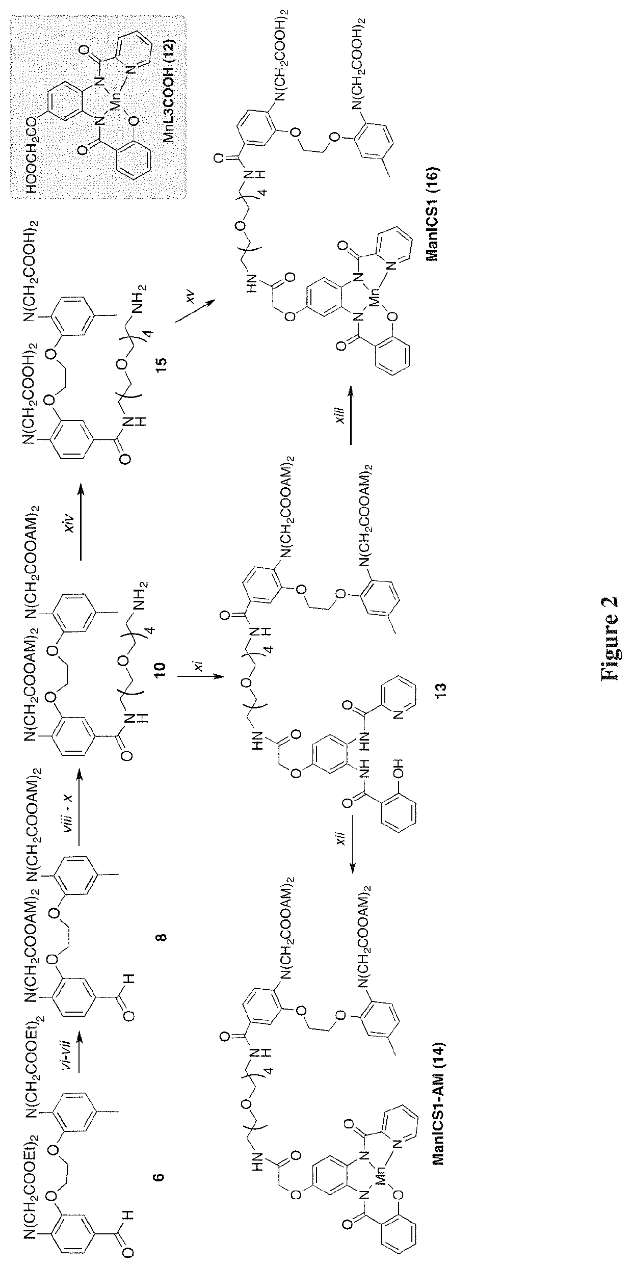
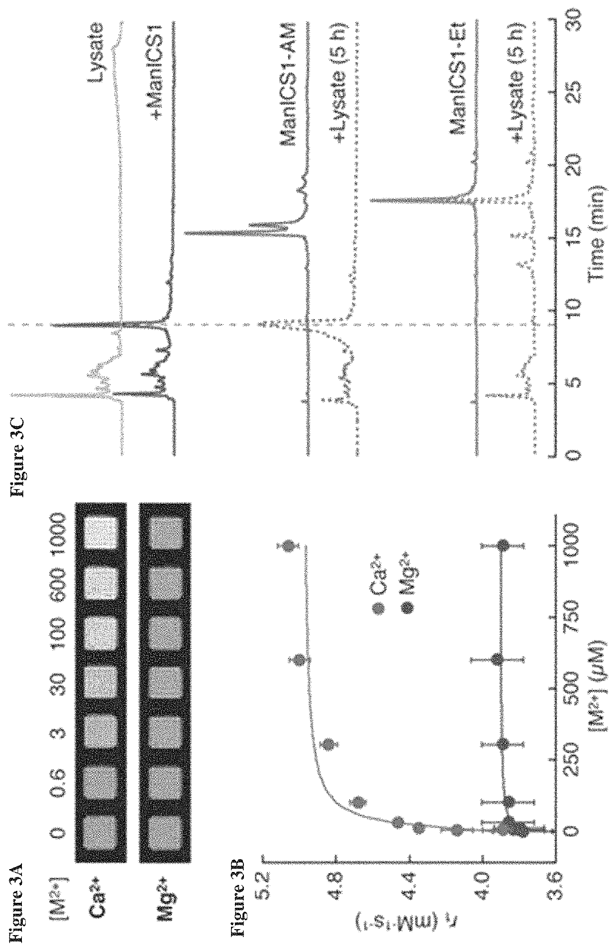
[0086]To test the ability of ManICS1-AM to enable readouts of intracellular calcium by T1-weighted MRI, its propensity was first examined to accumulate within cells. Cultured HEK293 cells were incubated with 10 μM ManICS1-AM or ManICS1 for 30 minutes, followed by washing and MRI analysis (FIG. 7A). It was found that cells incubated with the cell permeable ManICS1-AM underwent a substantial increase in R1 that persisted above basal levels for up to 24 h, while cells labeled with ManICS1 experienced a somewhat lesser increase in R1 that returned to baseline within 5 h. To assess the subcellular localization of manganese following ManICS1-AM or ManICS1 labeling, cytosolic fractions were isolated from cell lysates and the samples were analyzed by inductively coupled plasma mass spectrometry (ICP-MS). The ICP-MS results indicated that only ManICS1-AM labeling produced cytosolic elevations in manganese content (FIG. 7A...


PUM

PropertyMeasurementUnit
depthsaaaaaaaaaa
dissociation constantaaaaaaaaaa
timeaaaaaaaaaa

Abstract

The disclosure relates in some aspects to imaging agents, and in particular, imaging agents for sensing of calcium signaling. According to some embodiments of the disclosure, contrast agents for magnetic resonance imaging and related technologies are provided, and methods of making and using the contrast agents.

Description

RELATED APPLICATIONS[0001]This application claims priority under 35 U.S.C. § 119(e) to U.S. Provisional Application No. 62 / 597,875, filed Dec. 12, 2017 and entitled “CELL-PERMEABLE IMAGING SENSORS AND USES THEREOF,” which is incorporated herein by reference in its entirety for all purposes.GOVERNMENT FUNDING[0002]This invention was made with Government support under Grant Nos. R01 DA038642 and U01 NS090451 awarded by the National Institutes of Health. The Government has certain rights in the invention.FIELD OF THE INVENTION[0003]The invention relates in some aspects to imaging agents.BACKGROUND OF THE INVENTION[0004]Available imaging techniques for measuring large-scale signaling dynamics in intact organisms have been limited. With optical techniques it is possible to perform functional imaging of signaling dynamics at depths of up to about two millimeters in tissue, but for most vertebrate species this only represents a small fraction of the volumes of experimental interest. Implan...

Claims


Application Information

Patent Timeline
Patent Type & Authority Patents(United States)
IPC IPC(8): A61K49/00A61K49/08A61K49/10G01N33/68
CPCA61K49/085A61K49/0002A61K49/103G01N33/6872
Inventor JASANOFF, ALAN PRADIPBARANDOV, ALIBARTELLE, BENJAMIN B.
Owner MASSACHUSETTS INST OF TECH
Why Eureka
Try Eureka