Prosthetic cardiovascular valves and methods for replacing native atrioventricular valves with same

a technology of native atrioventricular valves and cardiovascular valves, which is applied in the field of prosthetic cardiovascular valves and methods for replacing native atrioventricular valves with same, can solve the problems of heart failure, adversely affecting organ function, stenosis and insufficiency, etc., and achieves the effect of rapid heart ra

Active Publication Date: 2021-03-16
CORMATRIX CARDIOVASCULAR INC
View PDF4 Cites 0 Cited by
  • Summary
  • Abstract
  • Description
  • Claims
  • Application Information

AI Technical Summary

Benefits of technology

[0047]In some embodiments of the invention, during the prosthetic valve implant procedures described herein, a rapid heart rate is induced, wherein blood flow to and through the valve to be replace is reduced, more preferably, abated.

Problems solved by technology

Stenosis and insufficiency can also occur concomitantly in the same valve or in different valves.
Both of the noted valve abnormalities can adversely affect organ function; especially, heart function, and result in heart failure.
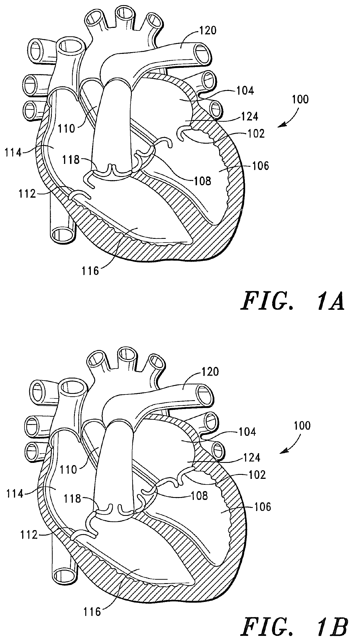
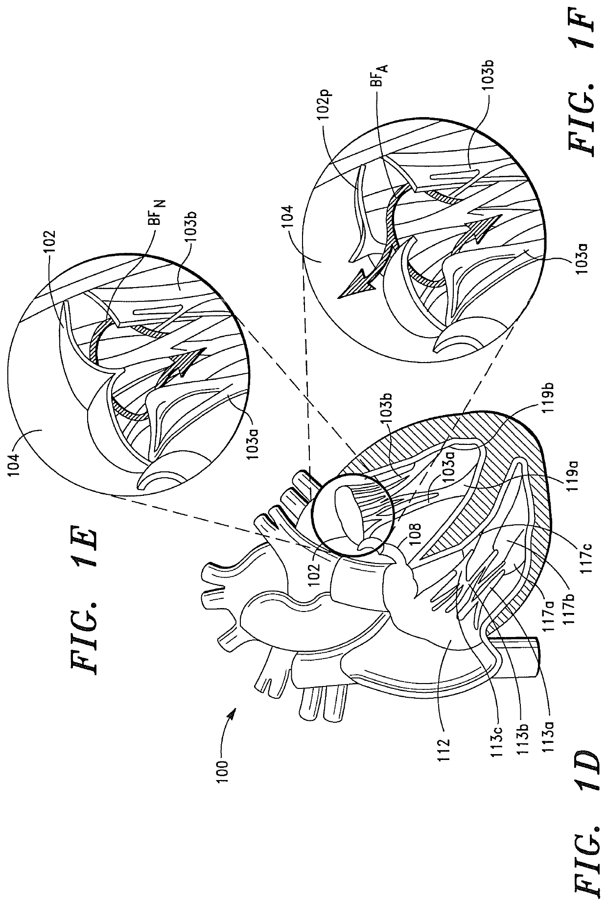
As illustrated in FIG. 1F, the regurgitated blood “BFA” flows back into the left atrium, which can, if severe, result in heart failure.
If such vegetation is present on a valve on the left side (i.e., the systemic circulation side) of the heart, embolization can, and often will, result in sudden loss of the blood supply to the affected body organ and immediate malfunction of that organ.
Likewise, bacterial or fungal vegetation on a tricuspid valve can embolize to the lungs.
The noted embolization can, and in many instances will, result in lung dysfunction.
A further disadvantage associated with mechanical heart valves is that such valves also have a propensity to cause the formation of blood clots after implantation in a patient.
If such blood clots form on the mechanical valve, they can preclude the valve from opening or closing correctly or, more importantly, can disengage from the valve and embolize to the brain, causing an embolic stroke.
In addition to being expensive, these anticoagulant drugs can themselves be dangerous in that they can cause abnormal bleeding in the recipient or patient that can lead to a hemorrhagic stroke.
A further disadvantage associated with mechanical heart valves is that such valves often have large and cumbersome skirt attachments that partially extend into the left atrium and the left ventricle when implanted in a mitral valve region.
The skirt attachment can, and often will, impair aortic valve function by obstructing the outflow tract of the aortic valve and preventing the leaflets of the adjacent aortic valve from coapting.
The risks and complications associated with impaired aortic valve function typically include left ventricular hypertrophy with fibrosis, systolic dysfunction (a decrease in the ejection fraction), diastolic dysfunction (elevated filling pressure of the LV), and in severe cases, congestive heart failure.
Further, mechanical heart valves with and without the skirt attachments are notoriously difficult to implant and often require large and cumbersome catheter assemblies for percutaneous or transapical implantation.
These large catheter assemblies are excessively difficult to operate during a percutaneous or transapical implantation procedure.
However, there are still several drawbacks and disadvantages associated with allograft tissue valves.
A major drawback of allograft tissue valves is that such valves are not available in sufficient numbers to satisfy the needs of all patients who need new heart valves.
A further drawback of allograft tissue valves is that recipients of allograft tissue valves, i.e. patients, are typically required to take systemic antirejection and / or immunosuppressive drugs for a predetermined period of time and, in some instances, for a lifetime.
Although antirejection and / or immunosuppressive drugs increase the possibility that a patient will accept an allograft without complications, the drugs will often leave the recipient vulnerable to a plurality of other infectious diseases, including bacterial infections, fungal infections, viral infections and the like.
Various significant complications have also been encountered when allograft tissue valves have been used to replace native atrioventricular (AV) valves.
The complications include, for example, the recipient's rejection of the allograft tissue valve leading to transplant rejection and further complications resulting therefrom.
Another complication is the onset of Graft-Versus-Host disease.
Further, allograft tissue valves are generally more difficult to implant than mechanical valves or xenograft tissue valves.
Because of the implantation difficulties, the operative risk associated with allograft tissue valves is often greater than the operative risks associated with mechanical valves and xenograft tissue valves.
Conventional xenograft tissue valves are, however, prone to calcification and lack the long-term durability of mechanical valves and, consequently, require frequent replacement as compared to mechanical valves.
Without the chemical treatment(s), conventional xenograft tissue valves can, and in many instances will, induce an adverse inflammatory response when implanted in a patient.
Implantation of a prosthetic valve, including mechanical valves and bioprosthetic valves, requires a great deal of skill and concentration given the delicate nature of the native cardiovascular tissue and the spatial constraints of the surgical field.
A major drawback of the noted attachment of a prosthetic valve to a cardiovascular structure; more particularly, a papillary muscle is that the attachment of the distal end of the valve to the papillary muscle can, and in most instances will obstruct the outflow tract of the adjacent aortic valve.

Method used


Image

  • Prosthetic cardiovascular valves and methods for replacing native atrioventricular valves with same
  • Prosthetic cardiovascular valves and methods for replacing native atrioventricular valves with same
  • Prosthetic cardiovascular valves and methods for replacing native atrioventricular valves with same

Examples


example 1

Assessment of Acute Hemodynamic Function of a Porcine Heart

[0434]A study was performed using a porcine model in order to evaluate the acute hemodynamic function of a porcine heart when the mitral valve of the porcine heart is replaced with a prosthetic tissue valve comprising a base ribbon structure, such as the prosthetic tissue valve shown in FIGS. 8A and 8B.

[0435]The study included a single treatment group consisting of one (1) adolescent pig (sus domesticus). The pig received anesthetic premedication, followed by anesthetic induction via intravenous injection. After the pig was anesthetized, a prosthetic tissue valve comprising a base ribbon structure was implanted into the mitral valve region of the porcine heart.

[0436]During the implantation procedure, the distal end of the prosthetic tissue valve was secured to a predetermined region of the left ventricle myocardium between the anterior and posterior papillary muscles of the porcine heart.

[0437]Approximately two (2) hours aft...


PUM


Abstract

A percutaneous transseptal surgical implantation method for replacing a defective atrioventricular (AV) valve with a conical shaped prosthetic valve formed from a polymeric composition. When the method is employed to replace a native mitral valve, the method positions the prosthetic valve in the mitral valve region, whereby the valve does not obstruct the outflow tract of the aortic valve and prevents the leaflets of the aortic valve from coapting.

Description

CROSS-REFERENCE TO RELATED APPLICATIONS[0001]This application is a continuation-in-part of U.S. application Ser. No. 16 / 238,730, filed on Jan. 3, 2019, which is a continuation-in-part of U.S. application Ser. No. 16 / 193,669, filed on Nov. 16, 2018, which is a continuation-in-part of U.S. application Ser. No. 16 / 129,968, filed on Sep. 13, 2018, which is a continuation-in-part of U.S. application Ser. No. 15 / 206,833, filed on Jul. 11, 2016, now U.S. Pat. No. 10,188,510, which is a continuation-in-part application of U.S. application Ser. No. 14 / 960,354, filed on Dec. 5, 2015, now U.S. Pat. No. 9,907,649, which is a continuation-in-part application of U.S. application Ser. No. 14 / 229,854, filed on Mar. 29, 2014, now U.S. Pat. No. 9,308,084, which claims priority to U.S. Provisional Application No. 61 / 819,232, filed on May 3, 2013.FIELD OF THE INVENTION[0002]The present invention generally relates to prosthetic valves for replacing defective cardiovascular valves. More particularly, the...

Claims


Application Information

Patent Timeline
Patent Type & Authority Patents(United States)
IPC IPC(8): A61F2/24
CPCA61F2/2418A61F2/2409A61F2/2412A61F2/2433A61F2/2436A61F2/2454A61F2220/0008A61L2430/20A61F2220/0016A61F2230/0067
Inventor MATHENY, ROBERT G
Owner CORMATRIX CARDIOVASCULAR INC
Why Eureka
Try Eureka