
Method for denoising time series images of a moved structure for medical devices

a technology of moving structure and time series images, which is applied in the field of denoising time series images of moving structure for medical devices, can solve the problems of loss of contrast within the regions of moving structure, noise impression, and different artifacts, and achieve the effect of optimizing the contrast-to-noise ratio

Active Publication Date: 2021-12-28
SIEMENS HEALTHCARE GMBH
View PDF1 Cites 0 Cited by
  • Summary
  • Abstract
  • Description
  • Claims
  • Application Information

AI Technical Summary

Benefits of technology

The method effectively improves the visibility and contrast-to-noise ratio of moved structures by dynamically adjusting the denoising process, reducing noise and artifacts, and enhancing image quality in medical devices like X-ray and infrared recording systems.

Problems solved by technology

Averaging over the time series images leads to a loss of contrast within the regions of the moved structure.
Furthermore, local differences in the noise impression, for example, different artifacts may occur due to the reduced averaging in the moved regions.

Method used


Image

  • Method for denoising time series images of a moved structure for medical devices
  • Method for denoising time series images of a moved structure for medical devices

Examples


Embodiment Construction

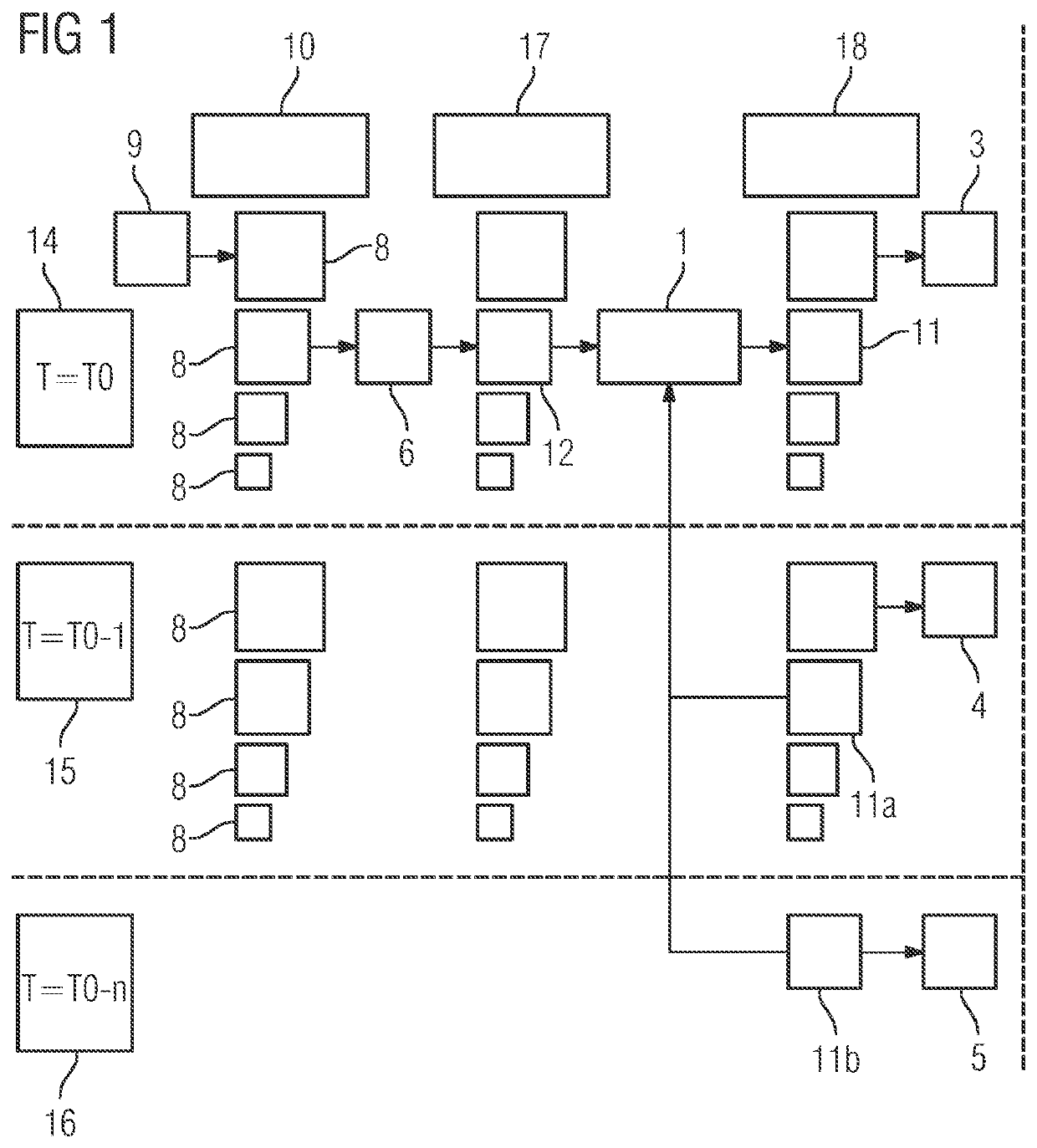
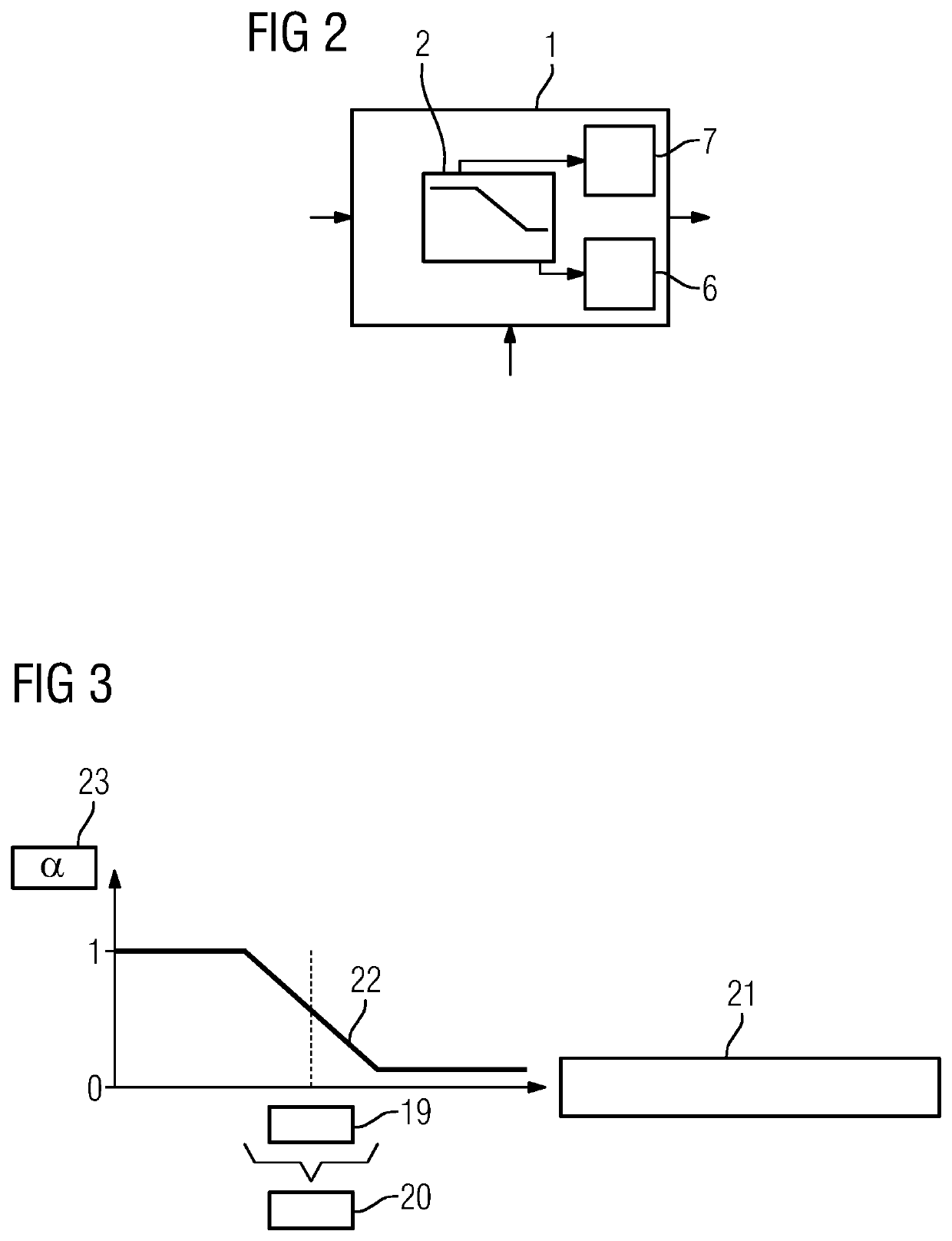
[0024]FIG. 1 depicts a block diagram of how a denoised time series image 3, 4, 5 is achieved from an image of a moved structure 9. The method corresponds to a corresponding apparatus having denoising device and movement detector. The moved structure 9 may be segmented into bandpass signals 8. The segmentation may use, for example, a Laplace segmentation or by the application of an à trous segmentation by an adaptive edge-preserving kernel. A bandpass segmentation may include a mean freedom with bandpass signals 8. Segmentation in a segmentation plane 10 into bandpass signals 8 has the property that noise, for example, in X-ray systems, is Poisson distributed and convoluted with the system modulation transfer function. The noise, but also other signals, may be analyzed and processed in different spatial frequencies. Band passes may be used. In FIG. 1 the method is carried out, for example, on only one bandpass signal 8. The method may additionally or alternatively also be applied to ...


PUM


Abstract

Embodiments provide a method for denoising time series images of a moved structure for a medical device. A movement detector detects the moved structure. The movement detector obtains a measurement of the similarity of two images that each represent the same section of the moved structure. The two images originate from two different time series images. A ratio between spatial and temporal denoising is defined for the section as a function of the measurement of the similarity.

Description

CROSS REFERENCE TO RELATED APPLICATIONS[0001]This application claims the benefit of DE 102016205603.4 filed on Apr. 5, 2016, which is hereby incorporated by reference in its entirety.TECHNICAL FIELD[0002]Embodiments relate to a method for denoising time series images of a moved structure for medical devices. Embodiments further relate to time series images of X-ray devices.BACKGROUND[0003]Medical applications images may be averaged over time in order to increase the image quality. This averaging over time involves a method in which images from the same section are placed one above the other. By placing the different time series images from the same section one above the other, individual image points are overlaid, so an intensification may occur that leads to an improvement in the contrast-to-noise ratio. The improvement in the contrast-to-noise ratio contributes to an increase in the image quality. An increase in the contrast-to-noise ratio may be achieved with time series images w...

Claims


Application Information

Patent Timeline
Patent Type & Authority Patents(United States)
IPC IPC(8): A61B6/00G06T5/00G06T7/00A61B5/00G06T7/11
CPCA61B5/7214A61B5/0075A61B5/725A61B5/7225A61B6/487A61B6/5264G06T5/002G06T7/0012G06T7/11A61B2576/00G06T2207/10016G06T2207/10048G06T2207/10116G06T2207/20182G16H30/40G06T5/70
Inventor BÖHM, STEFANSTOWASSER, BORIS
Owner SIEMENS HEALTHCARE GMBH
Learn More