
Method to produce protein in Penicillium amagasakiense's sleeping spores by transformation of ssRNA

a technology of penicillium amagasakiense and protein, applied in the field of biological technologies, can solve the problems of single-stranded molecule, short half-life, unstable rna, etc., and achieve the effect of avoiding killing cells, high uniformity, and intense electric field

Active Publication Date: 2022-03-29
FUZHOU UNIV
View PDF1 Cites 0 Cited by
  • Summary
  • Abstract
  • Description
  • Claims
  • Application Information

AI Technical Summary

Benefits of technology

[0054]In the present invention, the above technical solution is employed. It is very simple to use non-germinated spores as a starting material for introducing exogenous molecules because the complicated step of germination of spores can be omitted. More importantly, polyploidy might have occurred in the germinated spores, whereas non-germinated spores are guaranteed to be haploid. In many applications of genetic engineering, the host cells have to be haploid in order to achieve the desired result or efficiency. Meanwhile, the HDEN electro-transformation technique is utilized in the present invention to introduce exogenous RNA into resting spores of Penicillium amagasakiense. In the HDEN electro-transformation technique, a high-density matrix electrode that produces a highly uniform and intense electric field is used. Each cell within the electric field receives an electroporation condition which is almost exactly the same. During operation, the cells are placed in a common container, such as a cell culture plate, and followed by inserting an electroporator head with a matrix electrode into the container, and energized. In the traditional electroporation technique, a special electroporation cuvette is generally used, cells are placed between two metal plates positioned in parallel and subsequently the metal plates are electrified to form an electric field for electroporation. Therefore, the discharge modes in the two methods are completely different. In the former one, the electrode is inserted into the cell suspension and an electric field is generated within, whereas in the latter one, an electric field is generated outside of the whole cell suspension. In the former one, the electrode head is formed by many metal pins, and a voltage is generated between the pins, whereas in the latter one, only two metal plates are present, i.e. a positive electrode plate and a negative electrode plate, and a voltage is generated between the two plates. The HDEN technique also basically eliminates the cathode effect existing in the traditional electroporation technology, to avoid the generation of large amounts of hydroxide ions, so as to avoid killing the cells and improve the cell survival rate after electroporation. However, the cathode effect is difficult to be eliminated in the traditional electroporation technology. Furthermore, in traditional electroporation methods, the electroporation is performed only once. This is because if multiple electroporations are performed, the cell death rate will be increased greatly. However, in the HDEN method of the present invention, multiple electroporations may be performed, and the effects of the multiple electroporations can be superimposed without significantly increasing the cell death rate.
[0055]The HDEN electro-transformation technique is an electro-transformation technique developed specifically for the characteristics of mammalian cells, to improve the efficiency of delivery of exogenous DNA and drug to mammalian cells. It is well known in the art that the characteristics and the culturing methods of microbial cells are vastly different from those of mammalian cells. The structures of mammalian cells and microbial cells are different. Mammalian cells have no cell walls. Microbial cells (such as most fungi, including Penicillium amagasakiense) have cell walls. The cell membrane of mammalian cells is also different from the cell membrane of microbial cells. Even in the same organism, the structures, states, and chemical compositions of respective cell membrane and cell wall of different tissues, different organs or different cell types are also different. Even the structures, states, and chemical compositions of respective cell membrane and cell wall of the same tissue, the same organ, and the same cell type of the same organism are also different in different stages of growth and development or in different external environments. The cell wall and cell membrane are barriers that block exogenous molecules to enter the cell. Depending on different barriers (with different structures and different chemical compositions), the methods to break through the barriers are different. In addition, the methods for different exogenous molecules breaking through the barrier are also different due to that different exogenous molecules with various structures, molecular weights, volumes and chemical compositions encountering the same barrier. While encountering with different barriers, the methods and mechanisms of different exogenous molecules breaking through the different barriers are even vastly different.
[0058]By using the method of the present invention, the RNA of encoded protein is introduced directly into the dormant spores of Penicillium amagasakiense and the protein is expressed for the first time in history, and the steps are very simple and fast, in addition, the effect is excellent, and the transformation rate of at least 90% can be obtained.

Problems solved by technology

RNA, as a single-stranded molecule, is unstable, has a short half-life compared to the DNA of a double-stranded structure, and in nature, RNAse are widespread and RNA is highly susceptible to degradation.
However, since the spores are generally dormant, their cell walls are very thick, and the state of the cell wall and cell membrane of resting spores are different from those of germinated spores and mycelia.
Therefore, it is very difficult to introduce exogenous RNA molecules directly into the interior of sleeping spores.
At present, there is no way to directly introduce exogenous RNA molecules into dormant (un-germinated) fungal spores without medium-mediated conditions.
This is also a technical problem that has not been tackled in this field.
However, it is well known that the life activity of sleep spores is in the least exuberant state, the vitality of various protein translation factors and enzymes in cells is very low, the amount of various factors and enzymes is also very rare, even if exogenous RNA enters the inside of un-germinated spores, it also needs a relatively long interaction time, in order to translate the target protein.
There are already ready-made technical solutions in this field to germinate fungal spores, but no matter which germination method, it takes time, energy, manual operation, and chemical reagents.
Unfortunately, so far, there has been no precedent for the introduction of exogenous encoded RNA for spores (whether germinated or un-germinated).
Fungal spores collected from cultures carry a large amount of RNAse, and it is difficult to thoroughly clean the RNAse without killing spores (wash with DEPC or other compounds will certainly kill fungal spores).
Therefore, deliver exogenous single-stranded RNA to fungal spores is very difficult, single-stranded RNA will be degraded by RNAse before into spores.
In conclusion, introducing the RNA molecules that encode proteins directly into fungi (whether mycelium or spores, whether germinated spores or un-germinated spores) are a technical problem that has not been tackled and no report in this field.
Currently, there is no reports on the use of this technique in species other than mammalian cells at present.
Therefore, whether the HDEN electro-transformation technique can be applied to species other than mammalian cells is still unknown.

Method used


Image

  • Method to produce protein in <i>Penicillium amagasakiense</i>'s sleeping spores by transformation of ssRNA
  • Method to produce protein in <i>Penicillium amagasakiense</i>'s sleeping spores by transformation of ssRNA
  • Method to produce protein in <i>Penicillium amagasakiense</i>'s sleeping spores by transformation of ssRNA

Examples


example 1

[0064]Expression of green fluorescent protein (GFP) in Penicillium amagasakiense cells:

[0065]1. Construction of plasmid for in vitro transcription

[0066]The GFP gene encoding sequence is shown in SEQ ID No. 5,

[0067]The GFP protein sequence is shown in SEQ ID No. 6,

[0068]PCR primers for amplification of GFP gene:

[0069]

(SEQ ID NO: 1)(SEQ ID NO: 2)

[0070]Among them, the GCTAGC sequence in the primer F near the 5′ end is used to promote binding of eukaryotic translation initiation factor and RNA which transcribed from DNA in vitro, this sequence adjacent to the target gene's start codon ATG. After PCR with above primers, the GFP gene can be fused with GCTAGC in the upstream, and restriction enzyme cutting sites in the upstream and downstream.

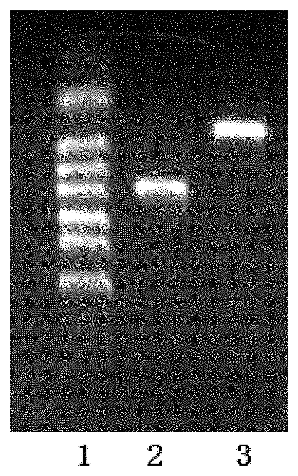
[0071]PCR system (50 μl): GFP template 1 μL, primer F 2 μL, primer R 2 μL, 2× Taq PCR mix 25 μL, and ddH2O top up to 50 μL. PCR program: 94° C. for 2 min, 30 cycles of (94° C. for 30 s, 61.6° C. for 30 s, and 72° C. for 60 s), and 72° C. for 5 min.

[00...

example 2

[0101]Expression of red fluorescent protein (RFP) in Penicillium amagasakiense cells:

[0102]1. Construction of plasmid for in vitro transcription

[0103]The RFP gene encoding sequence is shown in SEQ ID No. 7,

[0104]The RFP protein sequence is shown in SEQ ID No. 8,

[0105]PCR primers for amplification of RFP gene:

[0106]

RFP-F: (SEQ ID NO. 3)5′ CGGAATTCGCCACCATGGCCTCCTCCGAGGACGT 3′, with EcoR I site. RFP-R: (SEQ ID NO. 4)5′ TCGAGCTCGTTAGGCGCCGGTGGAGTGG 3′, with Sal I site. 

[0107]In RFP-F, the GCCACC sequence near the 5′ end is used to promote binding of eukaryotic translation initiation factor and RNA which in vitro transcribed from DNA, this sequence adjacent to the target gene's start codon ATG.

[0108]RFP-R′ 5′ end contains Sal I site.

[0109]In accordance with example 1, the RFP gene is amplified by PCR, ligated to the PGEM-T easy plasmid, then in vitro transcription, and the resulting RNA will be transformed into the host spores.

[0110]2. Using the above-mentioned red fluorescent protein e...

example 3

[0125]Expression of yellow fluorescent protein (YFP) in Penicillium amagasakiense cells:

[0126]1. Construction of plasmid for in vitro transcription

[0127]The YFP gene encoding sequence is shown in SEQ ID No. 9,

[0128]The YFP protein sequence is shown in SEQ ID No. 10,

[0129]In accordance with example 1, the YFP gene is amplified by PCR using the same primers as GFP, ligated to the PGEM-T easy plasmid, then in vitro transcription, and the resulting RNA will be transformed into the host spores.

[0130]2. Using the above-mentioned yellow fluorescent protein encoded RNA to express yellow fluorescent protein in Penicillium amagasakiense dormant spores, the steps are as follows:

[0131]1) Culture of Penicillium amagasakiense and Collection of Spores

[0132]In a 15-cm Petri dish, a solid agar medium (PDA medium) was prepared.

[0133]Penicillium amagasakiense CICC 40341 was inoculated onto the surface of the solid agar medium and cultured at a temperature of 40° C. with 60-85% humidity for 3 days, to ...


PUM

PropertyMeasurementUnit
voltageaaaaaaaaaa
humidityaaaaaaaaaa
temperatureaaaaaaaaaa

Abstract

The present invention discloses A METHOD TO PRODUCE PROTEIN IN PENICILLIUM AMAGASAKIENSE'S SLEEPING SPORES BY TRANSFORMATION OF SSRNA. The method includes three steps of culture of Penicillium amagasakiense and collection of spores, pretreatment of Penicillium anagasakiense spores, and electroporation of Penicillium anagasakiense spores by using HDEN method. In the present invention, non-germinated spores are used as a starting material for introduction of exogenous molecules. The exogenous protein coding single stranded RNA is introduced into the resting spores of Penicillium amagasakiense by employing the HDEN electrotransformation technique to express protein. The method of this invention is simple and fast, the effect is excellent, and the transformation rate reaches more than 90%.

Description

CROSS-REFERENCE TO RELATED APPLICATIONS[0001]This application is a continuation of International Patent Application No. PCT / CN2017 / 096980 with a filing date of Aug. 11, 2017, designating the United States, now pending, and further claims priority to Chinese Patent Application No. 201610813618.9 with a filing date of Sep. 9, 2016. The content of the aforementioned applications, including any intervening amendments thereto, are incorporated herein by reference.BACKGROUND OF THE INVENTION1. Field of the Invention[0002]The present invention relates to the field of biological technologies, and specifically to a method to produce protein in Penicillium amagasakiense's sleeping spores using ssRNA.2. Description of Related Art[0003]The central dogma of molecular biology refers to the process by which genetic information is transmitted from DNA to RNA and then from RNA to proteins, that is, to complete the transcription and translation of genetic information. In modern genetic engineering, h...

Claims


Application Information

Patent Timeline
Patent Type & Authority Patents(United States)
IPC IPC(8): C12N15/87C12N15/80C12N5/10C12N15/82C12N13/00C12N1/14C12N3/00C07K1/13C12R1/80
CPCC12N15/87C12N5/10C12N15/80C12N15/8206C07K1/13C07K2319/00C07K2319/60C12N1/14C12N1/145C12N3/00C12N13/00C12R2001/80
Inventor LIN, JUN
Owner FUZHOU UNIV
Learn More