System and methods for aggregating features in video frames to improve accuracy of AI detection algorithms

a technology of video frames and features, applied in the field of real-time imaging of the body cavity, can solve the problems of ensuring that the entire internal surface of the colon has been imaged, difficulty in identifying a polyp or adenoma, and about half of screened patients with adenoma that is missed

Active Publication Date: 2022-08-23
SATISFAI HEALTH INC
View PDF67 Cites 1 Cited by
  • Summary
  • Abstract
  • Description
  • Claims
  • Application Information

AI Technical Summary

Benefits of technology

[0024]The systems and methods of the present invention enable an AI system to recognize and group portions of an area of interest in a multiple video frames generated by an endoscope, thereby enabling analysis of the subareas the multiple video frames together. In this manner, as an endoscopist continues to examine an area of interest, the AI algorithm is able to analyze additional information to ultimately compile a full data picture for the tissue under examination.
[0026]In accordance with one aspect of the invention, systems and methods are provided for generating high quality images for submission to AI detection algorithms used in endoscopic medical procedures, to thereby yield better outcomes. The inventive systems and methods are expected to provide essentially seamless performance, as if the AI detection algorithms were running in their canonical form.
[0028]As colonoscopy is an imaging technique that affords limited anatomical guidance, it is not uncommon for the endoscopist to become spatially disoriented regarding the precise location of the anatomy under examination. When this happens, it is possible for the endoscope to become oriented in a way that is physically difficult to unwind, thereby resulting in limited view of the anatomy and limiting the quality of data provided to the AI module for a specific area or lesion. In accordance with another aspect of the invention, the inventive software may guide the endoscopist where to next move the endoscope to collect additional information for processing by the AI module and to further visualize the area of interest.
[0029]Display of the first and the second display windows may be performed in a parallel or as a multi-threaded process. Parallel processing advantageously allows the system to display the video data received from the endoscope in real-time, and also display the graphical indications in the second window at a frame rate that may be lower than or equal to the frame rate of the first window. In this manner, the present invention provides visual clues that improve the quality and quantity of the information provided to the detection algorithms. Systems constructed in accordance with the inventive principles also enable the detection algorithm to determine if there are enough features extracted based on the real time images available to assess an area under examination, or if more data is required, thereby greatly improve the efficacy of the detection algorithms.

Problems solved by technology

This means that about half of screened patients have an adenoma that is missed.
One factor is the difficulty of identifying a polyp or adenoma, even though it may be in the visual field of the colonoscopy image.
Another factor that contributes to the lower than ideal ADR is the difficulty of ensuring that the entire internal surface of the colon has been imaged.
It is difficult for a colonoscopist to remember what has been imaged, and “integrate” those images mentally to conclude that the entire internal surface has been looked at, and thus it is extremely challenging for the endoscopist to assure that the entire internal surface of the colon has been visualized.
Failure to visualize the entire internal surface incurs a risk of missing potentially harmful polyps or cancers.
One of the challenges of using AI for endoscopy is that because of the large variance in tissue, anatomy and motion dynamics during the procedure, the AI must be trained over a very large set of data which can be hard to collect.
Another challenge is that previously-known AI processes the video feed in real time, and thus operates prospectively during a procedure.
Another challenge in endoscopy with AI is that the tip of the endoscope is controlled by a series of knobs and switches located on the end piece held and manipulated by the endoscopist.
However, the endoscopist's attention is focused on the screen (or monitor) and not the controls.
It is quite difficult for many people to correlate the image on the screen into hand movements necessary to direct the tip of the endoscope.
Hence, control reference between the tip of the endoscope and the series of controls on the handpiece often is lost.
This means many frames fed to the AI technology are ultimately not useful or carry very little information.
Further, some of the frames may contain only partial information that limits the extraction capability of the AI algorithms.
Accordingly, the AI may not be able to detect the entire abnormality in a single frame.
Although a wide angle lens provides a wide field of view, e.g., suited for outdoor photography, such lenses also introduce a high degree of distortion at the periphery.
One drawback of this approach is that the algorithm has no knowledge if it is stitching together images that belong to the same object or different objects.
In addition, none of the foregoing methods operate in real time, but rather require post-processing of the information.
Accordingly, none are suitable for real time applications, such as endoscopy.
This application does not address the problems that arise when attempting to stitch together images taken on different focal planes that provide only partial information of an object.
The patent does not address features extraction and stacking images based on similarity of the extracted features.

Method used


Image

  • System and methods for aggregating features in video frames to improve accuracy of AI detection algorithms
  • System and methods for aggregating features in video frames to improve accuracy of AI detection algorithms
  • System and methods for aggregating features in video frames to improve accuracy of AI detection algorithms

Examples


Embodiment Construction

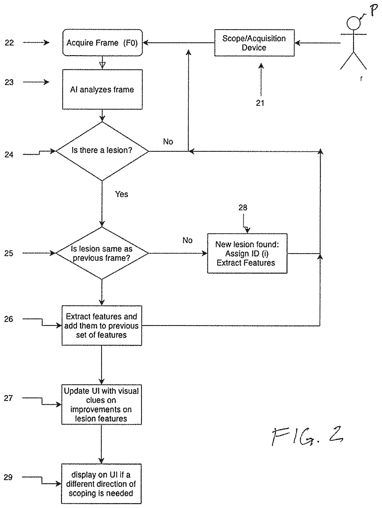
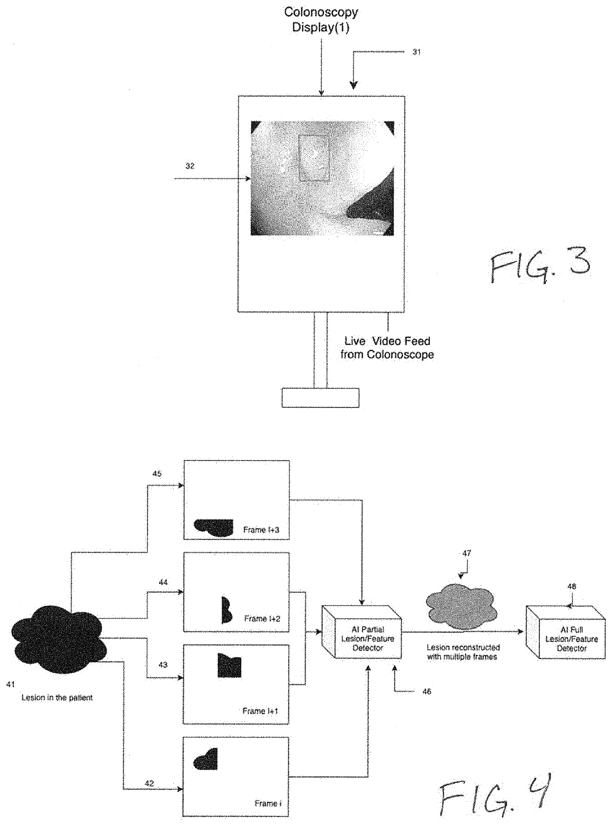
[0036]The present invention is directed to systems and methods for analyzing multiple video frames imaged by an endoscope with an artificial intelligence (“AI”) software module running on a general purpose or purpose-built computer to aggregate information about a potential tissue feature or abnormality, and to indicate to the endoscopist the location and extent of that feature or abnormality on a display viewed by the endoscopist. In accordance with the principles of the present invention, the AI module is programmed to make a preliminary prediction based on initially available information within a video frame, to aggregate additional information for a feature from additional frames, and preferably, to provide guidance to the endoscopist to direct him or her to move the imaging end of the endoscope to gather additional video frames that will enhance the AI module detection prediction.

[0037]Referring to FIG. 1, exemplary colonoscopy system 10 configured in accordance with the princi...


PUM


Abstract

Methods and systems are provided for aggregating features in multiple video frames to enhance tissue abnormality detection algorithms, wherein a first detection algorithm identifies an abnormality and aggregates adjacent video frames to create a more complete image for analysis by an artificial intelligence detection algorithm, the aggregation occurring in real time as the medical procedure is being performed.

Description

I. REFERENCE TO RELATED APPLICATIONS[0001]This application is a continuation-in-part of co-pending U.S. patent application Ser. No. 16 / 855,592, filed Apr. 22, 2020, which is a continuation application of U.S. patent application Ser. No. 16 / 512,751, filed Jul. 16, 2019, now U.S. Pat. No. 10,671,934, the entire contents each of which are incorporated herein by reference. This application is also a continuation-in-part application of U.S. patent application Ser. No. 16 / 931,352, filed Jun. 16, 2020, the entire contents of which are incorporated herein by reference.II. FIELD OF THE INVENTION[0002]This invention relates generally to the field of real-time imaging of a body cavity, with particular application to endoscopy such as colonoscopy and upper endoscopy.III. BACKGROUND OF THE INVENTION[0003]Endoscopy refers to a medical procedure in which an instrument is used for visual examination of an internal body part. A common example of endoscopy is colonoscopy, during which a flexible tube...

Claims


Application Information

Patent Timeline
Patent Type & Authority Patents(United States)
IPC IPC(8): G06K9/00G06N5/04H04N19/115G06N20/00H04N19/186G06K9/62G06V20/40
CPCG06N5/04G06K9/6267G06N20/00G06V20/41H04N19/115H04N19/186A61B1/0005A61B1/31G06T7/0012G06T17/00G06T19/00G06T2200/24G06T2207/10068G06T2207/30028G06T2210/41G06T2219/028A61B1/000094A61B1/000096G06T7/0014G06T7/70G06T2207/30096G06T2207/30032G06T2207/10016G06T2207/30101G06V2201/031G06V10/25G06V10/62G06F18/24
Inventor ZINGARETTI, GABRIELEREQUA, JAMES
Owner SATISFAI HEALTH INC
Why Eureka
Try Eureka