Localizing intravascular lesions on anatomic images

an anatomic image and intravascular technology, applied in the field of localizing intravascular lesions on anatomic images, can solve the problems of opacification of body lumen image, diffuse diffusion of media into body lumen,

Inactive Publication Date: 2002-08-22
IMETRX
View PDF12 Cites 247 Cited by
  • Summary
  • Abstract
  • Description
  • Claims
  • Application Information

AI Technical Summary

Problems solved by technology

Unfortunately, the contrast medium delivered into the body lumen will d

Method used


Image

  • Localizing intravascular lesions on anatomic images
  • Localizing intravascular lesions on anatomic images
  • Localizing intravascular lesions on anatomic images

Examples


Embodiment Construction

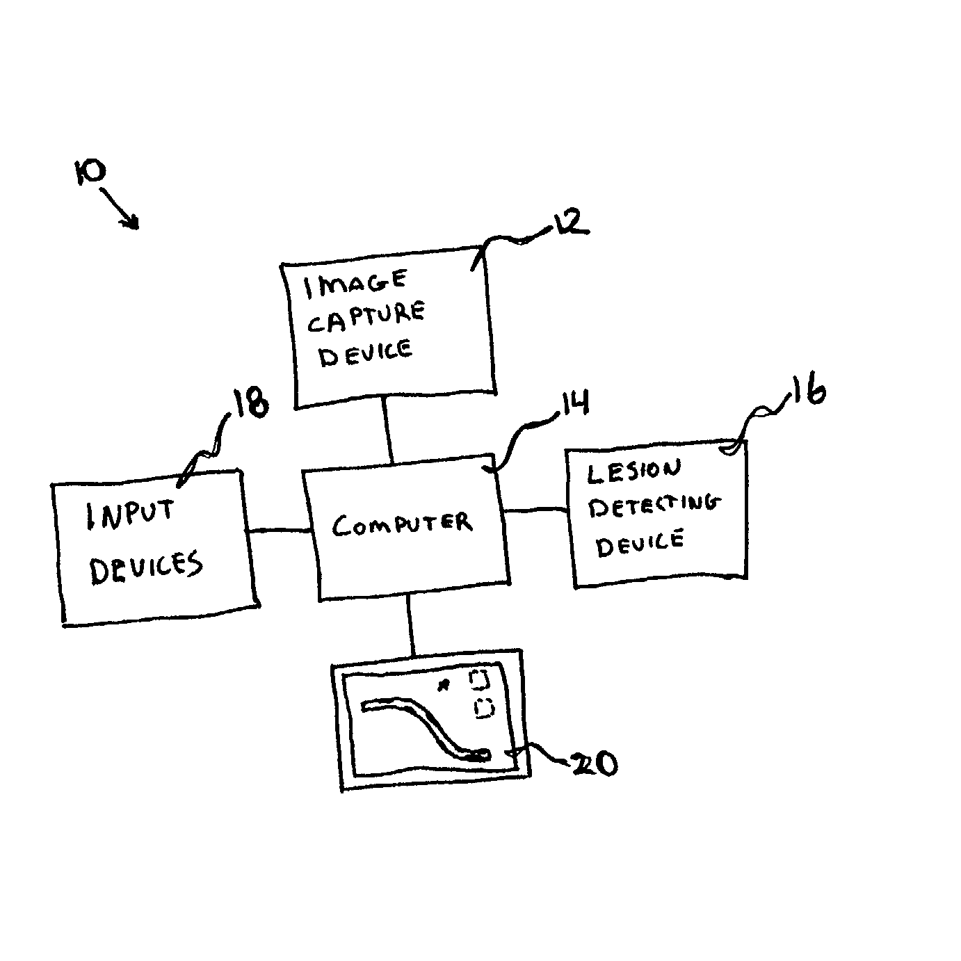
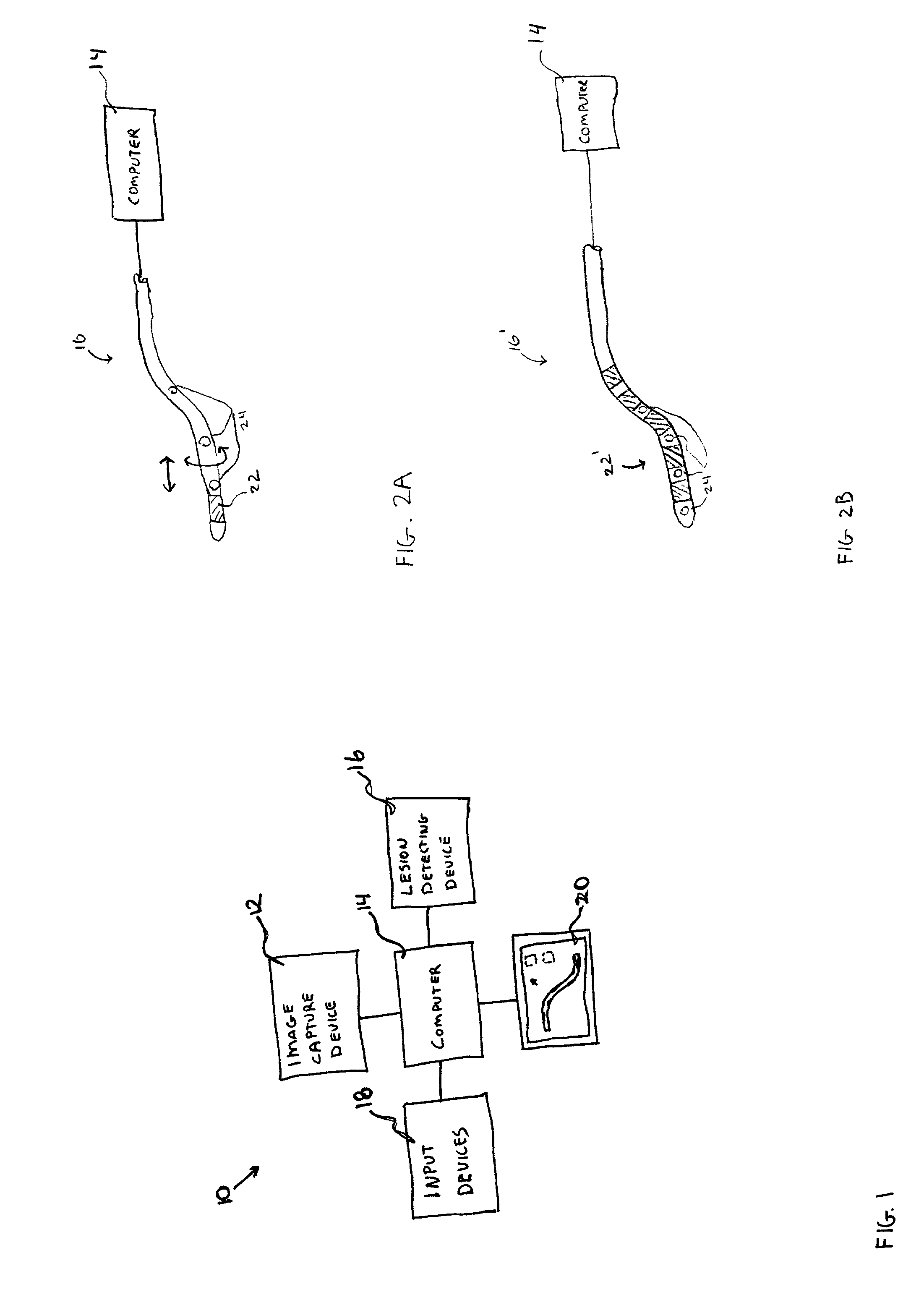
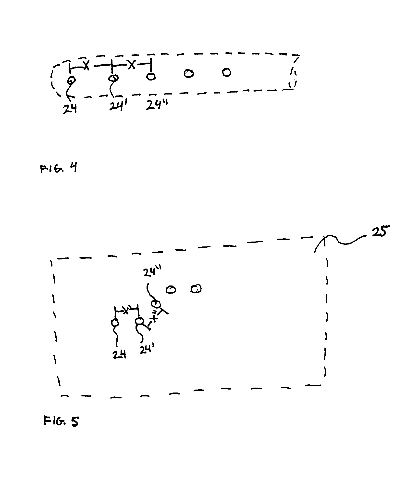
[0045] The present invention provides improved methods and apparatus for localizing and displaying lesions in body lumens, and in particular for displaying the distribution of vulnerable plaque in blood vessels. The methods of the present invention rely on acquiring a separate image of at least the target portion of the body lumen and superimposing information obtained about the lesion over the separately generated anatomic image. The information about the lesion can include, azimuthal distribution of the lesion, longitudinal distribution of the lesion in the body lumen, concentration or severity of the lesion, the type of lesion, biological activity occurring in the body lumen, temperature of the lesion, radiation counts, MRI parameters (signal, T1, T2, Hydrogen density, lipid content, water content, susceptibility, diffusion coefficient, and so on), x-ray density, paramagnetic, ferromagnetic or iodinated contrast agents, ultrasound signal, infrared or optical signature, and the li...


PUM


Abstract

Methods and systems for localizing intravascular lesion in a body lumen. In exemplary embodiments, the methods comprise providing or acquiring an image of the body lumen and displaying information about the lesion with or on the image. In some configurations the information is obtained with an intravascular catheter.

Description

CROSS-REFERENCES TO RELATED APPLICATIONS[0001] The present application is related to patent application Ser. No. 09 / 670,412, filed Sep. 26, 2000, entitled "Methods and Apparatus for Characterizing Lesions in Blood Vessels and Other Body Lumens," patent application Ser. No. 09 / 754,103 filed Jan. 3, 2001, entitled "Intravascular Imaging Catheter," patent application Ser. No. 09 / 754,074, filed Jan. 3, 2001, entitled "Position Sensitive Imaging Catheter," and patent application Ser. No. 09 / 754,822, filed Jan. 3, 2001, entitled "Position Sensitive Imaging Catheter Having Scintillation Detector," the complete disclosures of which are incorporated herein by reference.[0002] The present invention relates generally to medical devices and methods. More particularly, the present invention relates to devices and methods for displaying information about intravascular lesions over an anatomic image of the body lumen.[0003] Coronary artery disease resulting from the build-up of atherosclerotic pla...

Claims


Application Information

Patent Timeline
IPC IPC(8): A61B5/055A61B5/06A61B6/00A61B19/00A61M25/01A61M31/00
CPCA61B5/055A61B5/06A61B6/00A61B6/5247A61B19/5244A61B2019/5238A61B2019/5251A61B2019/5272A61B2019/5291A61B2019/5454A61B2019/5483A61M25/0108A61M25/0127A61M31/005A61B2090/3983A61B2034/2072A61B34/20A61B2090/376A61B2034/2051A61B2090/365A61B2090/3954A61B6/12
Inventor STRAUSS, H. WILLIAMKAUFMAN, LEONGHAZAROSSIAN, VARTAN
Owner IMETRX
Why Eureka
Try Eureka