
Methods and reagents for identifying rare fetal cells in the maternal circulation

Inactive Publication Date: 2003-09-04
ROCHE DIAGNOSTICS OPERATIONS
View PDF1 Cites 75 Cited by
  • Summary
  • Abstract
  • Description
  • Claims
  • Application Information

AI Technical Summary

Benefits of technology

0027] The fetal cell detection and diagnosis methods and related kits of the present invention utilize probes having sequences that hybridize to RNAs in fetal cells to a greater extent than RNAs found in non-fetal, e.g., maternal cells in a mixed cell population. Such probes comprise a nucleotide sequence having 20-30 nucleotides with at least 80% sequence identity to a corresponding portion of a fetal cell specific or selective transcript, e.g., SEQ ID NO:10, 11, 12, 13, 14, 15, 16, 17, 18, 19, 20, 21, 22, 23, 24, 25, 26, 27, 28, 29, 30, 31, 32, 33, 34, 35, 36, 37, 38, 40, 41 or 42. In other embodiments, the nucleotide sequence has at 30-40, 40-60, 60-80, 80-100, 100-150, 150-200, or greater than 200 nucleotides with at least 80% sequence identity to corresponding portion of a fetal

Problems solved by technology

However, both of these procedures are invasive and are accompanied by a small risk (on the order of 1%) of fetal death.
These low levels of fetal cells make even minimal levels of non-specific binding problematic for affinity separation of fetal cells from maternal cells.
Trophoblast cells have been considered undesirable due to concerns that these cells may be subject to confined placental mosaicism, rendering them unrepresentative of the fetus.
The isolation of nucleated fetal erythroid cells from maternal blood has, however, been fraught with difficulty.
The low abundance of these cells in maternal blood renders separation extremely difficult, as even extremely low non-specific binding by a separation reagent will result in large numbers of maternal cells if the reagent positively selects for the fetal cells, and unacceptably low yields if the antibody negatively selects for maternal cells.
However, these methods suffer from a number of deficiencies.
However, these techniques co-enrich large numbers of maternal nucleated erythroid cells, and so do not provide the level of enrichment required for reproducible genetic screening of fetal cells.
These methods suffer from the same problems as immunoaffinity methods, due to the lack of fetal cell specific probes.

Method used


Image

  • Methods and reagents for identifying rare fetal cells in the maternal circulation
  • Methods and reagents for identifying rare fetal cells in the maternal circulation
  • Methods and reagents for identifying rare fetal cells in the maternal circulation

Examples


example 5

6.5. Example 5

Erythrocyte-Specific Antibodies Obtained by Both Positive and Negative Selection

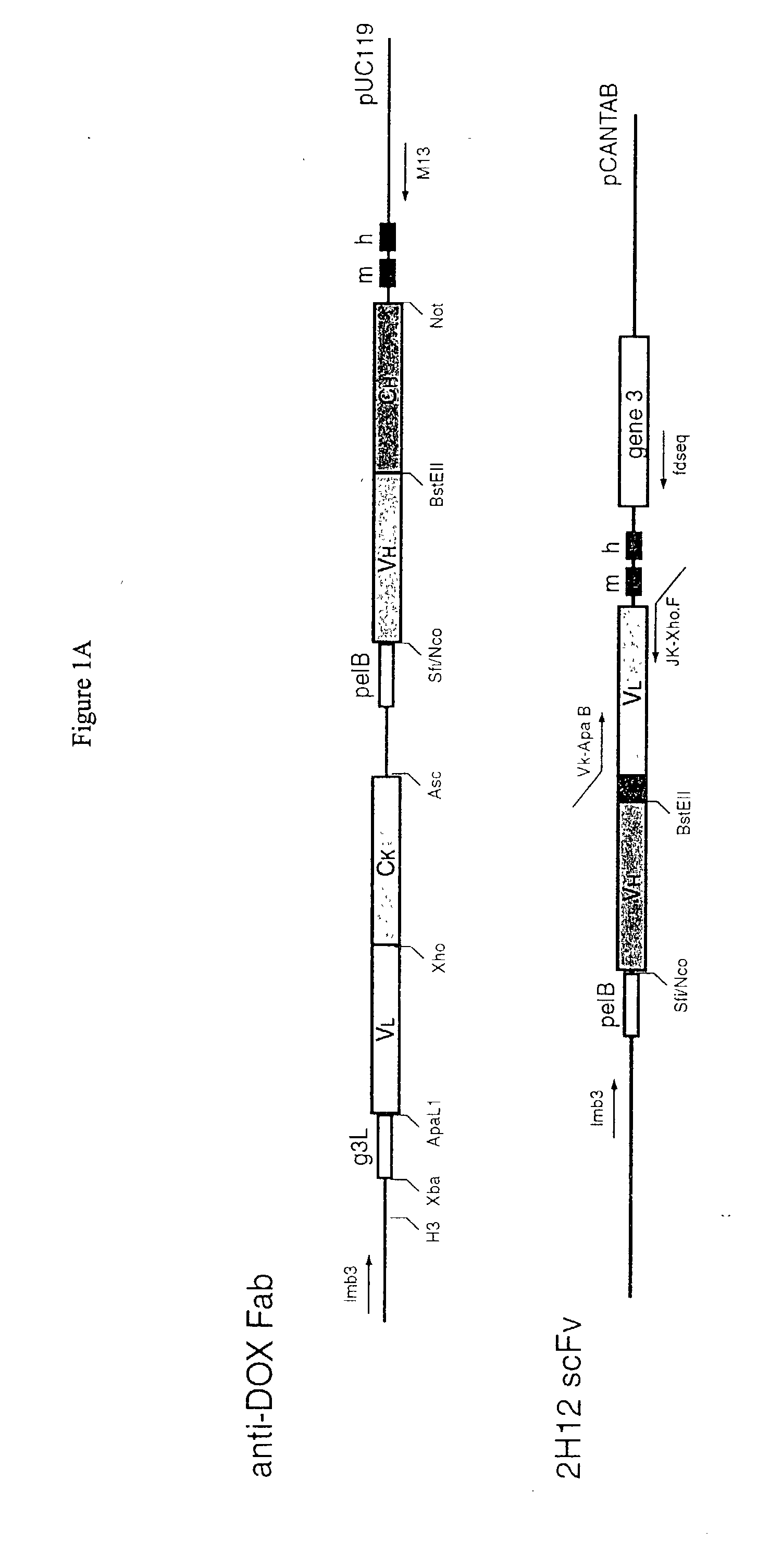
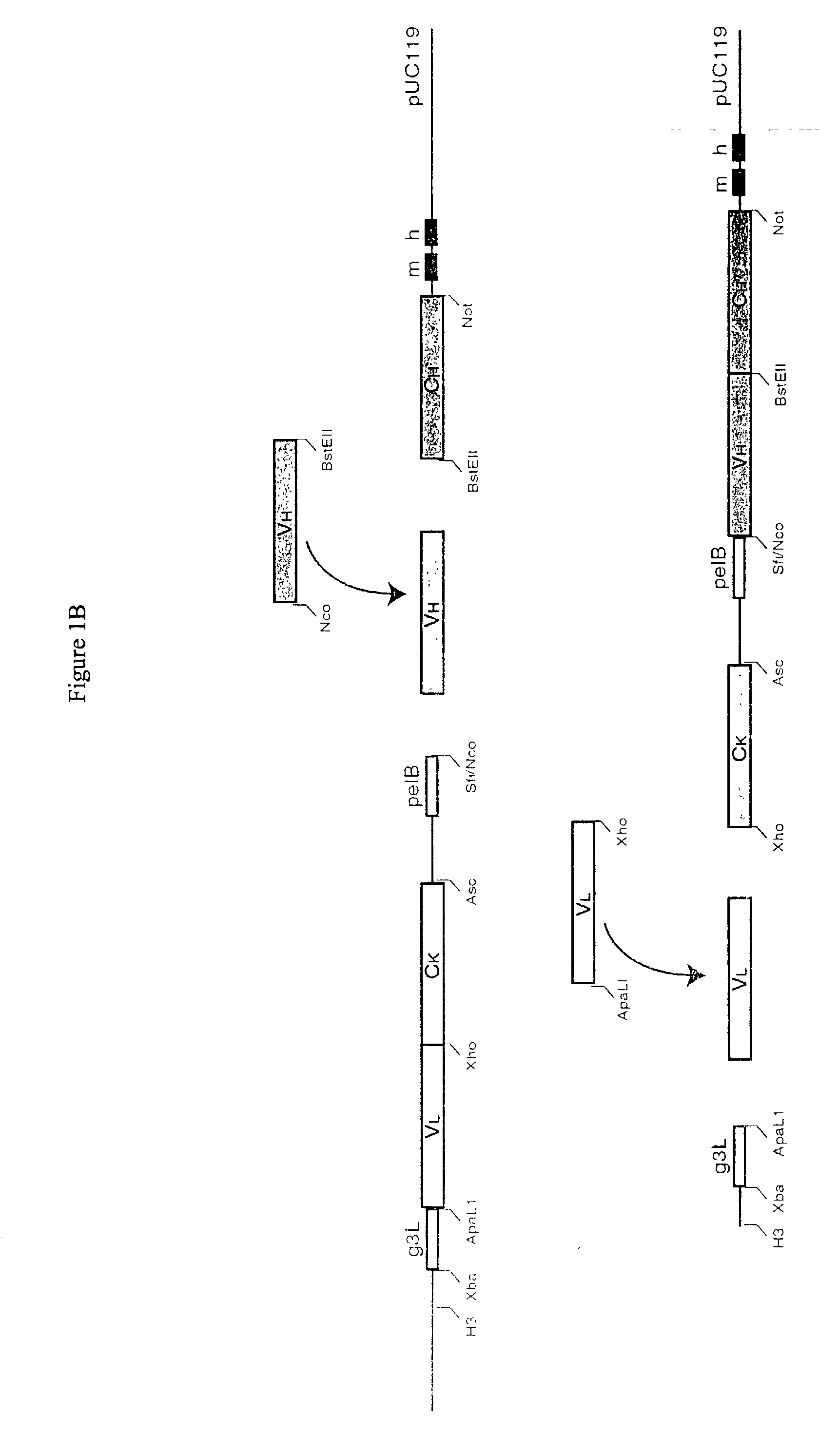
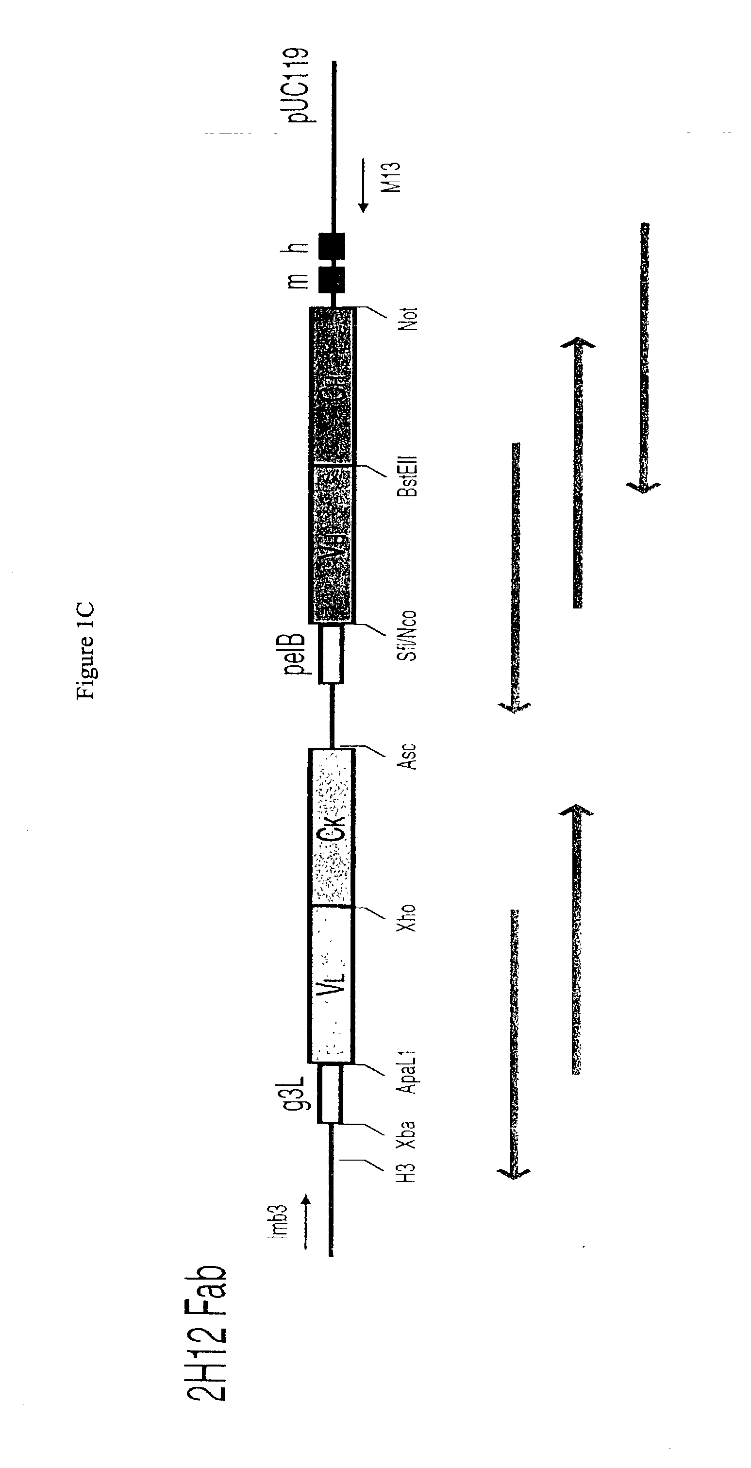
[0316] Additional erythrocyte-specific antibodies were obtained using a modified scFv library. A nave library of Fab expressing phagemids (about 10.sup.10 species) was converted so as to express the variable regions as scFv. The heavy chain CDR3 regions were scrambled to provide additional diversity.

[0317] Specific anti-erythroblast antibodies were obtained by a combination of positive and negative selection. Erythroblasts from fetal liver that had been cultured for 1-2 weeks were used for positive selection. Adult peripheral blood leukocytes (PBL) (pooled Ficol.TM. separated white cells) were used for negative selection. Briefly, the phagemid library was mixed with the erythroblasts. The bound phagemids were then recovered from the cells by adding 0.1 M glycine buffer pH 2.2, inclubating for 5 min, and centrifuging out the cells. The supernatant was neutralized by adding concentrated Tris ...

example 1

7.1. Example 1

Construction of Subtracted cDNA Libraries

[0320] This example is directed at identifying nucleic acid sequences that are expressed at the mRNA level in fetal cells appearing in the maternal circulation, but not in any type of circulating maternal cells that might be present in a test sample after antibody enrichment. Where the target fetal cell is a blood cell precursor such as an erythroblast, the sequence should be able to distinguish fetal cells from maternal cells at the same stage of differentiation.

[0321] To accomplish this, a number of cDNA subtraction libraries were prepared in which sequences specifically expressed in fetal cell precursors are enriched. The libraries were prepared by Suppression Subtraction Hybridization (SSH), a PCR-based method that combines normalization (the matching of mRNA levels) and subtraction (obtaining differentially expressed mRNA) in a single procedure, and requires less mRNA than other subtraction methods.

[0322] Different tissues ...

example 2

7.2. Example 2

Identification and Characterization of Short Fragment cDNAS (Tags) from Subtracted cDNA Libraries

[0328] Random clones from the subtracted libraries were picked and grown to provide sufficient material for characterization. The cDNA insert was PCR amplified for further testing using primers (T3 and T7) corresponding to the flanking sequences of the pCR-Script.TM. (SK+) vector (Cat #211189, Stratagene, La Jolla, Calif.) as designated in the package insert. The PCR products were purified according to manufacturers instructions using the PCR purification kit (Cat#28106) from Qiagen (Valencia, Calif.). The tags were single pass sequenced from one end using Dye-Terminator chemistry on a 377 ABI fluorescent DNA sequencer (PE Biosystems, Inc. Foster City, Calif.). Using either the BLAST (Altschul, S. F., Gish, W., Miller, W., Myers, E. W. & Lipman, D. J. (1990) "Basic local alignment search tool." J. Mol. Biol. 215:403-410.) or the BLAST2 algorithm (Altschul, Stephen F., Thoma...


PUM

PropertyMeasurementUnit
Fractionaaaaaaaaaa
Fractionaaaaaaaaaa
Fractionaaaaaaaaaa

Abstract

This invention provides methods and compositions useful for identifying and diagnosing rare fetal cells in a mixed cell population such as a maternal blood sample. The methods entail the use of specific nucleic acid probes that hybridize to fetal cell associated RNAs to identify the rare fetal cells or antibodies that bind to polypeptides encoded by the fetal cell associated RNAs for fetal cell detection. The cells detected by the methods of the present invention are useful for diagnosing the fetal cells for a genetic trait of interest, such as trisomy 21. Novel methods for simultaneous screening for fetal cells and diagnosing the fetal cells are also provided. Compositions comprising the fetal cell associated nucleic acids of the invention and their encoded proteins are also provided. The present invention further provides kits useful for practicing the present methods.

Description

[0001] The present application claims priority of U.S. provisional application No. 60 / 248,882 under 35 U.S.C. .sctn.119(e), which application is incorporated by reference herein in its entirety.1. FIELD OF THE INVENTION[0002] This invention relates generally to the fields of cell purification, cell identification, and prenatal genetic analysis. More particularly, the invention provides methods and compositions for identifying individual cells of fetal origin in samples of maternal blood. The methods encompass the use of specific nucleic acid probes to identify the rare fetal cells in the maternal blood sample and optionally further diagnosing the detected fetal cells for a genetic trait of interest. Compositions comprising the nucleic acid probes and kits useful in the present methods are also provided.2. BACKGROUND OF THE INVENTION[0003] Amniocentesis and chorionic villus sampling are the currently accepted methods for prenatal testing for genetic abnormalities. However, both of th...

Claims


Application Information

Patent Timeline
IPC IPC(8): C12Q1/68C12Q1/6883G01N33/50G01N33/569G01N33/68
CPCC12Q1/6883C12Q2600/158G01N2800/368G01N33/56966G01N33/689G01N33/5002
Inventor SCHUELER, PAULA A.XU, HONGXIAFOLTZ, LISAWU, XINGYONGSHA, YEHSIUNGNAGY, ALEXANDRAMAHONEY, WALTER C.
Owner ROCHE DIAGNOSTICS OPERATIONS
Learn More