[0032] The method further comprises contacting a
body fluid or tissue sample at a location corresponding to each candidate blob represented in the subset of the first image
data set, with a
reagent to generate a medically significant signal. This method provides the
advantage of being able to remove from further
processing a
body fluid or tissue sample for which no subset of the first
data set representing a candidate blob is created. The signal can be measured to determine whether it is a significant
signal level. The first and / or the second image data subsets can be transformed into a representation that is more suitable for control and
processing by a computer as described herein. In a preferred embodiment, the image data is transformed from an RGB (Red Green Blue) signal into an HLS (
Hue Luminescence Saturation) signal. Filters and / or masks are utilized to distinguish those cells that meet preselected criteria and eliminate those that do not, and thus identify rare cells.
[0035] In general, a subset of a first image
data set can be created as described above. The method can further produce an image file of red, green and blue pixels representative of red, green and blue intensities at respective pixel locations within the
color image received. According to some aspects of the invention, the
processing further includes manually selecting a plurality of pixels within the background; determining
color intensity value ranges corresponding to the portion of the background; and identifying as the background those areas of the image having
color intensity values within the ranges determined. The method may include before the step of measuring, processing in the computer to filter the
color image to make
color intensity values of dark pixels in the
color image lighter and to make color intensity values of light pixels in the color image darker. Moreover, the filtering may include passing the color image through a hole filling filter; passing the filled color image through an
erosion filter; performing a separate operation on the eroded filled color image, to define outlines around areas; selecting pixels within the outlines by performing a logical NOT operation, and performing a logical AND operation between the selected pixels and the filled color image. The steps further encompass contacting a
body fluid or tissue sample at a location corresponding to each candidate blob represented in the subset of the first image data set, with a
reagent to generate a medically significant signal. This provides the
advantage of being able to remove from further processing a body fluid or tissue sample for which no subset of the first data set representing a candidate blob is created. There is an optional step by which the signal can be measured to determine whether it is of a significant level. Another optional step encompasses transformation of one or both of the first and the second image data subsets into a representation that is more suitable for control and processing by a computer as described herein. In a preferred embodiment, the image data is transformed from an RGB (Red Green Blue) signal into an HLS (
Hue Luminescence Saturation) signal. Filters and / or masks are utilized to distinguish those cells that meet pre-selected criteria and eliminate those that do not.
[0049] In an alternative embodiment, the first step of the process is performed using
fluorescence microscopy, which enables identification of the possible rare cell positions at even lower
magnification and higher processing speed compared with the method described above. The rare cells of interest are stained with a fluorescent
label or
fluorophore.
[0053] According to yet another aspect of the invention, there is provided a device for screening fetal cells contained within a smear of an unenriched sample of maternal blood containing a naturally present concentration of fetal cells, comprising: a flexible film having thereon a smear of at least 250 μl of maternal blood. In one embodiment, the flexible film has thereon a smear of at least 500 μl of maternal blood. In one important embodiment, the flexible film is an elongated film, the length being at least 10 times the width. It is particularly preferred that the flexible film include marking coordinates, whereby the computerized microscopic vision
system described herein can locate a cell relative to a point on the film, permitting that cell to be returned to at a later time, if desired.
[0054] According to yet another aspect of the invention, there is provided a device for screening rare cells contained within a sample of cells at a concentration of no greater than one rare cell for every 10,000 cells in the sample of cells. The device is a flexible film having fixed thereon the sample of cells, wherein the flexible film is at least five inches long. In one preferred embodiment the flexible film has a length at least 10 times its width. In another important embodiment, the flexible film includes marking coordinates, whereby the computerized microscopic vision
system described herein can locate a cell relative to a point on the film, permitting the cell to be returned to at a later time, if desired.
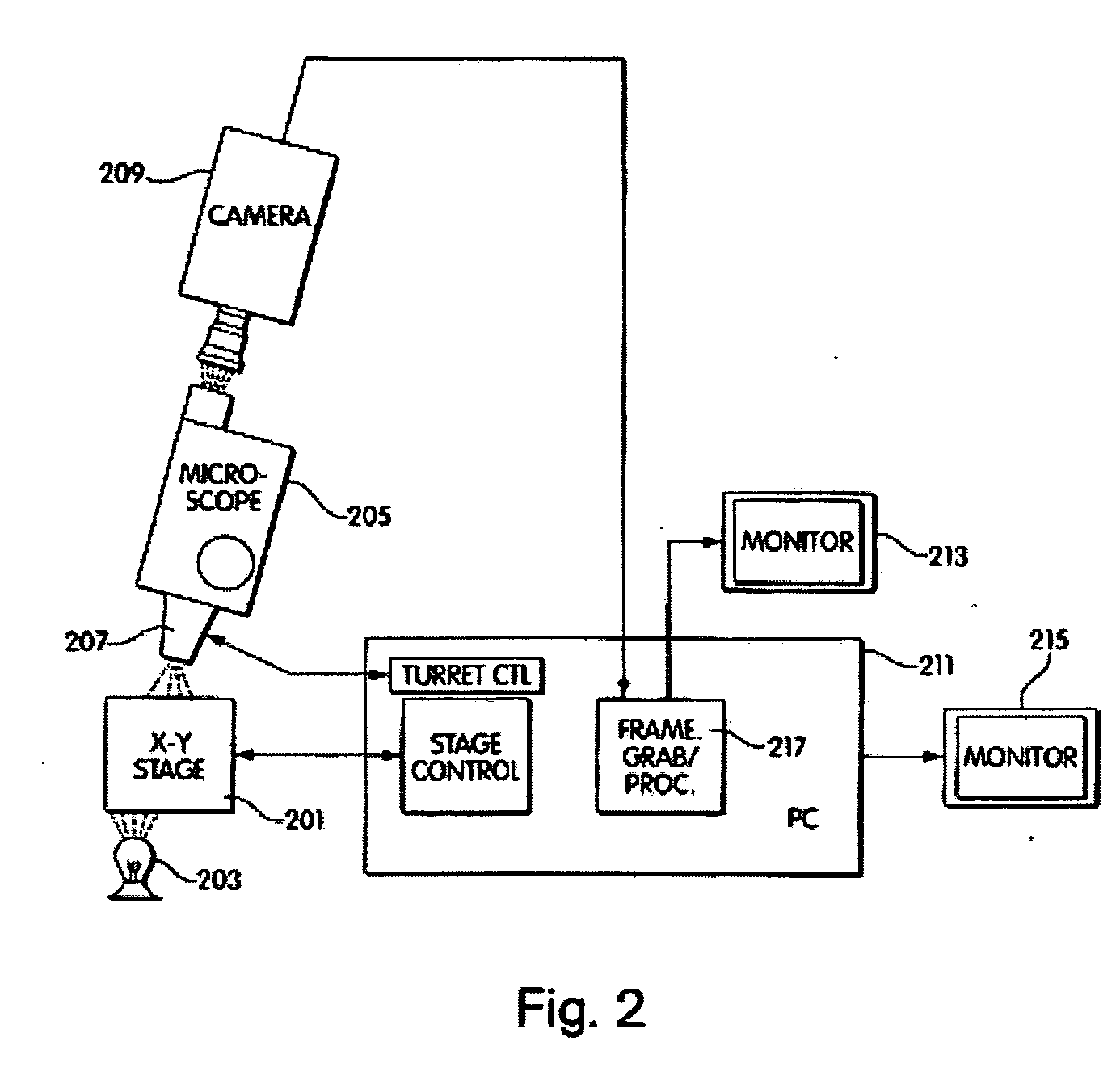
[0055] According to another aspect of the invention, there is provided a device for dispensing materials to a specific location on a slide. The device includes a microscopic vision
system for detecting a signal indicative of the presence of a rare cell in a sample of cells. The device also includes means for identifying the coordinates of the rare cell in an
optical field. The device further has attached to it a dispenser for dispensing a volume of material and means for moving the dispenser to the coordinates whereby the volume of material may be dispensed upon the rare cell. The material dispensed can be reagents such as a
label, PCR, primers, and the like. According to another important embodiment of the invention, the need for scanning large areas of microscopic preparations in the minimum possible amount of time is met by the use of an apparatus or system that provides a “composed” image. It is based on the simultaneous use of an array of computer controlled objective lenses, arranged on a
support system and having the capacity to focus on a microscopic preparation. Each of the objective lenses is connected to a
charge coupled device camera, herein referred to as a
CCD camera, being connected to
image acquisition hardware installed in a host computer. The images are stored in the
computer memory and they are combined in an appropriate side to side fashion, so that a “composed” image is formed in the
computer memory. The “composed” image can be further processed as a unity, using any kind of
imaging procedures to detect specific features that are in question. The significant
advantage of the described system consists in its capacity to acquire images simultaneously from a number of objective lenses, thus minimizing the time needed to process large areas of the sample in a manner that is inversely proportional to the number of objectives used.

