

Method and system for detecting electrophysiological changes in pre-cancerous and cancerous tissue
a technology of electrophysiology and cancer, applied in the field of detection can solve the problems of high mortality rate, requiring expensive, complex, invasive, and/or uncomfortable procedures, and detecting the presence of abnormal or cancerous tissue is difficul
- Summary
- Abstract
- Description
- Claims
- Application Information
AI Technical Summary
Problems solved by technology
Method used
Image

Examples
example 2
[0094] Breast Cancer
[0095] As mentioned above, impedance and DC electrical potential have been used separately at the skin's surface to diagnose breast cancer. In the current invention, the impedance characteristics of the overlying skin or epithelium are measured and factored in to the diagnostic interpretation of the data. For example the surface potential may be more positive (or less negative) than the reference site because of increased conductance of the overlying skin, rather than because of an underlying tumor.
[0096] The electrodes are placed over the suspicious region and the passive DC potential is measured. Then AC impedance measurements are made as discussed below. The variable impedance properties of the overlying skin may attenuate or increase the measured DC surface electropotentials. Alternatively, impedance measurements at different frequencies may initially include a superimposed continuous sine wave on top of an applied DC voltage. Phase, DC voltage and AC voltage...
example 3
[0117] Nasopharyngeal Cancer
[0118] Using methods similar to those described with respect to colon cancer, it is possible to use pharmacological and hormonal agents to enhance electrophysiological alterations caused by nasopharyngeal cancer. One exemplary method would be a nasopharyngeal probe that would include wells providing for varying concentrations of K.sup.+ and would perform simple DC measurements.
example 4
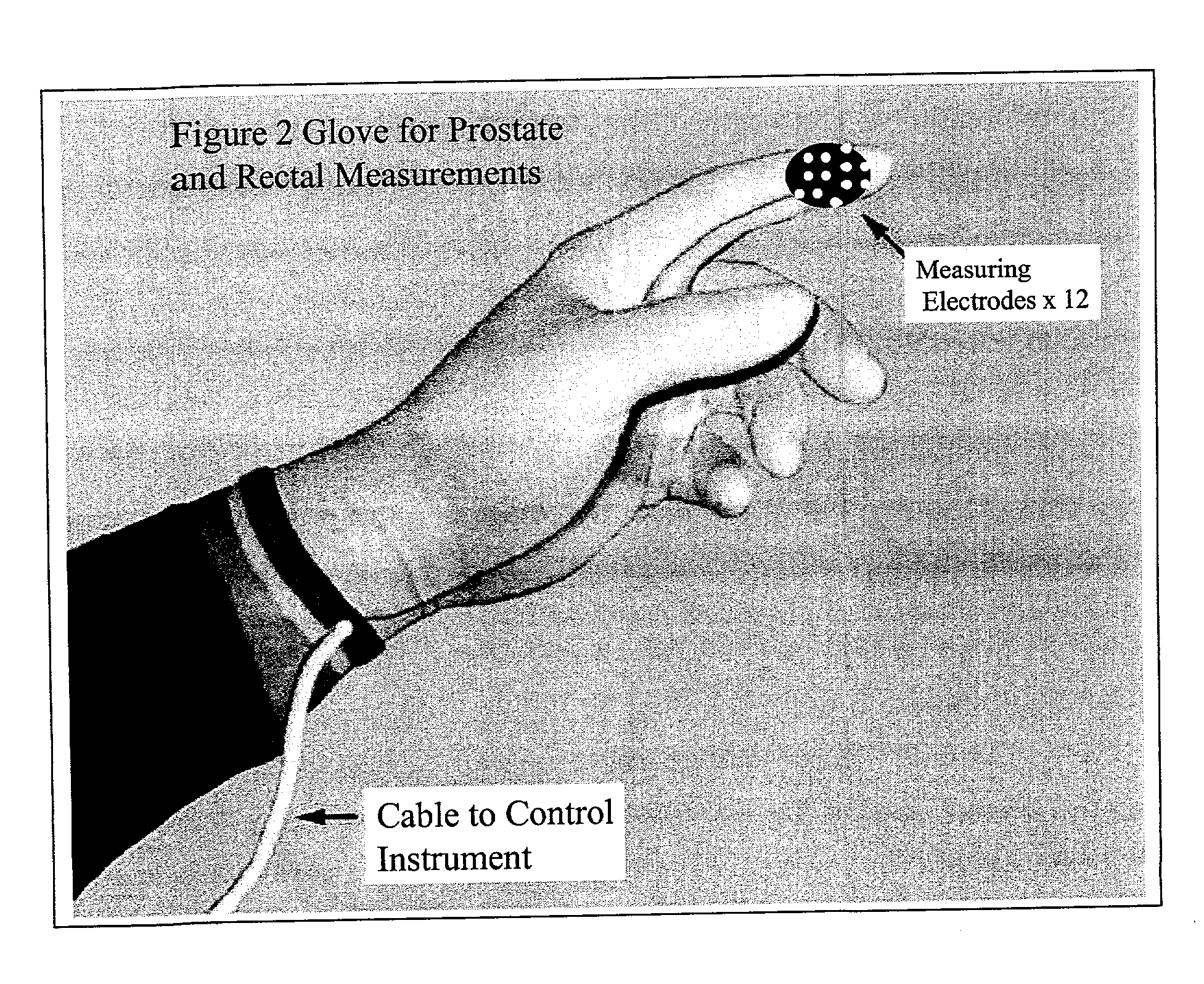
[0119] Prostate
[0120] FIG. 13 represent measurements of cell membrane potential (.psi.) in human prostatic epithelial cells under different growth conditions. A Voltage-sensitive FRET (fluorescent energy transfer) probe was used for potentiometric ratio measurements. It has two fluorescent components: CC.sub.2-DMPE (Coumarin) and DISBAC.sub.2(3) (Oxonol). The oxonol distributes itself on opposite sides of the cell membrane in a Nernstian manner according to the .psi.. The voltage sensitive distribution of oxonol is transduced through a ratiometric fluorescence signal via the coumarin which is bound to the outside surface of the cell membrane thereby amplifying the fluorescence. Measurements were made using a fluorescence microscope and a digital imaging system. The ratio measurements are calibrated using Gramicidin D to depolarize the cell membrane and then varying the external K.sup.+-concentration. The calibrated cell membrane potential in mV is depicted on the y-axis.
[0121] The f...
PUM
 Abstract
Description
Claims
Application Information