
Method for the selection and identification of peptide or protein molecules by means of phage display

a phage display and peptide technology, applied in the field of phage display for peptide or protein molecules, can solve the problems of not being able to achieve spatial assignment of beads, affecting the accuracy of phage display methods, and requiring a large amount of beads to be processed,

Inactive Publication Date: 2005-01-20
SANTHERA PHARMA SCHWEIZ
View PDF13 Cites 8 Cited by
  • Summary
  • Abstract
  • Description
  • Claims
  • Application Information

AI Technical Summary

Benefits of technology

[0006] The immobilisation of an interaction partner (ligand) is necessary to enable separation of the virus-ligand complexes from the non-interacting viruses. It furthermore facilitates conduction of the process such as, for example, the performance of washing steps, and may, in connection with a suitable detection method, give information about the presence and strength of the interactions between the interaction partners.
[0041] Infection of the host cells for the multiplication of the viruses preferably occurs by means of the viruses bonded to the surface of the solid phase carrier. The advantage of this is that the bond between the ligand and the virus-presented peptide or protein does not have to be removed.

Problems solved by technology

A big disadvantage of using these bead affinity columns is that no spatial assignment of the beads is possible.
This presents a considerable problem for the automation and miniaturisation of bead-based phage display methods.
The complex handling of the beads, for example when carrying out washing steps, is also disadvantageous.
A further disadvantage is that clearances form between the spherical beads, in which dead volumes of virus suspension collect.
A big disadvantage in using microtiter plates as the incubation vessel is that the cavities only allow a relatively small sample volume for selection (e.g. 0.2-1.2 ml volume per cavity in standard 96-cavity microtiter plates; 20 to 60 μl in 384-cavity microtiter plates).
Owing to this volume limitation of the cavities, it is often necessary to enrich the viruses before screening.
It can, however, be necessary for successful screening to bring a total number of phages between 1×10¹⁰ to 1×101 pfu / ml or more into contact with the affinity ligand, and thus the concentration of the viruses from the lysate cannot be avoided before microtiter plate-based screening owing to the limited sample volume of the individual cavities (see above) (T7Select® System Manual, Novagen, Madison (USA) (TB178 06 / 00), page 6 et seq.).
The disadvantage hereof is that the virus precipitates hereby forming can often only be re-dissolved with difficulty.
The screening process is thus negatively influenced by the solubility behaviour of the viruses found in the precipitate.
Furthermore, procedural steps connected with the precipitation of viruses are time-consuming and difficult to automate.
It is further disadvantageous that a competing, simultaneous selection of structurally similar ligands against the phage library is ruled out by the separated volumes since only one ligand can be immobilised per cavity.
The disadvantage of this method is that a common ELISA for identifying interacting virus clones is only possible when using peptidic affinity ligands derived from a common protein / polypeptide sequence.
A disadvantage of using membranes as the surface is that the local concentration of the ligand can only be controlled with a lot of effort.
This can lead to the formation of non-specific virus-ligand complexes owing to local avidity effects.
A very big disadvantage of the above method is that the spatial information of the array with regard to the ligands is lost during selection since the interacting viruses i) cannot be detected on the array and ii) cannot be eluted from the array in a site-specific manner.
If marker-based detection methods (e.g. ELISA assays) are used, the bonding assay forming the basis for selection will be restricted during performance to the conditions of the marking reaction, and thus a variation of the bonding assay which is advantageous for selection, e.g. a change in the pH value, the ionic strength or the use of detergents, is not possible.
A big disadvantage of marker-based detection methods as used in the method cited above is furthermore that the viruses identified in the selection process cannot be used for the further method steps.
This physical interaction can lead to a change in the bond between the ligand and the virus (weakening or strengthening) or even to impairment of the host-virus interaction (loss of infectiousness).
It must be expected when using marker-based detection methods that insoluble aggregates form, and thus the viruses contained therein are no longer available for further method steps.
A very big disadvantage of this method is that a flow system is used.
The disadvantage hereof is that it is precisely due to the use of a flow system that a two-dimensional sample arrangement (two-dimensional array) and the miniaturisation thereof is made considerably more difficult.
Competing affinity selection can furthermore only be laboriously realised in a flow system.
The marker-free selection of a plurality of virus clones, which is, in the end, the result of a massively parallel screening set-up, can furthermore not be accomplished with the available technology.

Method used


Image

  • Method for the selection and identification of peptide or protein molecules by means of phage display
  • Method for the selection and identification of peptide or protein molecules by means of phage display
  • Method for the selection and identification of peptide or protein molecules by means of phage display

Examples


Embodiment Construction

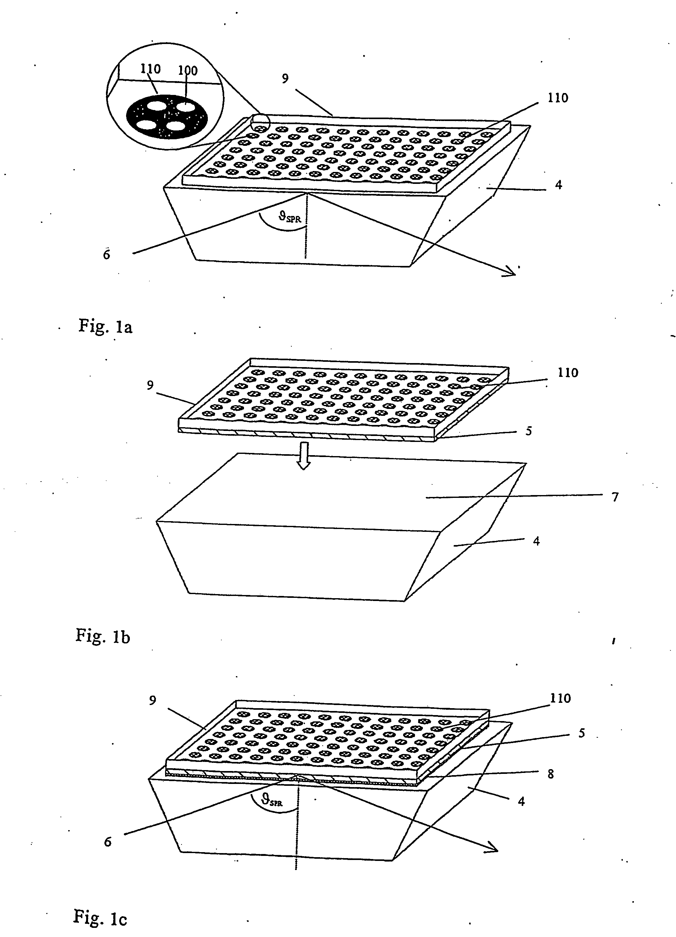
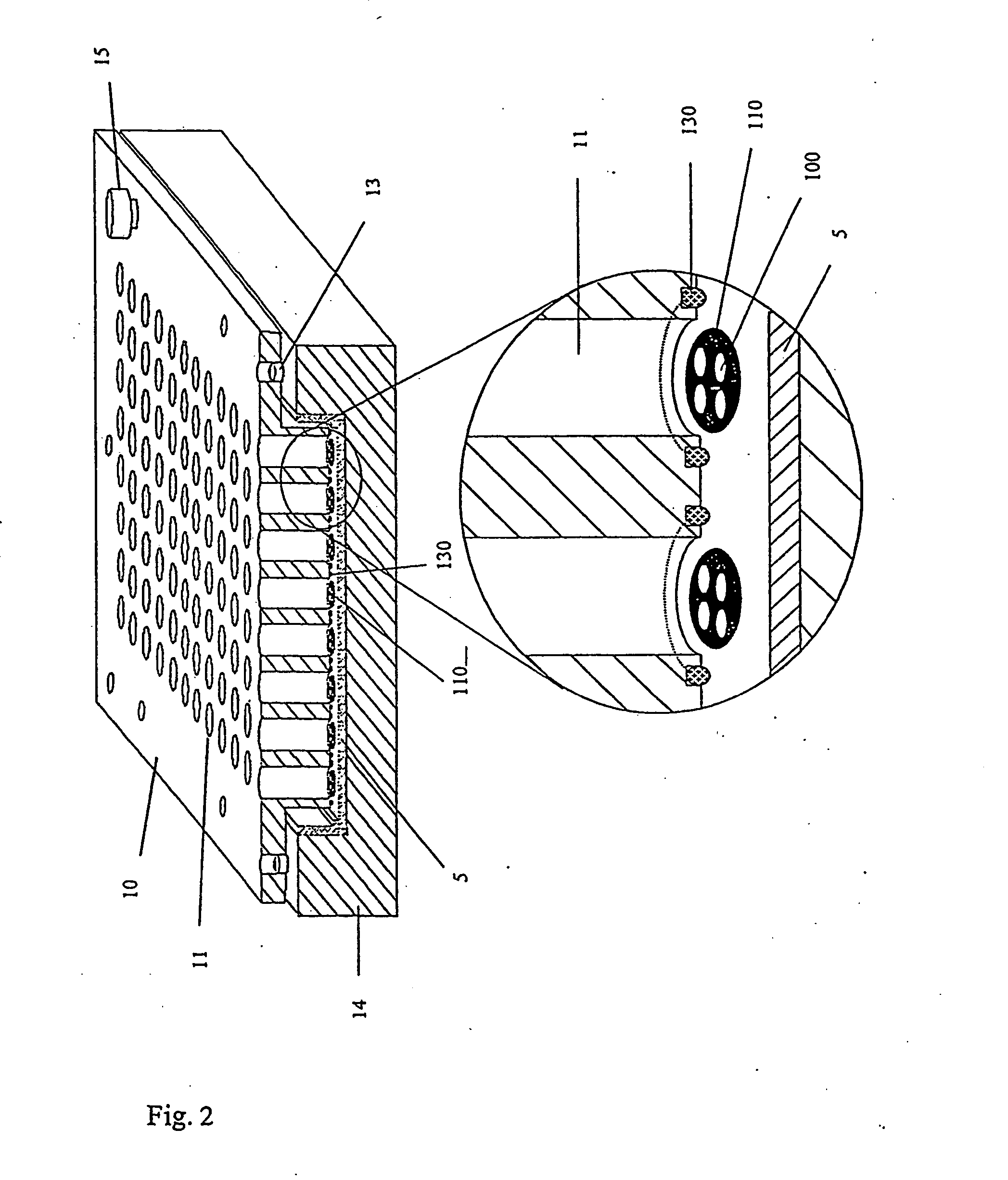
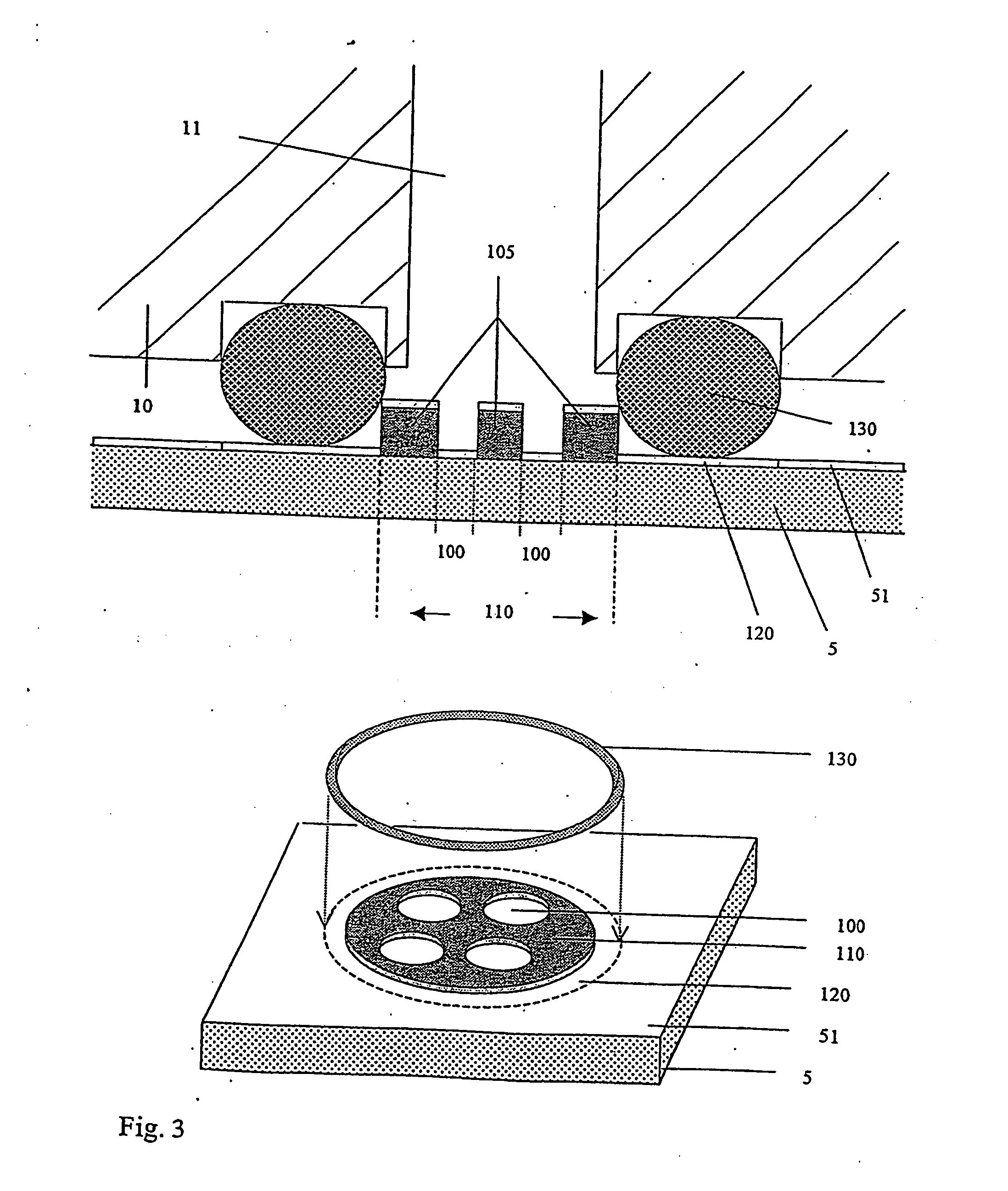
[0046] The method according to the invention allows the selection and identification of one or more representatives of peptide or protein molecules from a plurality of such molecules. In this context “representative” means that each different peptide or protein molecule in the plurality of molecules does not normally occur as an individual molecule, but is rather present in the protein mixture to a greater or lesser extent. The selection and identification principle is then based on the fact that the peptide or protein molecule sought can interact with one or more previously chosen “selection molecules” to form a bond. These selection molecules are not particularly limited as regards their nature and can have any structure provided that they can be used at all in such a test and are able to form a bond. According to the invention they are therefore also simply referred to as “molecules”. The expression “ligand” is also used within the context of the present invention for those molec...


PUM


Abstract

The invention relates to a method for the selection and identification of at least on representative (interaction partner) from a plurality of peptide or protein molecules, which can specifically interact with at least one representative from a plurality of target molecules, forming a bond. The inventive method comprises the following steps: (a) a virus system consisting of a plurality of viruses, wherein each virus respectively presents at least on representative from the plurality of peptide or protein molecules on the surface thereof, is brought into contact with the plurality of target molecules (ligands) which are immobilized on the surface of a solid phase carrier such that they are position addressable in a two-dimensional grade; (b) unbound viruses removed from the surface thereof; and (c) the interaction partner is identified by detection and determination of the position of the bond between the immobilized ligand and the interaction partner presented by the virus with the aid of a marker-free detection method. The described method makes it possible to concentrate viruses presenting interaction partners by means of an optionally cyclic repetition of selection. Optionally, selected interaction partners are recombinantly expressed after identification of the coding nucleotide sequence.

Description

[0001] The present invention relates to a method for the selection and identification of peptide or protein molecules by means of phage display. [0002] Selection and identification methods have led to the understanding of numerous biological processes. The conventional experimental approaches used therein to date were cell-based screening and affinity chromatography. Although both techniques are helpful for discovering peptide or protein molecules, a method which couples protein identification with gene isolation is very desirable. [0003] Phage display screening systems follow this approach. The combination of in vitro gene expression techniques with traditional biochemical approaches such as, for example, affinity chromatography, which is used therein offers the possibility of functional gene selection by creating a direct link between natural product affinity and gene structure. [0004] In phage display, genes encoding non-viral proteins or peptides are incorporated into the viral ...

Claims


Application Information

Patent Timeline
IPC IPC(8): B01L3/00G01N21/55
CPCB01L3/5085G01N21/553B82Y30/00
Inventor HILL, OLIVEROTTLEBEN, HOLGERDICKOPF, STEFANBURKERT, KLAUS
Owner SANTHERA PHARMA SCHWEIZ
Learn More