
Method for assembling PCR fragments of DNA

a technology of pcr fragments and dna, which is applied in the field of assembling pcr fragments of dna, can solve the problems of difficult assembly of specific ordered fragments much larger, limited size of current methods of manipulating dna fragments, and inability to define the orientation of the resulting fragment. to achieve the effect of facilitating the steps

Inactive Publication Date: 2005-03-03
RICE UNIV
View PDF1 Cites 7 Cited by
  • Summary
  • Abstract
  • Description
  • Claims
  • Application Information

AI Technical Summary

Benefits of technology

[0013] The useful features of this invention are that several fragments can be joined in a defined order allowing the PCR generated DNA fragments to be assembled into an ordered arrangement and formed into a replicating plasmid without use of restriction enzymes. The final product is in a form for transformation into cells and the use of immobilized DNA facilitates the steps and allows for scaling with automated devices.

Problems solved by technology

Current methods of manipulating DNA fragments are each limited by size.
Plasmid sized fragments of up to 10 kb can be easily assembled, but specifically ordered fragments much larger are difficult to assemble by current techniques and require careful handling to avoid breakage.
However, PCR fragments often have like ends, so that orientation of the resulting fragment is not defined.
Further, many restriction enzymes also cleave within a large PCR fragment and cannot be used in this way.

Method used


Image

  • Method for assembling PCR fragments of DNA
  • Method for assembling PCR fragments of DNA
  • Method for assembling PCR fragments of DNA

Examples


example 1

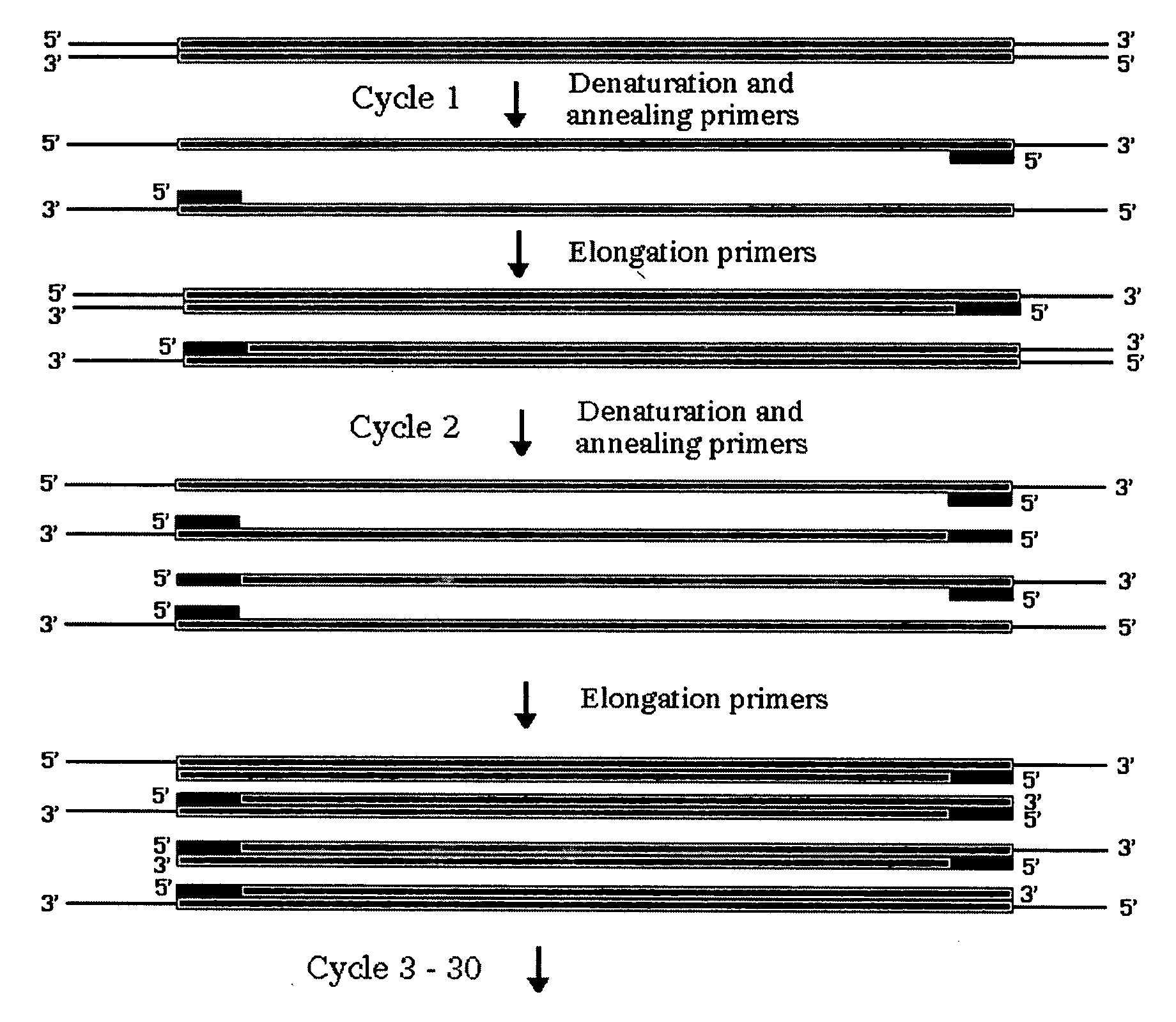
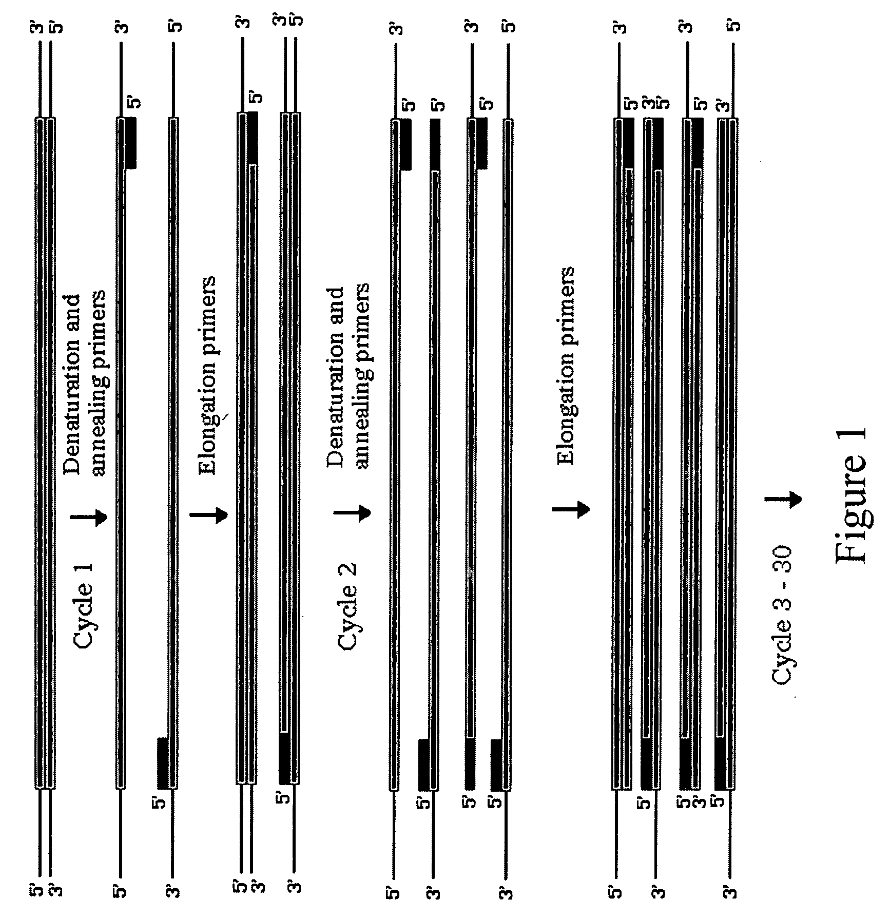
[0028] All PCRs were performed using Gene Amp® reagents (PERKIN-ELMER,™ Norwalk, Conn., USA) in 50 μL reaction mixtures with 2.5 U Taq DNA polymerase and 2.5 mM MgCl2 in a RoboCycler Temperature Cycler (STRATAGENE,™ La Jolla, Calif., U.S.A.). PCR primer sequences are shown in Table 1. Amplifications from pACYC184 (NEW ENGLAND BIOLABS,™ Beverly, Mass., U.S.A.) contained 50 ng of plasmid and 40 pmol of each PCR primer CAT3 and CAT5 or SacCAT3 and SacCAT5. Cycling was at 95° C. for 1 min. 55° C. for 1 min. 72° for 1 min for 30 cycles followed by 1 cycle at 72° C. for 3 min. Genomic amplifications from E. coli strain W3110 contained 100 ng genomic DNA, 40 pmol of LacST#1 and #2, LacMD#1 and #2, LacEN#1 and #2 (See Table 1.) and were carried out at 95° C. for 45 s, 55° C. for 45 s, 72° C. for 45 s for 30 cycles, then a final extension at 72° C. for 3 min.

TABLE 1PCR Primer SequencesCAT3(5′ AGCUCGGCAC GTAAGAGGTT CCAACTTTCA CC 3′ [32 nucleotide])CAT5(5′ AGCUCCAGGC GTTTAAGGGC ACCAATAACT GC...

example 2

[0035] Our first example demonstrated the viability of the method as used in solution. However, the method was cumbersome, tedious, and not applicable for large scale up. Thus, we now demonstrate a solid phase procedure that is suitable for scale-up and commercial use.

[0036] This example was based on the use of a plasmid which, intact, confers resistance to the antibiotic ampicillin and has the lacZ gene which allows for metabolism of the substrate Xgal; an E. coli host with a lacZ-containing plasmid grows as a blue colony on Xgal plates instead of a white colony which the E. coli host produces.

[0037] The first step is to design oligonucleotides so that the PCR product A will have an overhang on the 3 end opposite of the biotin and will leave a sticky 3′ end after enzymatic treatment with UDG and T4 endonuclease V AND so that there is a biotin attached to the 5 end AND so that the PCR amplified product includes a loxP site near the end where biotin is attached. In our example, thi...


PUM

PropertyMeasurementUnit
Digital informationaaaaaaaaaa

Abstract

A process for assembling a series of DNA fragments generated by PCR into an ordered circular arrangement for replication and genetic work in cells. The PCR fragments are made with a modified nucleotide in the primers that can be removed with a DNA excision repair enzyme to generate a 3′ overhang. The 3′ overhangs are designed to allow directional annealing and thus sequential PCR fragments can be assembled by annealing the overhangs and subsequent ligation. Sequential addition of PCR fragments is facilitated by growing the chain on a solid support, and the assembled chain can be removed with a site specific recombinase if the first and last primers contain the recombinase site. The circularized assembled fragment can be directly used for cell transformation if the appropriate sequences are included, such as an origin of replication and a selectable marker.

Description

PRIOR RELATED APPLICATIONS [0001] This patent application claims priority to U.S. Provisional Application Ser. No. 60 / 422,807, filed on Oct. 31, 2002, the disclosure of which is incorporated by reference in its entirety herein.FEDERALLY SPONSORED RESEARCH STATEMENT [0002] Not applicable. REFERENCE TO MICROFICHE APPENDIX [0003] Not applicable. FIELD OF THE INVENTION [0004] A process for assembling a series of DNA fragments generated by PCR into an ordered circular arrangement for replication and genetic work in cells. The method uses the 3′ overhangs created by excision repair enzymes to direct the sequential ligation of PCR fragments on a solid scaffold. The resulting assembled DNA is removed with a site specific recombinase. BACKGROUND OF THE INVENTION [0005] Current methods of manipulating DNA fragments are each limited by size. Plasmid sized fragments of up to 10 kb can be easily assembled, but specifically ordered fragments much larger are difficult to assemble by current techni...

Claims


Application Information

Patent Timeline
IPC IPC(8): C12P19/34C12Q1/68
CPCC12Q1/686C12Q2525/307C12Q2525/151C12Q2521/514
Inventor BENNETT, GEORGEHARRISON, MARY
Owner RICE UNIV
Learn More