Image processing apparatus and signal processing apparatus

a signal processing and image processing technology, applied in the field of image processing apparatus, can solve the problems of not easy to distinguish the difference, no proposal of a detection method in consideration of the shape of the curved surface, and no proposal of a detection method for abnormal shadow candidates

Inactive Publication Date: 2005-03-10
KONICA MINOLTA MEDICAL & GRAPHICS INC
View PDF7 Cites 25 Cited by
  • Summary
  • Abstract
  • Description
  • Claims
  • Application Information

AI Technical Summary

Benefits of technology

According to the above-mentioned apparatus, by using the feature value including curvatures, it is possible to detect a signal ar...

Problems solved by technology

However, since the Iris filter in general strongly responds to an area having a circled shape with lower density than the other part, if mammary gland which has high circularity appears in a massive fashion on an image, there is a case of detecting mammary gland of normal tissue as abnormal shadow by mistake.
Similarly, if a high frequency signal such as noise is generated on an image, since signal variation of noise is similar to that of clustered-microcalcification, it is not easy to distinguish the difference thereof.
Thereby, there is a case of ...

Method used


Image

  • Image processing apparatus and signal processing apparatus
  • Image processing apparatus and signal processing apparatus
  • Image processing apparatus and signal processing apparatus

Examples


first embodiment

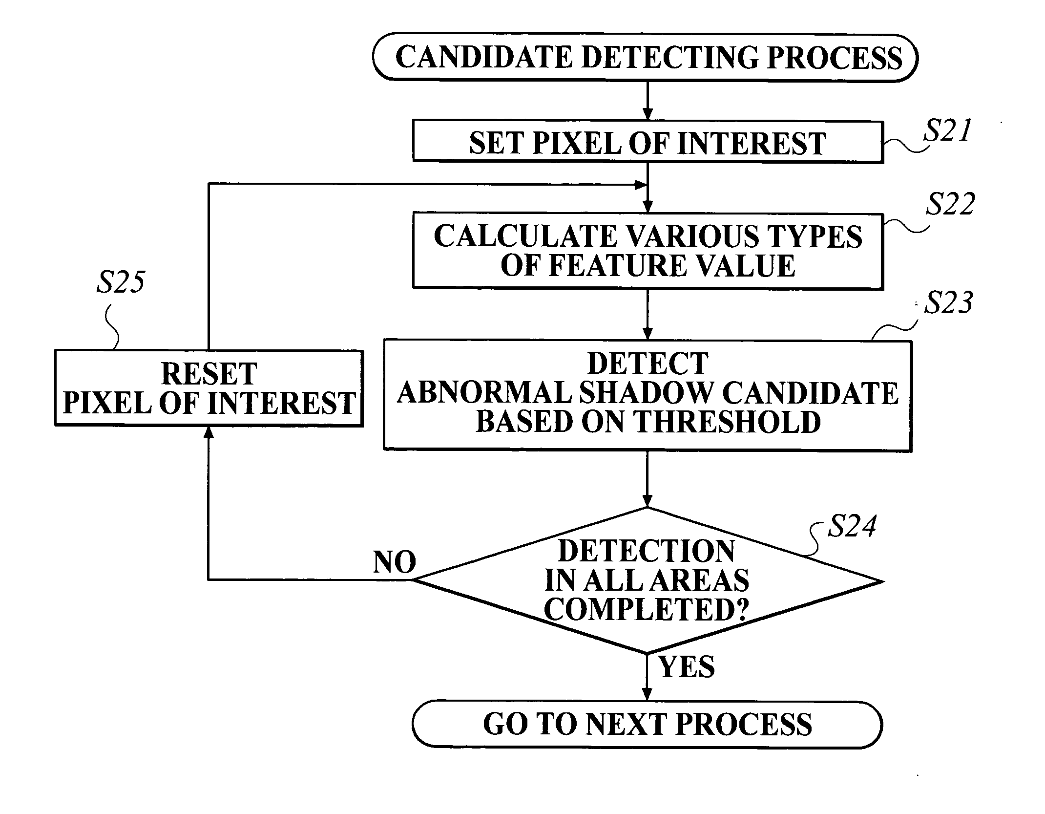
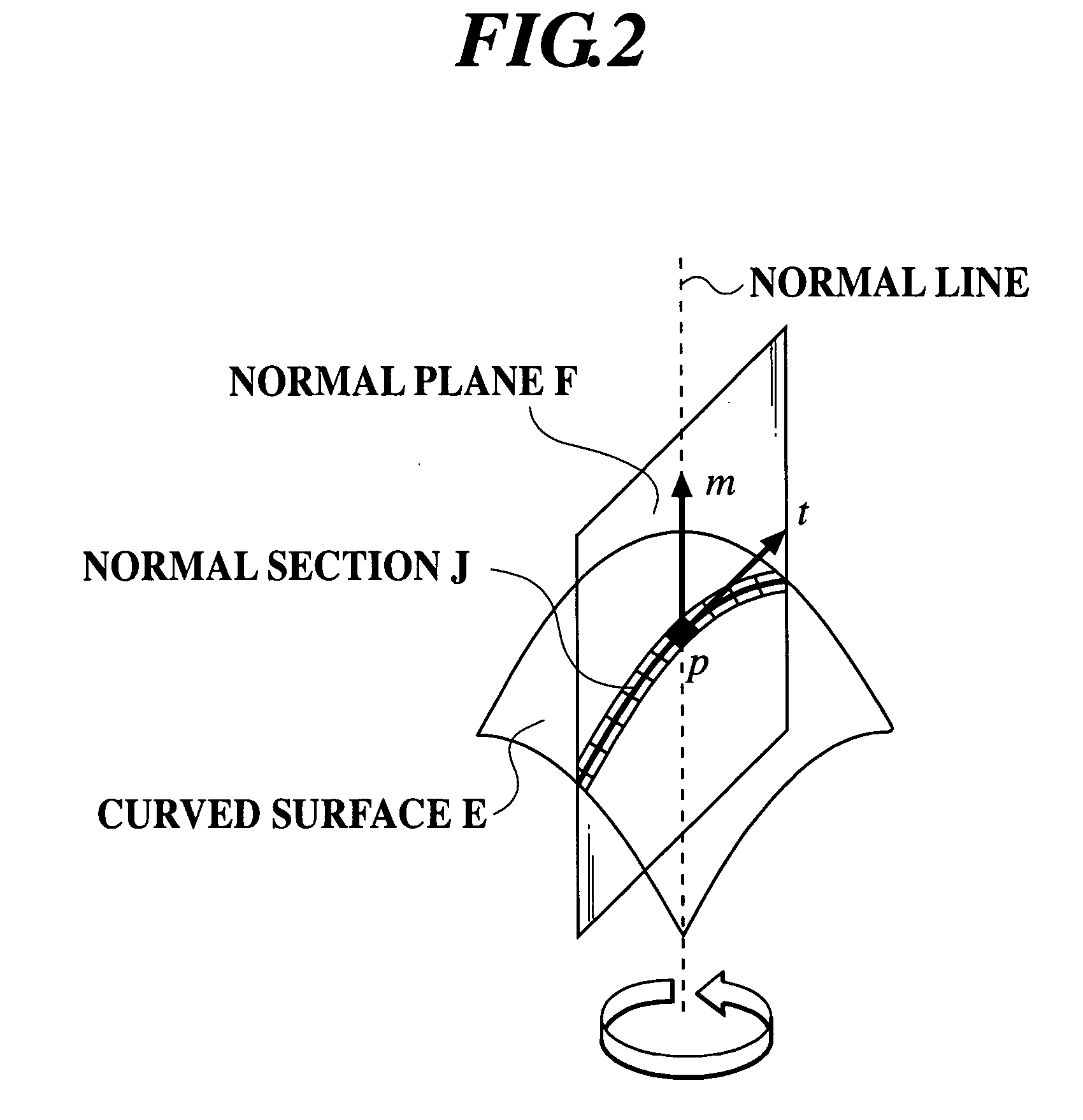
In the first embodiment, an example in which a pixel of interest is set in image signals of a digital medical image, the image signals forming a curved surface of density distribution, curvatures at the pixel of interest are calculated as a feature value according to image signals within a predetermined range around the pixel of interest, and a signal area of an abnormal shadow candidate is detected based on the calculated feature value will be described.

First, a structure of the first embodiment will be described.

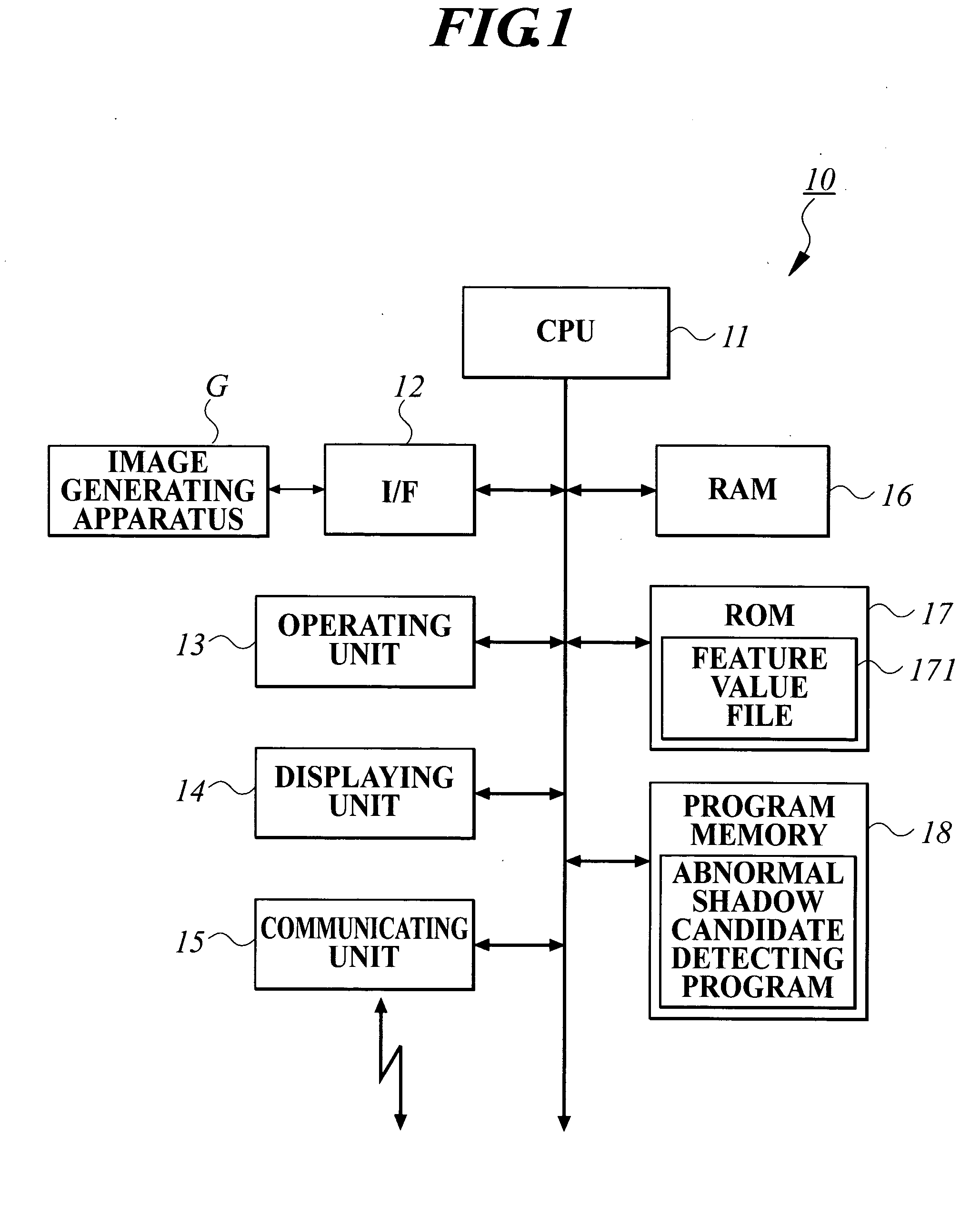
FIG. 1 shows a functional structure of an image processing apparatus 10 according to the first embodiment.

As shown in FIG. 1, the image processing apparatus 10 comprises a CPU (Central Processing Unit) 11, an I / F (InterFace) 12, an operating unit 13, a displaying unit 14, a communicating unit 15, a RAM (Random Access Memory) 16, a ROM (Read Only Memory) 17 and a program memory 18.

The CPU 11 develops a system program stored in the program memory 18 and an abnormal s...

second embodiment

In a second embodiment, what will be described is an example where an approximate function is calculated by approximating a curved surface of density distribution which is formed by medical image signals having a density direction according to the least squares method, and when curvatures of the curved surface are to be calculated as feature values by using coefficients determining the approximate function, a range of the curved surface within which the approximate function is calculated is changed, and an approximate function for each changed range is calculated for calculating the feature values.

First, a structure in the second embodiment will be described.

FIG. 13 shows a functional structure of a signal processing apparatus 20 in the second embodiment.

As shown in FIG. 13, the signal processing apparatus 20 comprises a CPU (Central Processing Unit) 21, an I / F (InterFace) 22, an operating unit 23, a displaying unit 24, a communicating unit 25, a RAM (Random Access Memory) 26,...

third embodiment

In a third embodiment, what will be described is an example where an approximate function by which a curved surface of density distribution comprising medical image signals including density direction is approximated is calculated according to the least squares method, and when a feature value is calculated by using coefficients determining the approximate function, the feature value is calculated by calculating approximate function at each changed degree.

First, a structure of the third embodiment will be described.

Since an internal structure of a signal processing apparatus in the third embodiment is the same as that of the signal processing apparatus 20 in the second embodiment, identical numerals are added to the same parts for omitting description thereof, and only parts having different function will be described. In other words, the signal processing apparatus 20 in the third embodiment comprises a CPU 21, an I / F 22, an operating unit 23, a displaying unit 24, a communica...


PUM


Abstract

An image processing apparatus includes a feature value calculating section for setting a certain pixel in a processing target image as a pixel of interest, and for calculating a curvature of a curved surface which is obtained from density distribution of adjacent pixels which are located within a predetermined range from the pixel of interest, as a feature value of an image area within the predetermined range from the pixel of interest.

Description

BACKGROUND OF THE INVENTION 1. Field of the Invention The invention relates to an image processing apparatus which calculates a feature value of an image having density distribution, and also relates to a signal processing apparatus which calculates a feature value of signals forming a curved surface. 2. Description of Related Art In a medical field, digitization of medical images is achieved. According to the digitization, medical image data generated by a CR (Computed Radiography) apparatus or the like is displayed on a monitor, and diagnosis is done with a doctor reading the displayed medical image on the monitor, to observe a state of a lesion and a change of the same with time. So far, for the purpose of reducing burdens of a doctor who reads images, what has been developed is a medical signal processing apparatus called Computer-Aided Diagnosis (hereinafter, it is referred to as CAD) which automatically detects a lesion which appears on the image as an abnormal shadow can...

Claims


Application Information

Patent Timeline
IPC IPC(8): G06T7/00G06T7/60G06V10/46
CPCG06K9/48G06T2207/30068G06T7/604G06T7/0012G06T7/64G06V10/46
Inventor KASAI, SATOSHIKOBAYASHI, TSUYOSHI
Owner KONICA MINOLTA MEDICAL & GRAPHICS INC
Why Eureka
Try Eureka