Therapeutic biological product and method for formation of new vascularised bone

a technology of biological products and vascularised bones, applied in the direction of powder delivery, peptide/protein ingredients, peptide/protein ingredients, etc., can solve the problems of complex action, unfavorable surgical repair of lost bone, and limited success of igfs as local stimulators of bone healing, so as to achieve clear osseoinductive activity and achieve regulatory approval.

Inactive Publication Date: 2005-03-10
CELLFACTORS
View PDF4 Cites 2 Cited by
  • Summary
  • Abstract
  • Description
  • Claims
  • Application Information

AI Technical Summary

Benefits of technology

It is an advantage of the invention that, despite only surprisingly small amounts of specific growth factors being present in the material, there is clear osseoinductive activity of the material if it is placed in the skeleton. Also, the invention demonstrates that the osseoinductive activity remains despite there being a surprisingly low level of type X collagen in the material. This is wholly unexpected in light of the substantial amount of type X collagen seen in growing bones that are undergoing endochondral ossification, and the amount of type X collagen in fracture callus, which is undergoing intramembranous and endochondral ossification, along with living hypertrophic chondrocytes.
It is a further advantage of the material of the invention that it can be produced as a cell-free therapeutic product. This can allow it to be physiologically acceptable for medical purposes and can allow it to more easily achieve Regulatory Approval than would be possible for a therapeutic biological product which comprises or consists of cells (whether dead or alive). A crude extract of the material of the invention can be purified by methods known to those in the art. Cell debris can be removed by suitable filtration. DNA can be removed or degraded by use of a suitable DNAase.
Furthermore, the method of the invention does not rely on the need for living cells to conduct the process of bone repair / regeneration, nor does it rely on trabecular bone / bone marrow as being the material from which the cells are sourced.
FIG. 1 is a 2-D gel electrophoresis analysis of “Skeletex” material according to the invention.

Problems solved by technology

The surgical repair of lost bone is a problem faced by dentists with patients suffering from periodontal disease, for example.
However its actions are complex and not yet fully understood.
Also, differences in osteogenic effects of BMPs have been demonstrated, but the results are conflicting.
However in vivo, IGFs have had limited success as local stimulators of bone healing.
However, due to limited autograft quantities, donor site morbidity, post-operative pain and other problems including variable biological performances there is a clear need for an alternative.
However because allografts are procured from humans, the transmission of disease from donor to the recipient is a concern.
Therefore, although allograft is valuable, it is not without its problems.
The abundant use of allograft, which has led to a shortage, is due actually to the lack of a safe, abundant alternative rather than its success.
This non-remodelled implant compromises the ultimate mechanical strength of the bone in some clinical situations.
The poor initial strength and handling characteristics are also a disadvantage.
Bone and cartilage growth on such implants is not optimal and offer little osteoconductive potential and no osseoinductive potential.
However, a large proportion of the proteins do not retain activity after release from the polymer carrier.
The development of an optimal biosynthetic matrix has yet to be achieved.
Biosynthetic matrices do not compare favourably to the performance of biologic materials used as osteoconductive implants, such as bovine collagen.
The inconsistent results from these clinical pilot studies suggest that certain factors negatively affect the BMP dependent bone induction process in humans, or that BMP-2 / 7 may have no true osseoinductive activity.
However, so far the study of in vivo enhancement of bone healing has been limited, to mainly the use of one growth factor.
The recognition of such growth factor interaction has received very limited attention in the field of fracture repair.
However, as a therapy, it does have its drawbacks.
The use of autologous cells for patient treatment on a worlwide scale would be a particularly arduous and expensive task.
It also, would not overcome the morbidity problems associated with patient sampling, nor the potential variation in the quality of the material being used.
Furthermore, such an approach would not have value in the immediate treatment of trauma cases, which is often required, particularly after road traffic accidents.

Method used


Image

  • Therapeutic biological product and method for formation of new vascularised bone
  • Therapeutic biological product and method for formation of new vascularised bone
  • Therapeutic biological product and method for formation of new vascularised bone

Examples


example 1

An immortalised cell line identified as a human hypertrophic chondrocyte cell line was used as the example in this study (ref: WO 96 / 18728).

Experiment to Determine the In Vitro Osteoinductive Properties of Hypertrophic Chondrocytes on Rat Bone Marrow Cells, Using a CFU-f Assay.

Using the cell culture techniques described in the materials and methods sections above the following experiment was set up. To enable statistical analysis, each treatment group contained 3-4 petri dishes (10 cm diameter); but, where possible, 4 dishes were used to allow for validation of results if one dish became infected and thus void.

Control group. Bone Marrow Cells (Bmcs).

To 4 petri dishes, 10 ml of bone juice was added to 0.5 ml of rat BMC suspension (5×10⁵ cells) and incubated at 37° C. and 5% CO2. No further additions were made and the medium was changed as specified in the method.

Treatment Group 1. Hypertrophic Chondrocytes—Mitomycin-C Treated Plus BMCs

3 petri dishes were seeded with 1.4×...

example 2

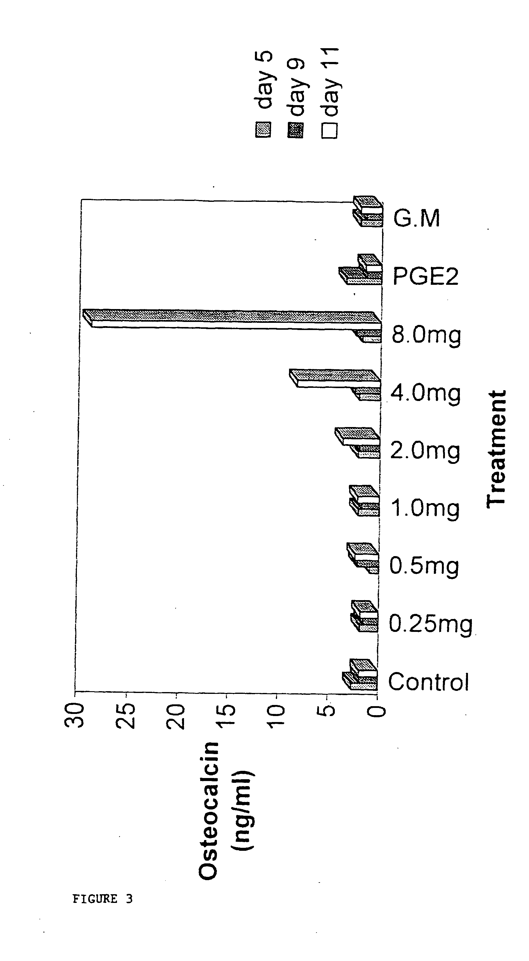
While grown in culture at either 33° C., and surprisingly at the permissive temperature of the active oncogene (37-39° C.), the immortalised human hypertrophic chondrocyte-like cells (HHC) expressed material which is released into the cell medium as, seemingly, cell debris or secreted extracellular matrix by the HHCs. The extracellular material produced is not restricted to a single clone but is a general characteristic of the skeletal cell lines that have been produced. A study was performed to see whether the material produced by these cells was capable of inducing the differentiation of marrow stromal cells into mineralising osteoblasts capable of elaborating a matrix, and secreting osteoblast marker proteins.

HHC cell “matrix” was collected as described in the materials and methods above and added to flasks containing bone marrow cells derived from rat femurs, or from human bone marrow biopsy material. In all cases the matrix—which was harvested from HHC medium, pelleted by ce...

example 3

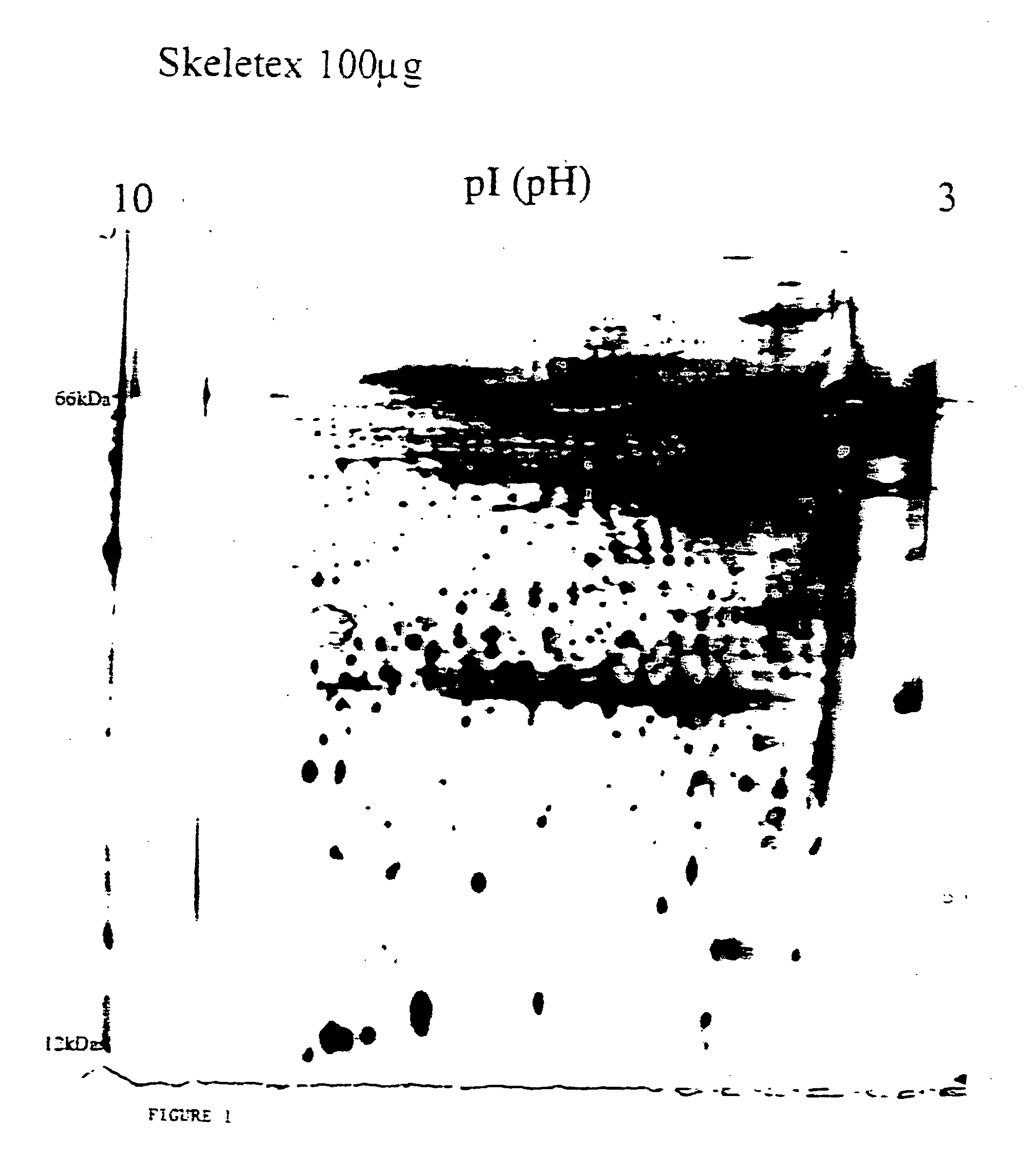
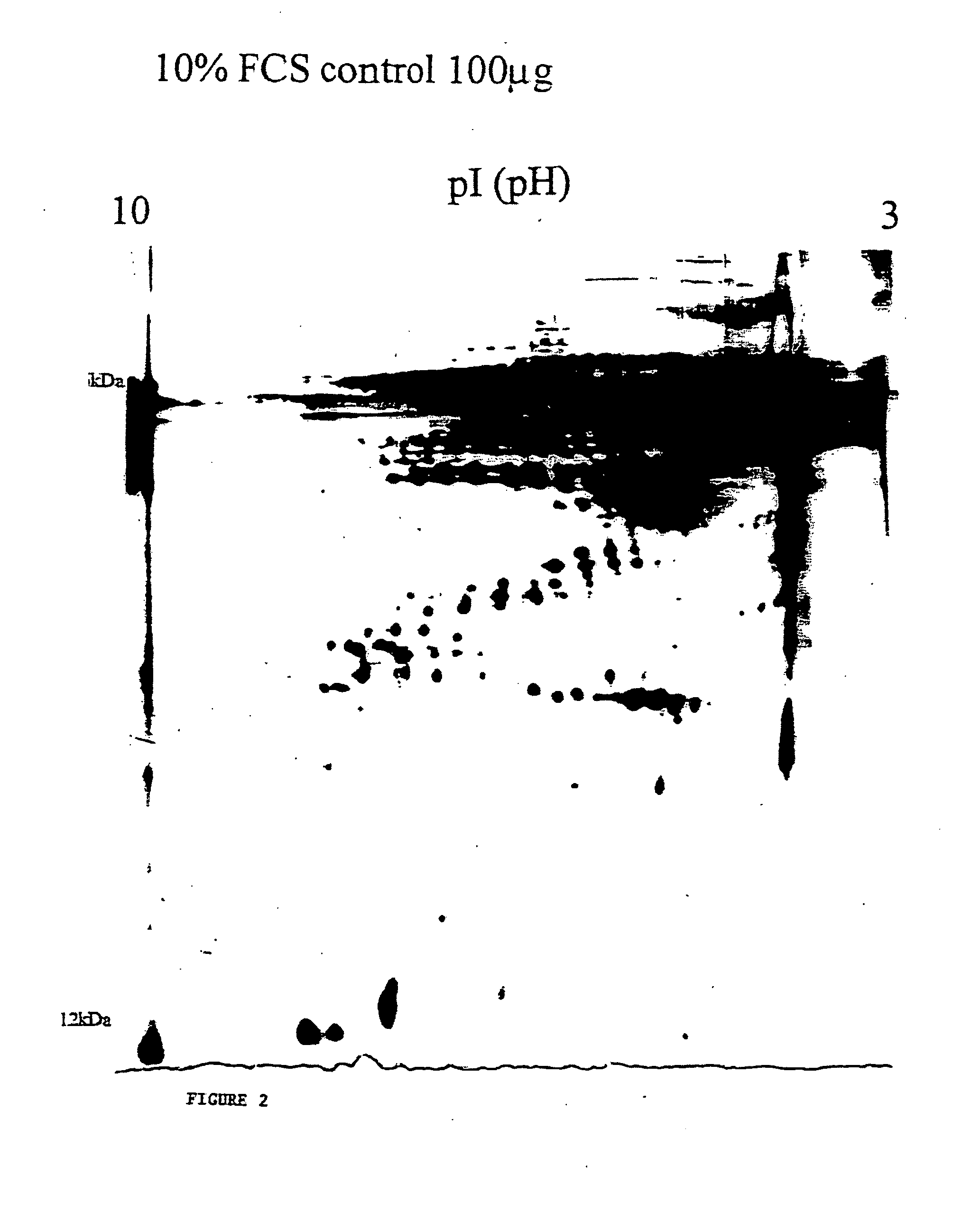
Electrophoretic Analysis of the Bioactive Matrix Material Harvested from Immortalised Human Hypertrophic Cartilage Cells

In order to determine the components of the HHC harvested material polyacrylamide gel electrophoresis was performed, and one and two dimensional gel analysis used to isolate the proteins present. Examples of the 2-D gel analysis are provided in FIGS. 1 and 2.

The results demonstrate that the extracellular matrix harvested from the HHC cells comprises a complex mix of noncollagenous and collagenous matrix proteins some of which are glycosylated, and, in addition, numerous cytokines and growth factors. It shows clearly a very complex mix comprised of potentially hundreds of proteins of various sizes and mobilities.

As described in the introductory sections, the natural processes of endochondral and intramembranous ossification require a complex myriad of signalling, over a sustained period of time, to be completed. This is performed by numerous factors, many unk...


PUM

PropertyMeasurementUnit
compositionaaaaaaaaaa
concentrationaaaaaaaaaa
pHaaaaaaaaaa

Abstract

An extracellular material in freeze-dried form obtained from skeletal cells which has osseoinductive bone repair and regeneration activity in vivo, and compositions and methods involving the material.

Description

BACKGROUND OF THE INVENTION Replacement of lost bone is the challenge facing orthopaedic surgeons, neurosurgeons, craniofacial surgeons, and periodontists all over the world today. The surgical repair of lost bone is a problem faced by dentists with patients suffering from periodontal disease, for example. Periodontal disease is one of the most prevalent afflictions, one consequence of which is alveolar bone loss, which in itself is a major disease entity. Presently periodontists and patients work together in treating the symptoms of periodontal disease, and effective techniques that predictably promote the body's natural ability to regenerate lost periodontal tissues (particularly alveolar bone) still need to be developed. In another field of dentistry, Dental Implantology, a great deal of biomaterial research is being conducted in an attempt to determine factors or substances that can improve the quality of bone to implant contact (osteointegration). In recent decades a surge ...

Claims


Application Information

Patent Timeline
Patent Type & Authority Applications(United States)
IPC IPC(8): A61K9/19A61K9/20A61K35/32A61K38/18
CPCA61K9/19A61K9/2095A61K35/32A61K38/1875A61K2300/00
Inventor STRINGER, BRADLEY MICHAEL JOHN
Owner CELLFACTORS
Why Eureka
Try Eureka