Methods for measuring peroxisome proliferation and peroxisomal induction

a technology of peroxisome proliferation and induction, which is applied in the direction of instruments, antinoxious agents, peptide/protein ingredients, etc., can solve the problems of sample destruction, oxidative damage to the cell, and inability to enable tru

Inactive Publication Date: 2005-03-17
PFIZER INC
View PDF0 Cites 8 Cited by
  • Summary
  • Abstract
  • Description
  • Claims
  • Application Information

AI Technical Summary

Problems solved by technology

This imbalance between H2O2 generation and degradation may lead to oxidative damage to the cell if any H2O2 escapes the peroxisome.
However, traditional biochemical methods have the following disadvantages: 1) they do not enable true determination of peroxisome proliferation, but only induction; 2) they are time consuming; 3) because tissue samples must be homogenized in order to obtain cell extracts for analysis, the samples are destroyed and tissue architecture is lost in the process, and re-examination of the same tissue is impossible; 3) the localization of specific tissue regions in which peroxisome induction has occurred cannot be determined; and 4) they often involve the use of hazardous substances, e.g., radioactive labels.
While this technique has the advantage (over biochemical methods) of preserving the architecture of the tissue samples, electron microscopy has the following disadvantages: 1) EM is time consuming because it requires special fixatives and techniques for processing the sample and preparing the slide for microscopy; 2) only a limited number of cells can be examined using electron microscopy; 3) complex, time consuming image analyses are needed in order to process the data and electron micrographs; 4) only peroxisome proliferation, and not peroxisomal induction, can be analyzed using electron microscopy; and 5) samples are consumed during the process of generating electron micrographs and thus cannot be archived and re-used later.

Method used


Image

  • Methods for measuring peroxisome proliferation and peroxisomal induction
  • Methods for measuring peroxisome proliferation and peroxisomal induction
  • Methods for measuring peroxisome proliferation and peroxisomal induction

Examples


example 1

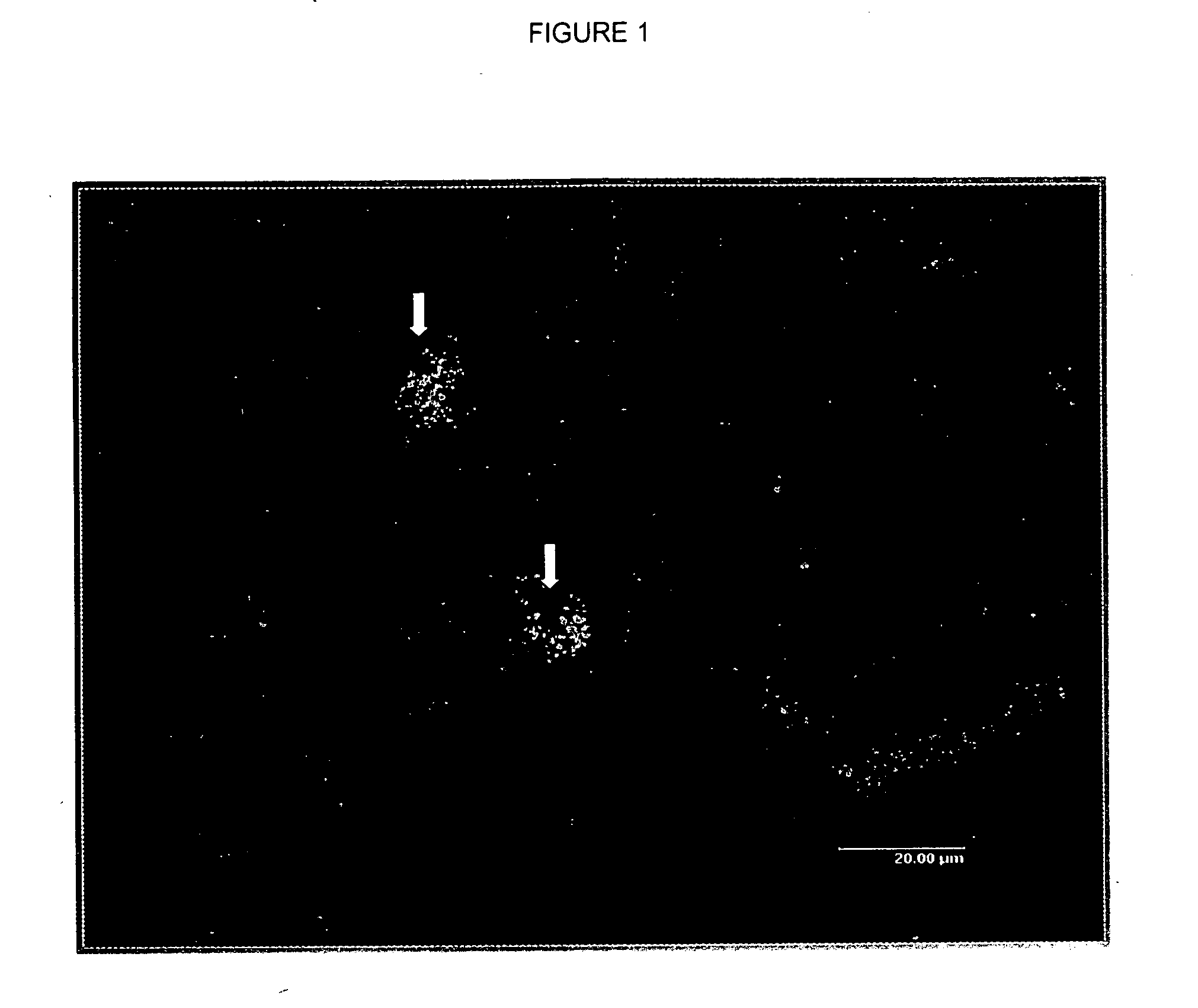
Analysis of Peroxisome Proliferation and Induction of Peroxisomal Enzymes in Rat Liver

[0038] Anti-AOX and PTS-1 Antibodies:

[0039] For the anti-PTS-1 antibody, a PTS-1 peptide with the sequence CRYHLKPLQSKL (one-letter amino acid designation) was synthesized and conjugated to keyhole limpet hemocyanine (KLH), as previously described (see Gould et al., cited above). Rabbit polyclonal antibodies were raised against the conjugated PTS-1 peptide and the serum with the highest titers was used for all subsequent immunofluorescence experiments.

[0040] The antibody specific for AOX was a generous gift from Dr. Nobuteru Usuda and was prepared by immunizing rabbits with AOX purified from rat liver tissues (Usuda et al., The Journal of Histochemistry & Cytochemistry, vol. 47(9), pp. 1119-26 (September 1999)).

[0041] Animals and Tissue Preparation:

[0042] Sprague-Dawley rats were treated with a potent peroxisome proliferator by oral gavage daily for 30 consecutive days. The rats were divided i...

example 2

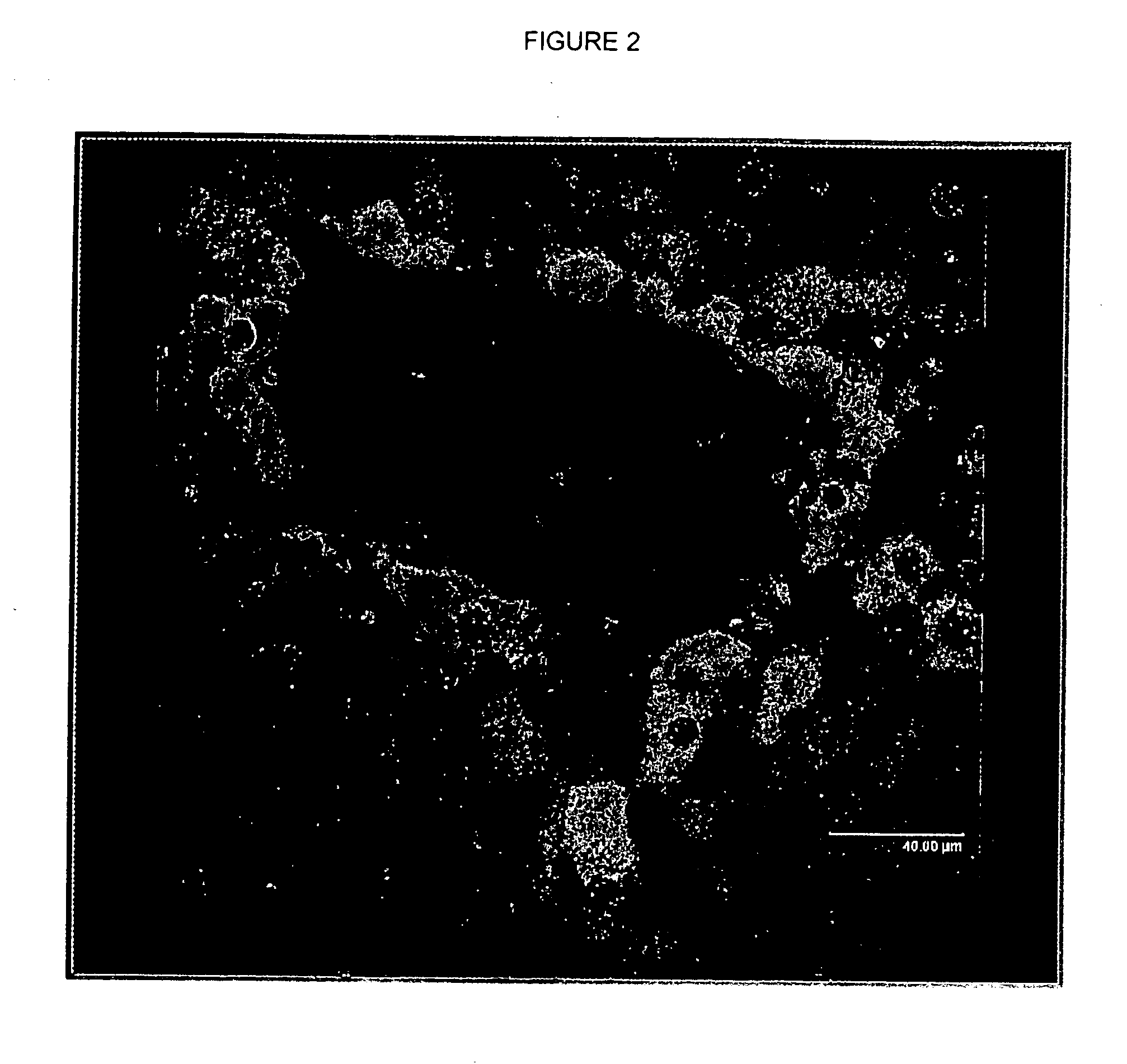
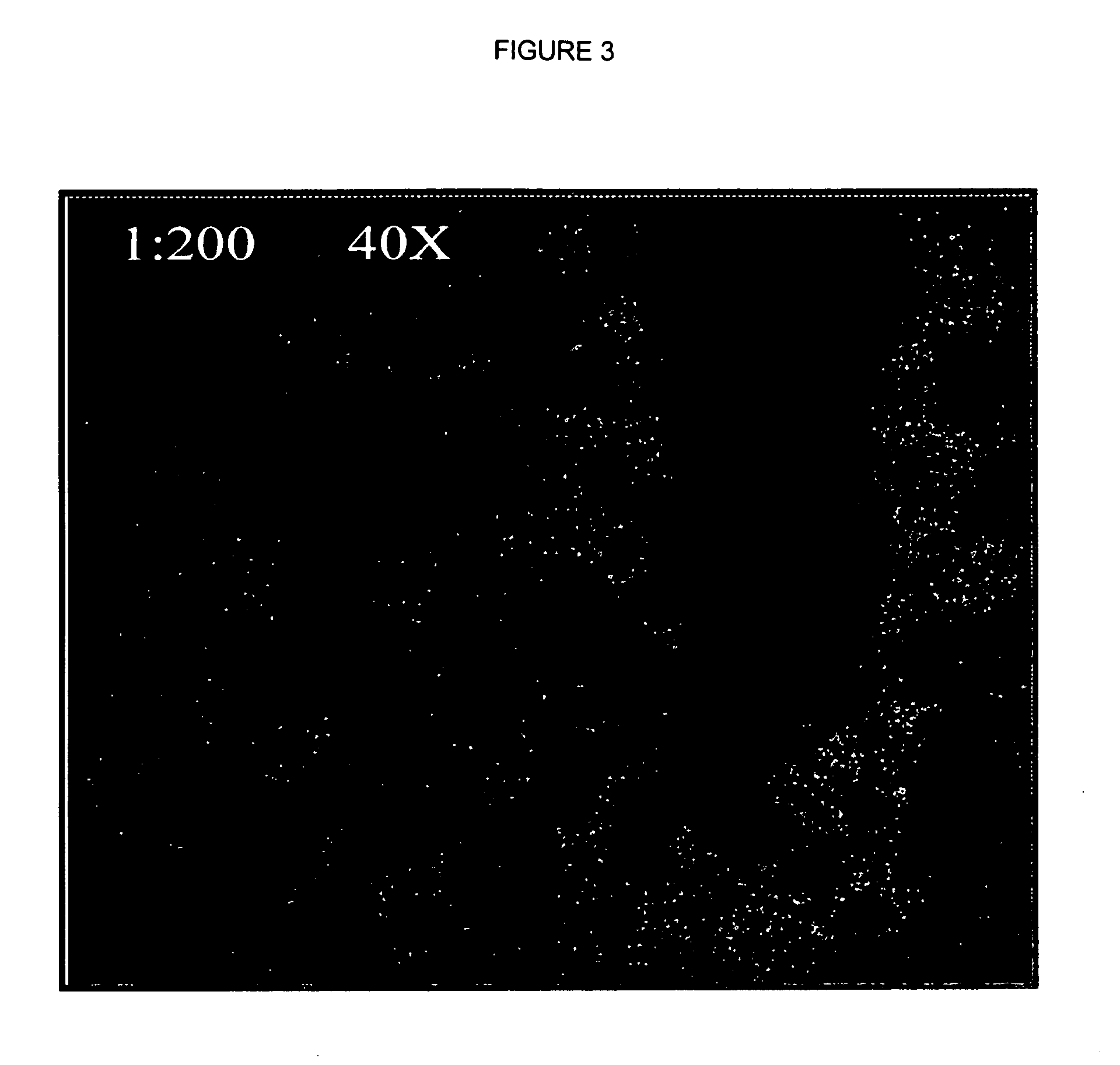
Analysis of Peroxisome Proliferation and Induction of Peroxisomal Enzymes in Dog Liver

[0057] Anti-AOX and Anti-PTS-1 Antibodies:

[0058] Anti-AOX and anti-PTS-1 antibodies were prepared as in Example 1, above.

[0059] Animals and Tissue Preparation:

[0060] Three beagle dogs were treated with 50 mg / kg of body weight of a potent peroxisome proliferator by oral gavage daily for 30 consecutive days. Three dogs remained untreated for use as control animals. After the 30 day period, the dogs were fasted overnight, anesthetized with CO2, and killed. Following necropsy, the right and left liver lobes were collected and fixed in 10% formalin buffer, processed for hematoxylin and eosin (H&E) staining and examined by light microscopy. Remaining liver tissues were frozen in paraffin blocks for later use.

[0061] Immunofluorescence:

[0062] 5-micron (μm)-thick liver sections were cut from the paraffin blocks, de-paraffinized using xylene rehydrated in alcohol, and washed in OptiMax wash buffer (OWB...


PUM

PropertyMeasurementUnit
thicknessaaaaaaaaaa
thickaaaaaaaaaa
body weightaaaaaaaaaa

Abstract

This invention provides methods for detecting peroxisome proliferation and peroxisomal beta oxidation in animals and tissue samples.

Description

FIELD OF THE INVENTION [0001] This invention relates to novel methods for the detecting peroxisome proliferation and peroxisomal beta oxidation in animals and tissue samples. BACKGROUND OF THE INVENTION [0002] Peroxisomes are single membrane-bound intracellular organelles that are contained in virtually all eukaryotic cells. All peroxisomes contain H2O2-catabolizing catalases and are therefore involved in protecting cells from this toxic byproduct. Peroxisomes also contain flavin-containing oxidases. One such oxidase is acyl-CoA oxidase (AOX). AOX is the rate-limiting enzyme of the peroxisomal β-oxidation of medium, long, and very long chain fatty acids. In rodents, peroxisomes characteristically increase in number and volume when exposed to so-called peroxisome proliferators. Such an increase is particularly relevant for rodent liver cells (hepatocytes). The effect of peroxisome proliferators is mediated by a class of nuclear receptors known as Peroxisome Proliferator Activated Rec...

Claims


Application Information

Patent Timeline
Patent Type & Authority Applications(United States)
IPC IPC(8): G01N33/50
CPCG01N33/5076
Inventor PRUIMBOOM-BREES, INGRID M.BREES, DOMINIQUE J.J.E.
Owner PFIZER INC
Why Eureka
Try Eureka