Multiplex binding and activity assays

a multi-binding and activity assay technology, applied in the field of assays, can solve the problems of detection of false positives or false negatives in the drug screen, limiting the sensitivity of luminescence-based assays,

Inactive Publication Date: 2005-03-24
LIFE TECH CORP
View PDF98 Cites 29 Cited by
  • Summary
  • Abstract
  • Description
  • Claims
  • Application Information

AI Technical Summary

Benefits of technology

The invention provides compositions, methods, apparatuses, and kits useful for monitoring molecular interactions, including competitive binding events and enzymatic activities. Accordingly, in one embodiment, the invention provides a method for measuring the effect of a test compound on binding between a first binding partner and a second binding partner. The method includes contacting a first binding partner, a second binding partner, and a test compound to form a

Problems solved by technology

Background luminescence (e.g., fluorescence or luminescence from assay components), non-specific interactions of assay components, and light scattering, however, can limit the sensitivity of luminescence-based assays, particularly when luminophores having short lifetimes are used, resulting in the detection of false positives or false negatives in a drug screen.

Method used


Image

  • Multiplex binding and activity assays
  • Multiplex binding and activity assays
  • Multiplex binding and activity assays

Examples


example 1

Labeling of Antibody with a Luminescent Metal Chelate

1 mg purified PY72 (anti-phosphotyrosine) IgG antibody, an antibody that preferentially binds amino acid sequences containing phosphorylated tyrosines (e.g., sequences phosphorylated by protein tyrosine kinases (PTKs)) and was dialyzed for 1.5 hours in a 100 mM sodium bicarbonate buffer, pH 9.5, using a 12-14,000 MWCO dialysis membrane. [PY72 hybridoma cells were obtained from the Salk Institute; the immunogen was phosphotyrosine conjugated to KLH. Ascites were produced by Harlan Bioproducts for Science, Indianapolis Ind. Ascites were purified with a protein G column (Pierce). Purified antibody is also available from Covance, Berkeley Calif. (Part # MMS414P).] The antibody was then removed from the dialysis membrane and concentrated to 48.8 uM (7.3 mg / mL) using a Centricon YM50 (Millipore) concentrator. 100 uL of this antibody solution was diluted to 5 mg / ml (33.4 uM) into the labeling reaction which consisted of 10 mM phenyl ph...

example 2

Binding Curve Experiment between Protein Tyrosine Kinase Product Tracer (PTK Tracer) and Anti-PTK Product (PY72) Antibody

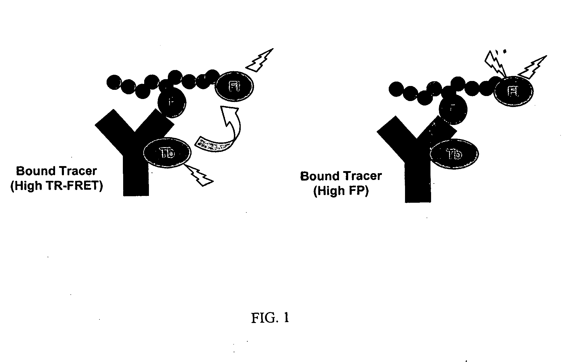
A direct binding curve (showing luminescent metal chelate -labeled PY72 antibody binding to fluorescent acceptor labeled tracer) was generated by incubating serial dilutions of the labeled antibody (10 nM to 9.8 pM in two fold dilutions) with 1 nM fluorescent acceptor-labeled tracer (PTK labeled tracer; sequence F-ADE(pY)LIPQQS, where F is fluorescein and pY is a phosphorylated tyrosine, SEQ ID NO:1; note that the tracer is a phosphorylated tyrosine derivative of a protein tyrosine kinase (PTK) substrate) in FP dilution buffer (PanVera, Madison WI part #P2839). After a 30 minute incubation, the fluorescence polarization of each composition in the plate was read on a Tecan Ultra plate reader using a 485 nm excitation filter (20 nm bandpass) and 535 nm emission filters (25 nm bandpass). Data was collected using 10 flashes per well and a 40 μs integration time. The...

example 3

Competition Curve between Labeled Kinase Product Tracer and Unlabeled Kinase Product

A competition curve to show that the disruption of the antibody-tracer interaction could be monitored by both fluorescence polarization and time-resolved RET from the same sample was performed by incubating serial dilutions (10 μM to 19.5 nM in two-fold dilutions) of an0 unlabeled phosphotyrosine-containing peptide competitor (ADE(pY)LIPQQS, where pY is a phosphorylated tyrosine, SEQ ID NO:3) in the presence of 10 nM Th-chelate labeled PY72 antibody and 1 nM labeled PTK labeled tracer, as described above. After a 30 minute incubation, the plate was read on a Tecan Ultra plate reader. Fluorescence polarization was measured using a 485 nm excitation filter (20 nm bandpass) and 535 nm emission filters (25 nm bandpass). Time-resolved RET was measured using a 340 nm excitation filter (35 nm bandpass) and 495 nm (10 nm bandpass) and 520 nm (25 nm bandpass) filters using a 200 μs integration window after ...


PUM

PropertyMeasurementUnit
Timeaaaaaaaaaa
Timeaaaaaaaaaa
Fractionaaaaaaaaaa

Abstract

Compositions, including antibodies, polypeptides, and organic molecules, kits, apparatuses, and methods for probing molecular interactions using fluorescence polarization (FP) and / or time-resolved resonance energy transfer (TR-RET) are provided.

Description

TECHNICAL FIELD This invention relates to assays employing fluorescence polarization (FP) and / or time-resolved resonance energy transfer (TR-RET) detection methods, and more particularly to methods for monitoring and measuring molecular interactions, such as competitive binding or enzymatic activity events, using the same. BACKGROUND Drug discovery can involve the systematic and / or high-throughput screening of diverse chemical libraries containing thousands of members. The size and complexity of these libraries, when coupled with the expense and length of the FDA approval process, have resulted in the need for simple, efficient, and homogeneous assays for probing molecular interactions. Luminescence-based techniques, including fluorescence polarization (FP), resonance energy transfer (RET), and luminescence resonance energy transfer methods (LRET) methods, are typically highly sensitive, homogenous methods for probing molecular interactions. Background luminescence (e.g., fluores...

Claims


Application Information

Patent Timeline
IPC IPC(8): C12Q1/00C12Q1/68G01N21/64G01N33/53G01N33/537G01N33/542G01N33/543G01N33/58
CPCG01N21/6428G01N21/6445G01N2500/02G01N33/573G01N33/582G01N33/542
Inventor VOGEL, KURT
Owner LIFE TECH CORP
Why Eureka
Try Eureka