
Tissue retractor and method for using the retractor

a tissue retractor and tissue technology, applied in the field of tissue retractors, can solve the problems of potentially fatal infection, inconvenient insertion of fasteners, and only providing sufficient plication for retraction of mucosa, so as to simplify mucosal resection and reduce tissue trauma. , the effect of reducing the trauma to the tissu

Inactive Publication Date: 2005-04-14
ID LLC (US)
View PDF22 Cites 85 Cited by
  • Summary
  • Abstract
  • Description
  • Claims
  • Application Information

AI Technical Summary

Benefits of technology

[0011] It is accordingly an object of the present invention to provide a tissue retractor and method for using the retractor that overcome the hereinafore-mentioned disadvantages of the heretofore-known devices and methods of this general type and that can effectively grab the tissue of the alimentary tract during operation and avoid reaching into the serosa, and can grab the tissue without compressing and / or tearing the tissue.
[0012] Various endoscopic procedures require manipulation of specific layers in the gastric wall. For instance, in the case of mucosal resection, the mucosa is tented away from the muscularis and resected away. Such a procedure is currently performed by injecting fluid beneath the mucosa to, thus, lift the mucosa from the muscularis. The mucosal tissue is, then, resected using electrocautery. The tissue retractor of the present invention can be used to selectively grasp the mucosa and lift it from the muscularis, thus enabling and simplifying mucosal resection. In the case of forming a full thickness plication in the stomach, the stronger muscular layer of the gastric wall must be grasped to ensure that the full thickness of the wall will be retracted when forming the plication. By tailoring the needles and the way in which they exit from the tip of the retractor, the retractor can be made to selectively grasp the different layers in the gastric wall. Being able to grasp a specific layer of the gastrointestinal wall is advantageous depending on the requirements of the specific procedure being performed.
[0013] The tissue retractor of the present invention has applications in laparoscopic and general surgery as well. It can be used to retract organs that are in the way of the surgical field, or to appose and hold tissue in-place during suturing. An advantage to an organ retractor or tissue apposition device according to the present invention is the ability to retain the tissue without having to clamp onto it. The tissue retractor atraumatically retains the tissue by penetrating it with fine needles. To further reduce the trauma to the tissue, the needles can be formed with a conical point instead of a faceted point. This is especially advantageous when retracting sensitive organs such as the pancreas. Currently available tissue graspers use more aggressive serrated articulating end effectors, which require clamping forces to retain the tissue and, therefore, potentially cause trauma in the process.
[0015] Also, a version of the retractor can be made that allows the distal tip of the retractor to be deployed in the tissue and, then, decoupled from the main shaft. In such an embodiment, the distal tip of the device is coupled removably to the shaft and the actuation wire is coupled removably to the needles. The needles are deployed on the target tissue and the shaft of the device is pulled proximally, thus allowing the actuation wire to slip free of the needles and the tip to slide free of the shaft. The released tip being firmly attached to the tissue has application as a marker, suture attachment points for a purse string closure, a tissue apposition suture, and an anchoring point for various things such as pH probes, miniature capsule cameras, and feeding tubes.
[0016] The device and method of the present invention allows the needles to be configured such that they can be made to penetrate deep through the mucosa and into the muscularis, making a more secure attachment to the tissue, while substantially reducing the possibility of puncturing the serosa, or penetrate less deep to grasp only the mucosal layer. The present invention, in the two-needle embodiment, engages the tissue at two opposing points, so that the tissue-engaging forces of each point react against the forces of the other; the result is that there is very little reaction load transmitted to the flexible shaft of the device. This deployment of the device does not require substantial torque or thrust loads to be supplied by the shaft. The present invention provides better visibility during placement of the retractor as no jaws are used that could obscure a view of the retraction site. It is also less traumatic to the tissue than a conventional articulating grasper due to the fine diameter and non-cutting points of the needles. The handle can be released from the user's grasp after the needles have been deployed, while still maintaining a secure attachment to the tissue, which frees the user to do other tasks after the tissue has been manipulated or is being manipulated. The tissue retractor is separate from an endoscope but sized to fit within a working channel of the endoscope.

Problems solved by technology

The mucosa is a relatively thin layer, loosely attached to the muscularis, and retraction of the mucosa only will not provide a sufficient plication for insertion of a fastener for the treatment of GERD.
It is not desirable to perforate the serosal layer.
One of the most significant reasons is that an unsealed perforation of the serosa, if sufficiently large, could allow leakage of gastric contents into the peritoneal or thoracic cavities causing a potentially fatal infection.
Currently available tissue graspers use more aggressive serrated articulating end effectors, which require clamping forces to retain the tissue and, therefore, potentially cause trauma in the process.

Method used


Image

  • Tissue retractor and method for using the retractor
  • Tissue retractor and method for using the retractor
  • Tissue retractor and method for using the retractor

Examples


Embodiment Construction

[0044] While the specification concludes with claims defining the features of the invention that are regarded as novel, it is believed that the invention will be better understood from a consideration of the following description in conjunction with the drawing figures, in which like reference numerals are carried forward.

[0045] Before the present invention is disclosed and described, it is to be understood that the terminology used herein is for the purpose of describing particular embodiments only and is not intended to be limiting. It must be noted that, as used in the specification and the appended claims, the singular forms “a,”“an,” and “the” include plural references unless the context clearly dictates otherwise.

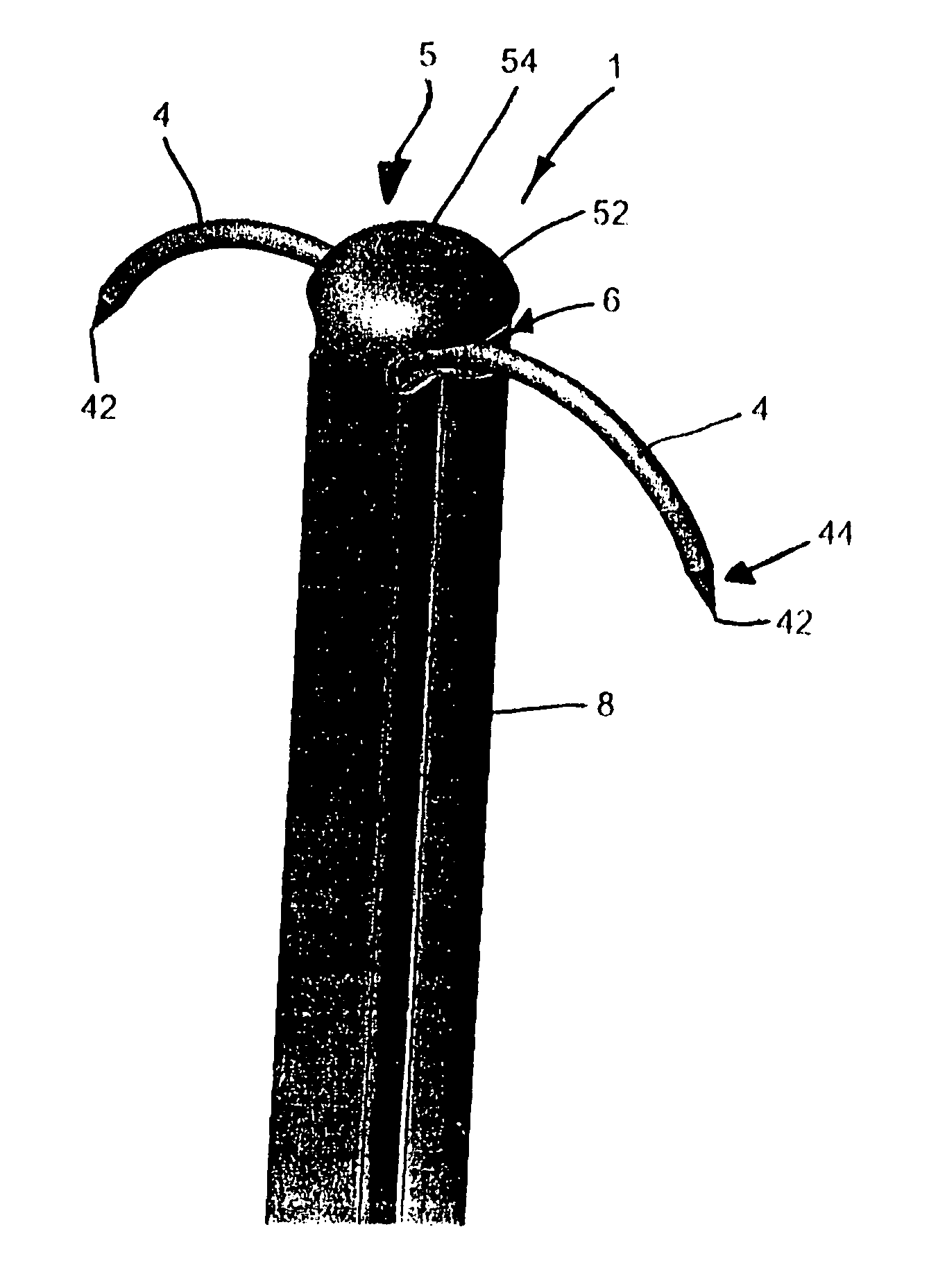
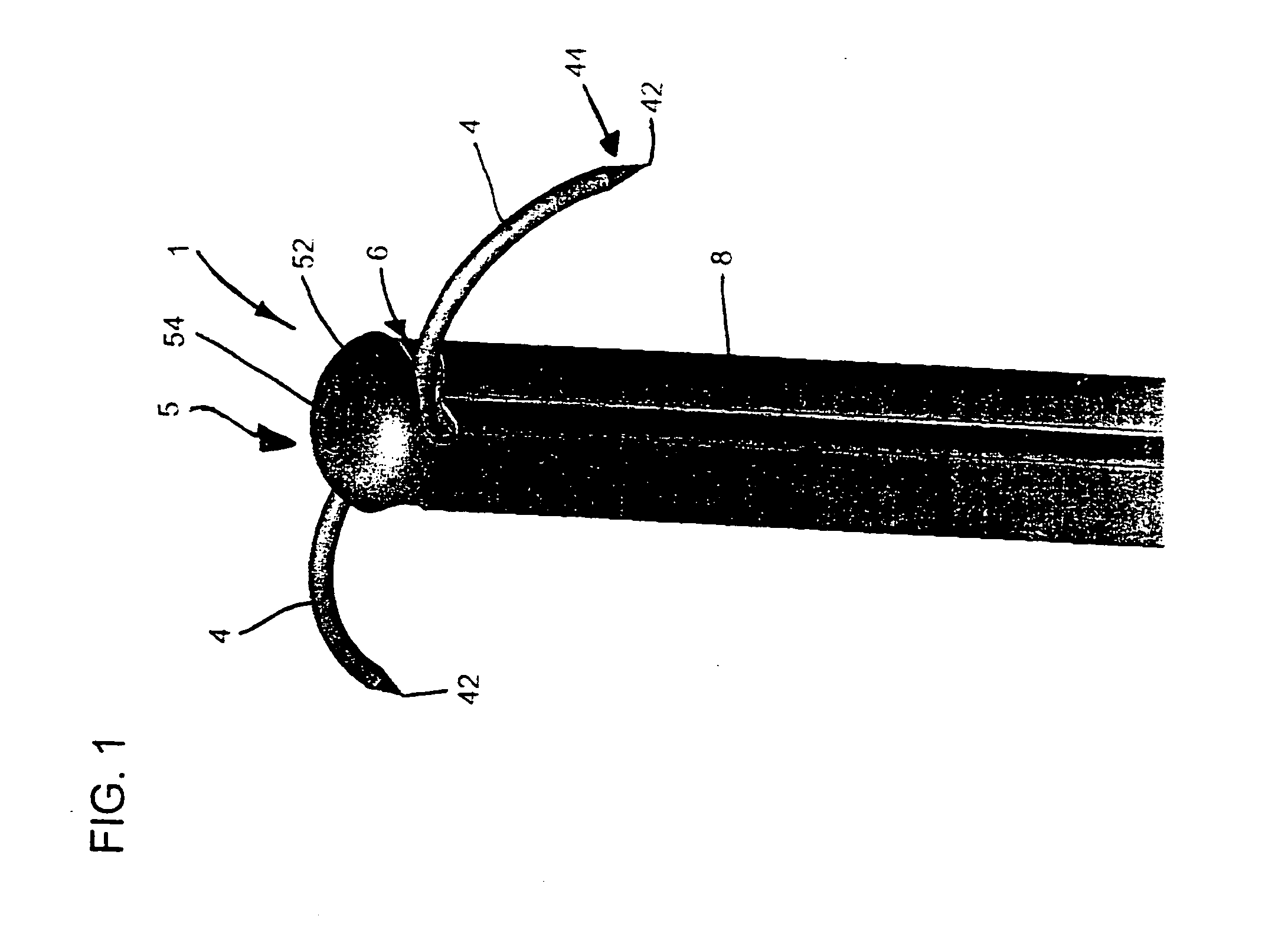
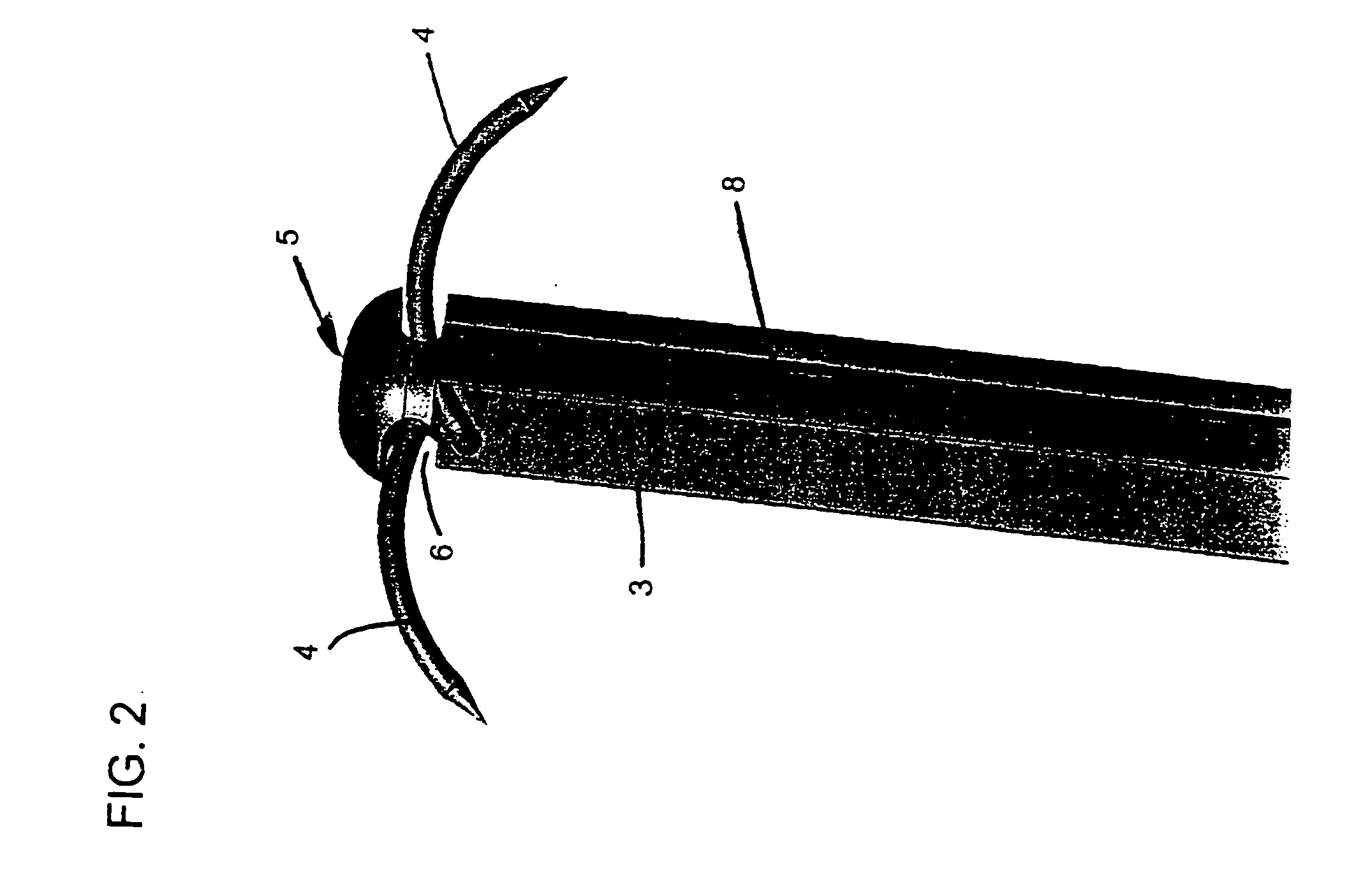
[0046] Referring now to the figures of the drawings in detail and first, particularly to FIG. 1 thereof, there is shown a perspective view of a distal portion of a flexible tissue retractor 1 according to the invention with needles 4 in a deployed or extended positi...


PUM


Abstract

A tissue retractor includes a body having proximal and distal ends. The retractor also includes a retraction device having a head connected to the distal end of the body and defining two opposing openings, an actuator movably disposed in the head, and two substantially rigid needles each pivotably connected to the actuator for traveling through a respective one of the openings. The needles have an arcuate shape. A one-handed actuation device is connected to the proximal end of the body and is operatively connected to the actuator through the body. Upon actuation of the actuation device, the actuator moves to selectively extend the needles out of the head and withdraw the needles into the head. Methods for using the tissue retractor, in particular for the treatment of Gastroesophageal Reflux Disease, are also provided.

Description

CROSS-REFERENCE TO RELATED APPLICATION [0001] This application claims the benefit under 35 U.S.C. § 119(e) of U.S. provisional applications Nos. 60 / 431,083 and 60 / 505,010, filed Dec. 5, 2002 and Sep. 22, 2003, and is a continuation in part of copending U.S. patent application Ser. No. 10 / 252,079, filed Sep. 20, 2002 and entitled “Surgical Fastener Particularly for the Treatment of Gastroesophageal Reflux Disease (GERD),” is a continuation in part of copending U.S. patent application Ser. No. 10 / 252,069, filed Sep. 20, 2002, and entitled “Instrument for Applying a Surgical Fastener Particularly for the Transoral Treatment of Gastroesophageal Reflux Disease (GERD),” and is a continuation in part of copending U.S. patent application Ser. No. 10 / 252,078, filed Sep. 20, 2002, and entitled “Method for the Surgical Application of a Fastener and the Endoluminal Treatment of Gastroesophageal Reflux Disease (GERD).”BACKGROUND OF THE INVENTION [0002] 1. Field of the Invention [0003] The presen...

Claims


Application Information

Patent Timeline
IPC IPC(8): A61B17/00A61B17/02A61B17/064A61B17/068A61B17/28A61B17/30
CPCA61B17/00234A61B17/0218A61B17/0643A61B17/068A61B17/0682A61B2017/301A61B2017/00349A61B2017/00867A61B2017/2905A61B2017/2926A61B17/29
Inventor SMITH, KEVINPALMER, MATTHEWKORTENBACH, JUERGENSIXTO, ROBERTPINOS, JORGE
Owner ID LLC (US)
Learn More