
Apparatus and methods for tissue preparation

a tissue and apparatus technology, applied in the field of apparatus and methods for tissue preparation, can solve the problems of preventing uniform slicing of tissue, affecting the quality of tissue samples, so as to prevent tissue sample deformation

Inactive Publication Date: 2005-05-05
THE INST OF CANCER RES ROYAL CANCER HOSPITAL +1
View PDF8 Cites 34 Cited by
  • Summary
  • Abstract
  • Description
  • Claims
  • Application Information

AI Technical Summary

Benefits of technology

[0011] Broadly, the present invention provides a tissue slicing apparatus comprising a tissue cutting means comprising a series of juxtaposed blades linked to a support. The apparatus may further comprise a cradle adapted to grip a tissue sample while it is cut with the tissue cutting means. The cradle can help to prevent the tissue sample deforming during sample preparation, even using an untreated or fragile sample, such as a prostate. As the tissue slices are produced together and in a defined orientation, the apparatus can also help to correlate tissue sections with diagnostic images.

Problems solved by technology

Treatment of prostate cancer has considerable morbidity, for example impotence and known side effects of radiotherapy.
Such tissues have a semi-solid consistency and tend to deform or even disintegrate when cut, preventing uniform slicing of the tissue.
Certain tissues may be difficult to cut owing to their structure.
For example, when the capsule of a prostate gland is nicked, the pressure of the hypertrophic nodules within may cause the tissue to bulge through the nick, making further slicing extremely difficult without destroying the integrity of the prostate gland.
A disadvantage, however, of the conventional method is that the treated tissue is unsuitable for certain research applications or diagnostic techniques.
In particular, the conventional treatment method destroys nucleic acids such as RNA in the tissue, so that the in-situ expression profile is lost.
Locating a piece of tissue that actually contains the suspected diseased tissue is also problematic.
Furthermore, it is not possible to correlate the tissue sections to medical images, usually radiological images, which may have been obtained earlier in the diagnosis process, to assist and / or confirm the diagnosis.
However, it is extremely difficult and time consuming to produce tissue slices that match the virtual slices.
No tissue support means is provided as the apparatus is designed to remove strips of tissue directly from a patient.

Method used


Image

  • Apparatus and methods for tissue preparation
  • Apparatus and methods for tissue preparation
  • Apparatus and methods for tissue preparation

Examples


Embodiment Construction

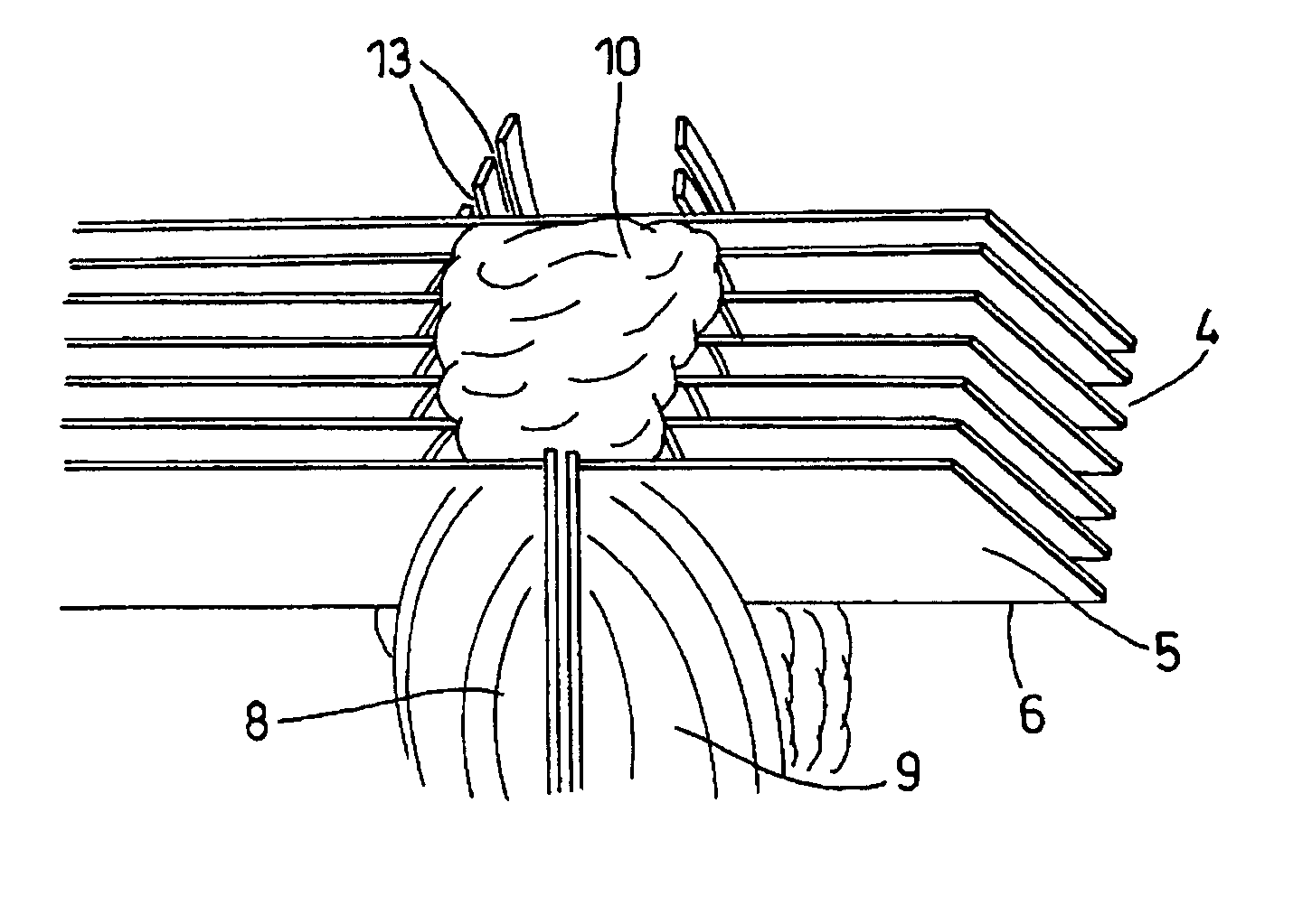
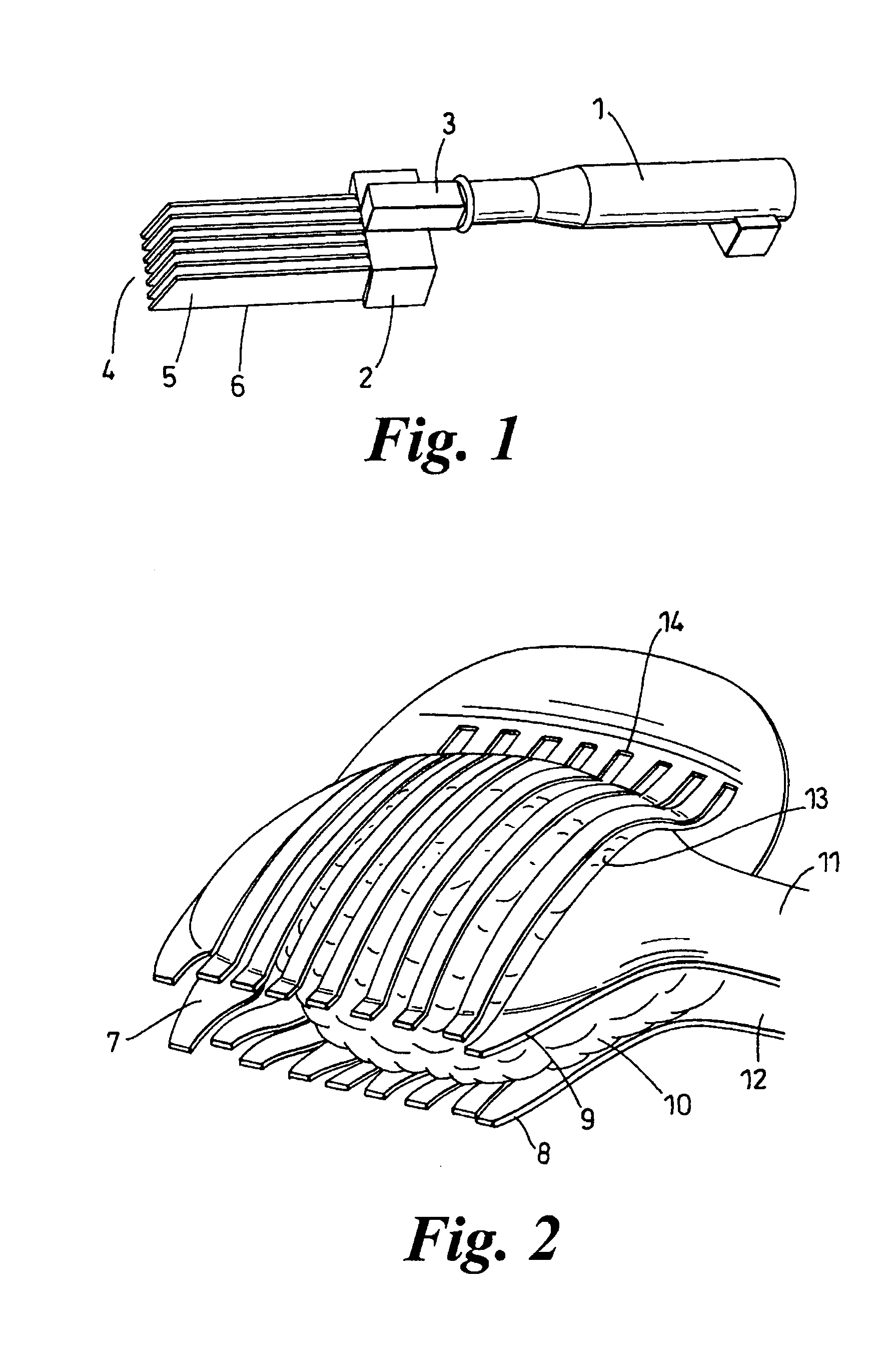
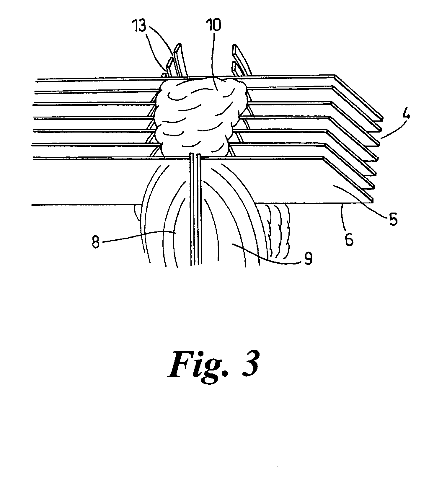
[0085] Cutting means of the present invention is shown in FIG. 1. The whole apparatus is of metal. The apparatus has an elongate handle 1, which thickens at one end to provide a handgrip. A blade support includes a support block 2 and a cuboid piece 3 attached at the other end of the handle. A series of blades, indicated generally at 4, extend from the support block in approximately the same direction as the axis of the handle.

[0086] The series of blades consists of seven planar blades, each planar blade 5 having a cutting edge 6. The blades are identical and their cutting edges lie in the same plane. The blades are individually detachable from the support block and the blade support is detachable from the handle.

[0087] The cutting edges 6 of the blades 5 are offset from the axis of the handle. When cutting edges abut an underlying surface, the offset provides, between the underside of the handle and the surface, some clearance for the user's fingers.

[0088] A cradle adapted to su...


PUM

PropertyMeasurementUnit
Lengthaaaaaaaaaa
Forceaaaaaaaaaa
Angleaaaaaaaaaa

Abstract

The present invention features a multi-bladed cutter and a cradle adapted to grip a tissue sample 10 during cutting. The cutter has a series 4 of juxtaposed blades 5 and the cradle has a series of slits 13 adapted to receive the blades during cutting. The cradle may have opposing curved walls 8,9 which may be brought together to clasp the sample. The apparatus is suitable for cutting tissue such as prostate tissue, which is difficult to section. The apparatus is particularly suited to cutting tissue where chemical fixing is undesirable because nucleic acids are to be sampled from the tissue.

Description

FIELD OF THE INVENTION [0001] The present invention relates to apparatus and methods for tissue preparation, in particular cutting tissue by slicing. The present invention is particularly useful where chemical fixing of the tissue is undesirable, for example where nucleic acids such as DNA or RNA are to be sampled from the tissue. The present invention may also be useful for preparing tissues that are difficult to section or slice without chemical fixing beforehand, such as prostate tissue. BACKGROUND OF THE INVENTION [0002] Prostate cancer is the second most common cancer in men and accounts for over 9000 deaths per year in the United Kingdom. Treatment of prostate cancer has considerable morbidity, for example impotence and known side effects of radiotherapy. Diagnosis can be accomplished by testing of an appropriate tissue sample, for example using histology. Additionally, techniques such as Magnetic Resonance Imaging (MRI) or other medical imaging techniques may be used in diagn...

Claims


Application Information

Patent Timeline
IPC IPC(8): A61B10/00A61B10/02A61B17/32G01N1/06G01N1/28G01N1/31
CPCA61B10/02A61B17/3211A61B2017/320052G01N2001/2873G01N1/31G01N2001/282G01N1/06Y10T83/869
Inventor JHAVAR, SAMEERREINSBERG, STEFANJACKSON, ANDREWCUMMINGS, CRAIGEELES, ROSALINDFALCONER, ALISONLEACH, MARTIN
Owner THE INST OF CANCER RES ROYAL CANCER HOSPITAL
Learn More