Vaccine against cancerous diseases which is based on mimotopes of antigens expressed on tumor cells

a technology of mimotopes and tumor cells, applied in the field of vaccines against cancerous diseases, can solve the problems of not being able to effectively prevent disease, and the side effects of chemotherapy treatment are very large for patients, and achieve the effect of good vaccine

Inactive Publication Date: 2005-05-12
BIOLIFE SCI QLD
View PDF4 Cites 4 Cited by
  • Summary
  • Abstract
  • Description
  • Claims
  • Application Information

AI Technical Summary

Benefits of technology

[0010] In a preferred embodiment, the inventive method uses antibodies that have as such already proved effective against cancerous diseases in clinical tests. Thus, administration of the vaccine induces the formation of endogenous antibodies against those antigens of the cancer cells against which the clinically tested antibodies are effective. This increases the effectiveness of the vaccine.
[0036] This expression method permits the mimotope proteins to be obtained easily in endotoxin-free form even on a large scale.

Problems solved by technology

These methods have the disadvantages that they do not permit effective prophylaxis against the genesis of the cancerous disease and that treatment for example by chemotherapy involves very considerable side effects for the patient.

Method used


Image

  • Vaccine against cancerous diseases which is based on mimotopes of antigens expressed on tumor cells
  • Vaccine against cancerous diseases which is based on mimotopes of antigens expressed on tumor cells

Examples


example 1

Identification of Herceptin Mimotopes

[0044] The wells of a Maxisorb plate from Nunc were coated with a monoclonal mouse antibody directed against the constant Fc region of human IgG, in a concentration of 10 μg / ml PBS for the duration of 16 hours.

[0045] After washing the wells and saturating free binding sites by BSA, the herceptin antibody was bound to the monoclonal mouse antibody by its Fc region. Herceptin was incubated in a concentration of 10 μg / ml PBS for two hours.

[0046] After further routine washing steps, the wells of the Maxisorb plate were covered with the phage library. This library contained filamentous M13 phages in whose pVIII gene coding for a phage coat protein, nucleotide sequences coding for peptides with a length of 9 amino-acid residues were inserted. This library contained 109 individual phage clones differing in the sequence of their inserts.

[0047] Phages that bound to the herceptin by these peptides were detached from the herceptin by pH lowering. Suitab...

example 2

Immunization Scheme

A) Immunization of Mice.

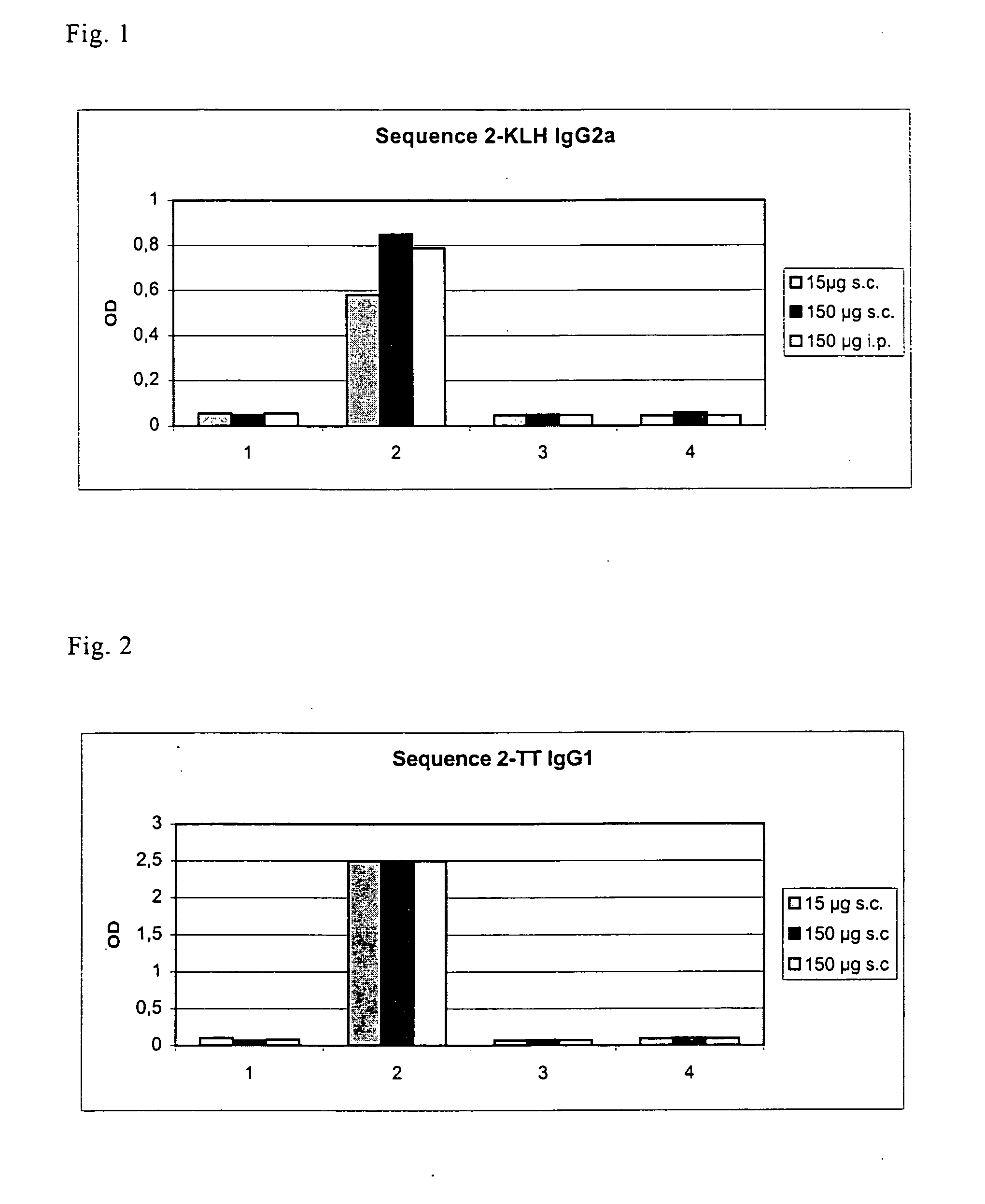
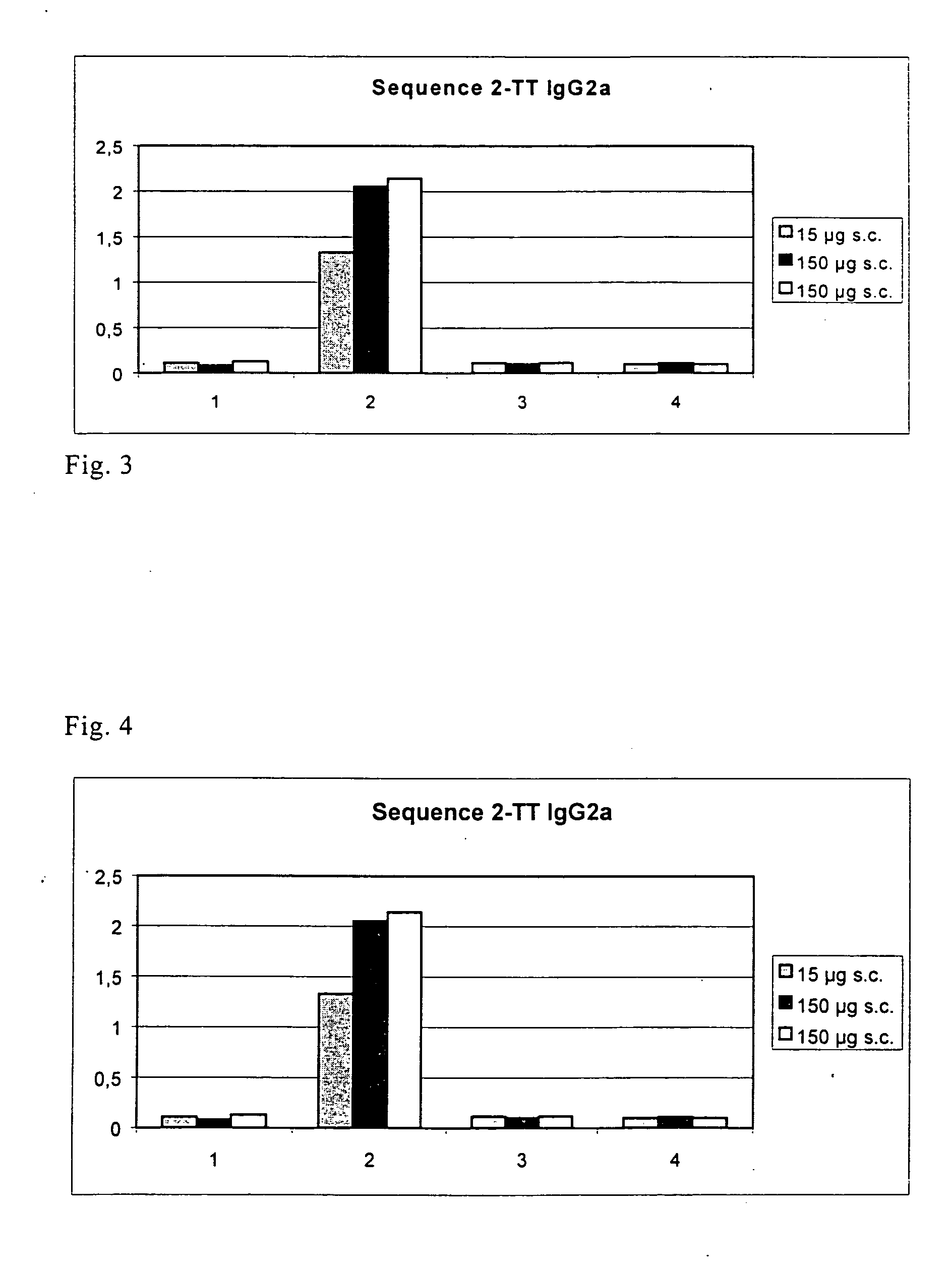
[0049] One of the mimotopes with the amino acid sequence ATLMQIISQ (SEQ ID NO: 3) found in the way described under 1 was used for immunization. At the N-terminus of this sequence there were 5 amino-acid residues of the pVIII protein, i.e., the residues AEGEF (SEQ ID NO: 4), as stated under 1.

[0050] Before coupling, the mimotope was provided at its C-terminus with the linker GGGGGC (SEQ ID NO: 5). The amino acid sequence coupled to the coupling partner was accordingly AEGEFATLMQIISQGGGGGC (SEQ ID NO: 1).

[0051] Two coupling partners (=carrier proteins) with SEQ ID NO: 1 were tested, namely KLH and tetanus toxoid. Two immunization routes were used. Subcutaneously, 15 μg of the particular conjugate was used, intraperitoneally 150 μg in each case. The adjuvant used was Gerbu adjuvant. Each subcutaneously immunized group had 5 mice, each i.p. immunized group 3 mice. The control groups (each with 3 mice) were only administered the conjugant w...

example 3

Detection of Specificity

A) Inhibition of Herceptin by Phage Mimotopes in ELISA.

[0070] For detecting the inhibition of the binding of herceptin and HER-2 / neu by mimotopes, one proceeds as follows:

[0071] Her-2 / neu is recognized in the cytomembranes of the cell line SKBR3 in ELISA by the humanized monoclonal antibody herceptin. For the ELISA inhibition assay one tries to prevent this bond by preincubation with mimotopes on phages or KLH as carrier.

[0072] The wells of a 96-well ELISA plate (Nunc) were coated with membrane fractions of the Her-2 / neu overexpressing cell line SKBR3 and as a control for non-specific antibody bonds with membrane fractions of the Her-2 / neu negative cell line HTB 132 in PBS overnight.

[0073] Herceptin (anti-Her-2 / neu antibody) was treated likewise overnight by preincubation with different antigens: mimotopes, control mimotopes or buffers.

[0074] The next day, non-specific binding sites were blocked by 3 percent BSA in PBS. Herceptin as well as herceptin a...


PUM

PropertyMeasurementUnit
concentrationaaaaaaaaaa
structureaaaaaaaaaa
chemical natureaaaaaaaaaa

Abstract

The present invention provides a method for producing a vaccine against cancerous disease, and the vaccine itself. This method involves first using one or more antibodies that are specifically effective against one or more antigens specially expressed by the tumor cells to select one or more mimotopes of said antigens from a phage peptide library. To obtain the vaccine, said mimotopes are conjugated to a macromolecular carrier singly or multiply in the form of their mono-, di-, tri- or oligomers. When administered, the inventively produced vaccine leads to a humoral immune response and thus to the formation of an active immunity as a consequence of vaccination.

Description

FIELD OF INVENTION [0001] The present invention relates to a method for producing a vaccine against cancerous diseases and to said vaccine itself. BACKGROUND OF INVENTION [0002] In recent years there has been a steady increase in cancerous diseases in Western industrial nations. For example, roughly 23,000 men and 29,000 women a year develop colonic and rectal cancer in Germany, the risk of disease rising gradually with age. Malignant lymphomas constitute about 5 percent of all cancer cases, about 9,000 persons a year developing non-Hodgkin's lymphoma in Germany—with an upward trend. Breast cancer even affects about 10 percent of all women in Western industrial nations. [0003] Methods hitherto known for treating cancerous diseases aim above all at early recognition of the illness and at surgical methods or selective destruction of tumor cells. These methods have the disadvantages that they do not permit effective prophylaxis against the genesis of the cancerous disease and that trea...

Claims


Application Information

Patent Timeline
Patent Type & Authority Applications(United States)
IPC IPC(8): A61K39/00A61P35/00A61P37/04C07K16/32
CPCA61K39/00C07K16/32A61K39/0011A61P35/00A61P37/04
Inventor ZIELINSKI, CHRISTOPHSCHEINER, OTTOJENSEN-JAROLIM, ERIKABREITENEDER, HEIMOPENAMBERGER, HUBERT
Owner BIOLIFE SCI QLD
Why Eureka
Try Eureka