
System for medical examination or treatment

Active Publication Date: 2005-05-12
SIEMENS HEALTHCARE GMBH
View PDF4 Cites 157 Cited by
  • Summary
  • Abstract
  • Description
  • Claims
  • Application Information

AI Technical Summary

Benefits of technology

[0013] The invention is based on the knowledge that an optimal diagnostic image quality can be achieved with a system which combines the advantages of intravascular ultrasound investigation (IVUS) with the advantages of optical coherence tomography (OCT). The medical investigation and treatment system in accordance with invention is optimized for artery investigations and removes the above-mentioned disadvantages of the two known methods.
[0028] To simplify the image processing of the medical investigation and / or treatment system there can be provision for the sensor of the optical coherence tomography and the sensor of the intravascular ultrasound imaging system to be able to be driven at the same speed. The two sensors can then be controlled so that the images that they deliver are automatically in the same phase, doing away with the need for expensive computational registration. In accordance with a further development of the invention there can however also be provision for the OCT sensor and the IVUS sensor to be operated at different speeds, if necessary with an intermediate microdrive.

Problems solved by technology

This disadvantage of this method however is that only the artery diameter usable by the blood flow or the restriction are shown as a silhouette.
This means that the method does not allow any information to be provided about the arteriosclerotic deposits such as their thickness or the presence of inflammation processes.
However the general disadvantage of ultrasound images is that their resolution is limited.
The disadvantage of this method lies in the low level of resolution of tissue further down.

Method used


Image

  • System for medical examination or treatment
  • System for medical examination or treatment
  • System for medical examination or treatment

Examples


Embodiment Construction

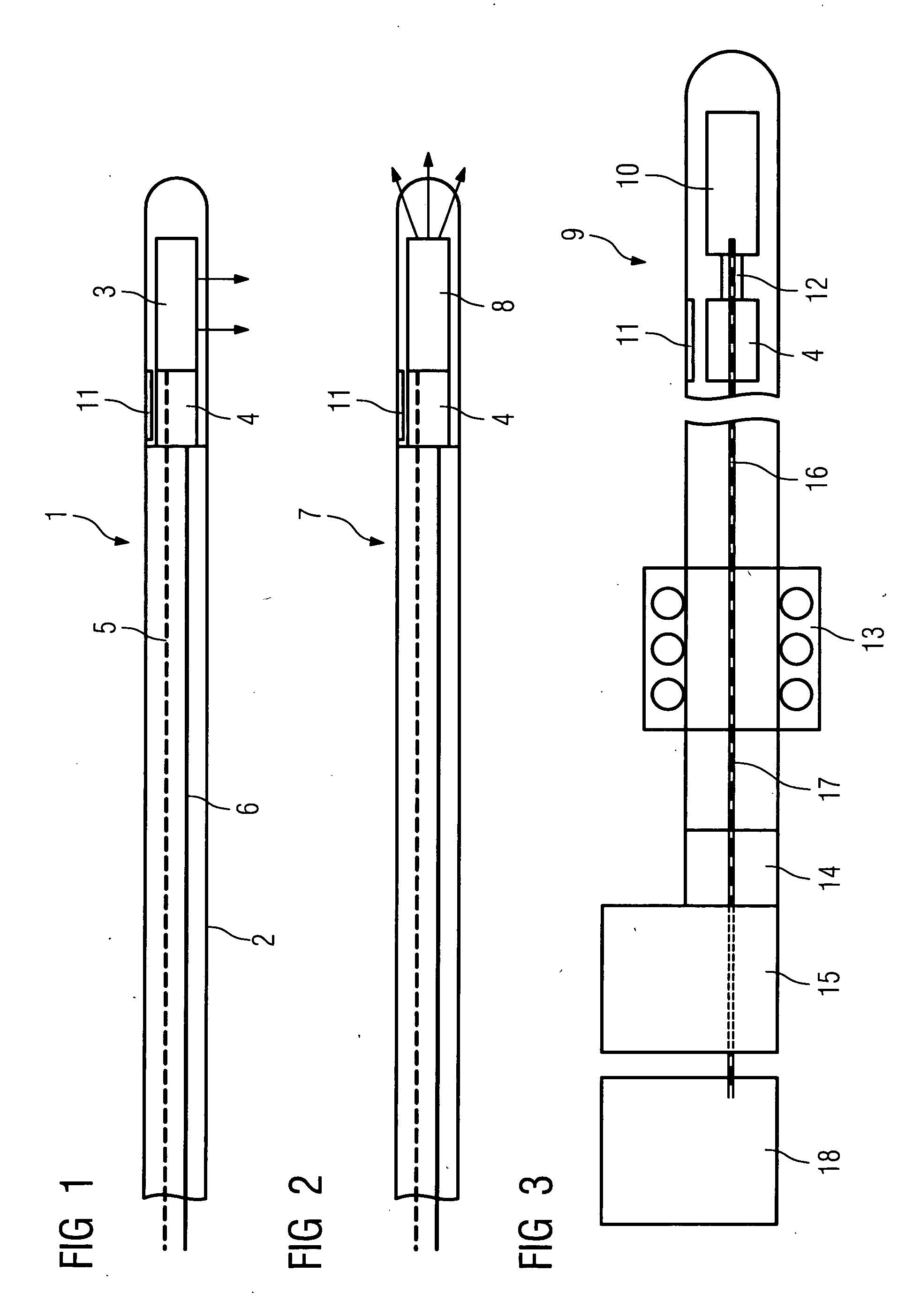
[0039]FIG. 1 shows a catheter 1, essentially consisting of a catheter envelope 2, an IVUS sensor 3 arranged in the area of the catheter tip which is part of an intravascular ultrasound imaging system and an OCT sensor 4 which is part of an imaging system for optical coherence tomography. The catheter envelope accommodating the sensors 3, 4 is transparent for ultrasound.

[0040] The IVUS sensor 3 is embodied so that the ultrasound is radiated and received in an approximately sideways direction. Since the IVUS sensor 3 is revolving at high speed, which can be 1,800 RPM, it delivers a 360° comprehensive cross-sectional image of the artery to be investigated. The reflected received sound waves are converted by the IVUS sensor 3 into electrical signals which are forwarded via a signal line 5 to a signal interface and thence to a pre-processing unit and an image processing unit.

[0041] The OCT sensor 4 is also directed to the side so that it generates a continuous image of the artery to be...


PUM


Abstract

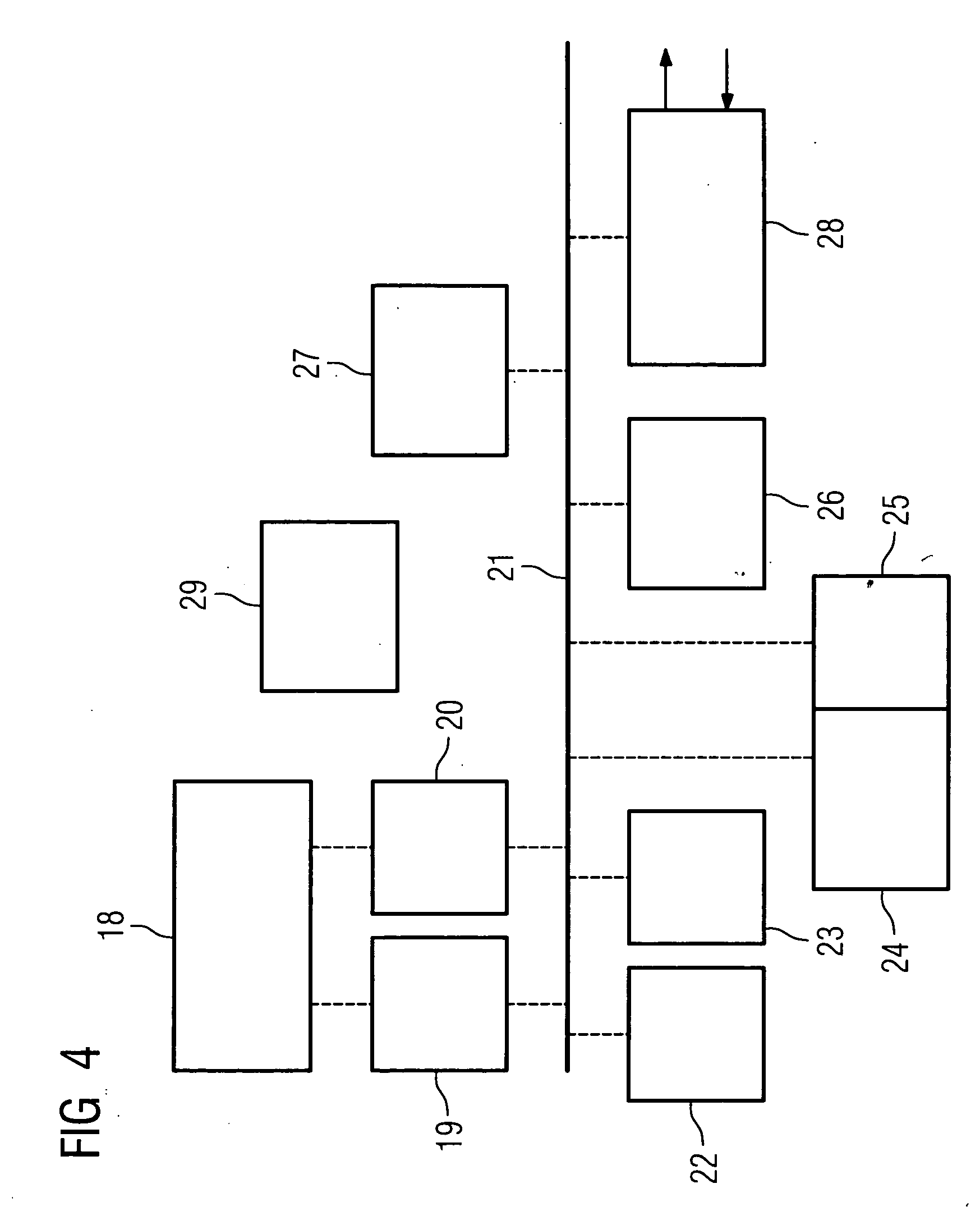
The invention relates to a medical investigation and / or treatment system comprising: a catheter system, comprising a catheter (1, 7, 9), with a first sensor (4, 11) of an imaging system for optical coherence tomography with an optical fiber via which the light is directed and is radiated in the area of a catheter tip introduced into an area to be investigated, via which fiber reflection light is directed from an illuminated investigation area to a first image processing unit (22), a second sensor (3, 8, 10) of an intravascular ultrasound imaging system to transmit and receive sound pulses which are fed as an electrical signal to a second image processing unit (23), und at least one display unit (24) for presenting the images of the first a nd second image processing unit (22, 23).

Description

CROSS REFERENCE TO RELATED APPLICATIONS [0005] This application claims priority to the German application No. 10343808.4, filed Sep. 22, 2003 and which is incorporated by reference herein in its entirety. FIELD OF INVENTION [0006] The invention relates to a medical investigation and / or treatment system. BACKGROUND OF INVENTION [0007] Vascular conditions, especially heart attacks, are among the most frequent illnesses resulting in death. These are caused by diseases of the coronary arteries (arterial sclerosis). In such conditions deposits, referred to as arterial plaque, result in “blocking” of coronary arteries. The latest knowledge shows that the danger of suffering a heart attack does not depend primarily on the reduction of the diameter of the arteries. It is far more a matter of whether the thin protective layer which covers the arteriosclerotic deposits is stable. If this layer breaks down, blood platelets are released which completely close off the artery within a short time,...

Claims


Application Information

Patent Timeline
IPC IPC(8): A61B1/00G01N21/17A61B5/00A61B8/12G01S15/89
CPCA61B5/0066A61B8/12A61B5/6852
Inventor MASCHKE, MICHAEL
Owner SIEMENS HEALTHCARE GMBH
Learn More