In situ bioprosthetic filler and methods, particularly for the in situ formation of vertebral disc bioprosthetics

a bioprosthetic and in situ technology, applied in the field of bioprosthetics, can solve the problems of sudden collapse of disc height, degeneration of vertebral disc function, and potential nerve root and/or spinal cord compression, and achieve the effect of discharging the energy of impact loading

Inactive Publication Date: 2005-05-12
CRYOLIFE
View PDF53 Cites 46 Cited by
  • Summary
  • Abstract
  • Description
  • Claims
  • Application Information

AI Technical Summary

Benefits of technology

[0003] The vertebral disc is a collagenous spacer positioned between the vertebral bones of the spinal column. The disc generically consists of a tough fibrillar outer annulus (annulus fibrosus) and a highly h...

Problems solved by technology

Degeneration of vertebral disc function in the lumbar portion of the spine is the leading cause of debilitating low back pain in adults over the age of 35.
DDD may also precipitate the formation of fissures within the annulus that allows extrusion of the disc nucleus (disc herniation) resulting in a sudden collapse in the disc height and the potential for nerve root and/or spinal cord compression.
Disc herniation may also result due to trauma related over compression of the spine, such as a heavy sitting fall.
Chronic diffuse low back pain results from irritation of pain receptors in the outer third of the disc annulus and surrounding soft tissues as the disc collapses.
The collapse of the disc space can lead to loss of motion and morbidity, as during the collapse of the disc spa...

Method used


Image

  • In situ bioprosthetic filler and methods, particularly for the in situ formation of vertebral disc bioprosthetics
  • In situ bioprosthetic filler and methods, particularly for the in situ formation of vertebral disc bioprosthetics

Examples


example 1

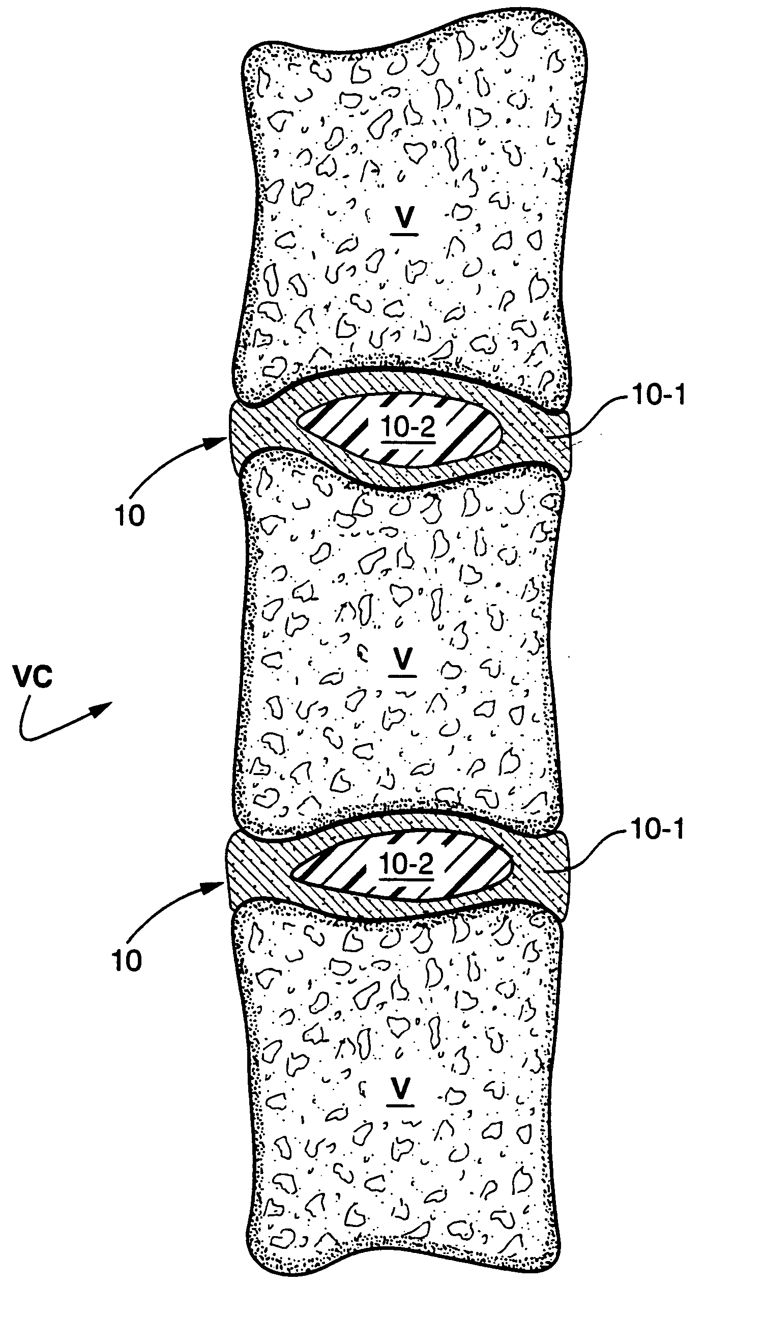
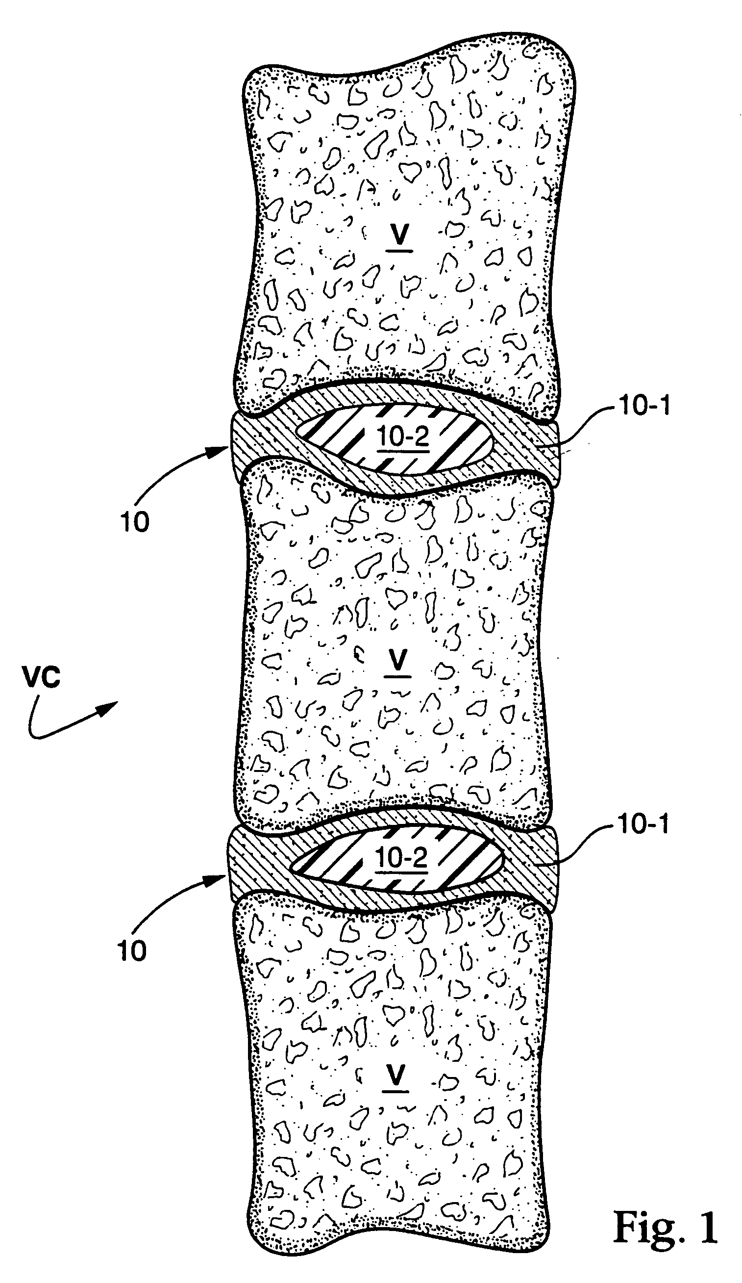
[0031] A formulation formed of a protein solution (serum albumin) and a cross linker (gluteraldehyde) was contained in the separate chambers of a delivery device. When the device is triggered, the two components are expelled from their respective chambers into a mixing tip that combines the two solutions and mixes them as they travel over the static mixing elements present in the tip. A medical needle was attached to the mixing tip and the formulation injected into the distal space between the vertebra of an explanted pig spine. The tip can be attached to a needle, catheter, or other hollow tubular device for delivery, for example. After 30 seconds, the needle was withdrawn from the injection site. The material that was injected had polymerized in place and did not exude out of the needle hole. After 2 minutes, the disc-vertebra plate was dissected and the presence of the biomaterial seen.

example 2

[0032] Bovine calf spines were obtained from a commercial slaughterhouse and cleaned by blunt and sharp dissection to expose the vertebral bodies and the discs. A 4 mm hole was made into the anterior face of the disc and the drill bit allowed to enter to the center of the nucleus. The nuclear material was removed using surgical forceps and curettes. The hollow space was filled with the formulation described in Example 1. The material that was injected polymerized in place and did not exude out of the hole. After 2 minutes the disc-vertebra plate was dissected and the presence of the biomaterial seen.

example 3

[0033] Bovine calf spines were obtained from a commercial slaughterhouse and cleaned by blunt and sharp dissection to expose the vertebral bodies and the discs. The top and bottom of the vertebral bodies were cut parallel to each other at mid-height using a miter box to yield a bone / disc / bone motion segment. A 4 mm hole was made into the anterior face of the disc and the drill bit allowed to enter into the center of the nucleus. The nuclear material was removed using surgical forceps and curettes. The hollow space was filled with the formulation described in Example 1. The material that was injected had polymerized in place and did not exude out of the hole.

[0034] Once polymerization had occurred, the construct could be compressed by hand in the front-back and left-right axes, indicating flexibility was retained after repair of this segment. Then, the construct was placed in a biomaterials testing device (Instron electromechanical test station) and compressed repeatedly to a load o...


PUM

PropertyMeasurementUnit
Flow rateaaaaaaaaaa

Abstract

Bioprosthetic devices include an exterior biological tissue member which at least partly defines a cavity, and a proteinaceous biopolymer which fills the cavity, and intercalates and is chemically bound (fixed) to the tissue of the surrounding biological tissue member. In preferred forms. the bioprosthetic device is a bioprosthetic vertebral disc having a fibrillar outer annulus which surrounds and defines an interior cavity and is formed by removal of at least a substantial portion of the natural gelatinous core therefrom. The cavity defined by the fibrillar outer annulus may then be filled with a flowable proteinaceous biopolymer. Preferably, the proteinaceous biopolymer is a liquid mixture comprised of human or animal-derived protein material and a di- or polyaldehyde, which are allowed to react in situ to form a cross-linked biopolymer within the cavity. The liquid mixture may be formed in advance of being introduced into the cavity, or may be formed simultaneously during introduction into the cavity.

Description

CROSS-REFERENCE TO RELATED APPLICATION [0001] The present application is based on, and claims domestic priority benefits under 35 USC §119(e) from, copending U.S. Provisional Application Ser. No. 60 / 242,457 filed on Oct. 24, 2000, the entire content of which is expressly incorporated hereinto by reference.FIELD OF THE INVENTION [0002] The present invention relates generally to bioprosthetics. In especially preferred embodiments, the present invention is related to bioprosthetics formed in situ. BACKGROUND AND SUMMARY OF THE INVENTION [0003] The vertebral disc is a collagenous spacer positioned between the vertebral bones of the spinal column. The disc generically consists of a tough fibrillar outer annulus (annulus fibrosus) and a highly hydrated gelatinous core (nucleus pulposus). The vertebral disc serves as a shock absorber to dissipate the energy of impact loading on the back, as well as a joint, allowing flexion and extension of the human torso. [0004] Degeneration of vertebral...

Claims


Application Information

Patent Timeline
IPC IPC(8): A61F2/44A61L27/18A61L27/00A61L27/22A61L27/36A61L27/40
CPCA61F2002/4445A61F2002/445A61L27/18A61L27/22A61L27/227A61L27/3604A61L27/40A61L27/3658A61L27/3691C08L71/02
Inventor YUKSEL, K. UMITWALSH, STEVEN P.BLACK, KIRBY S.
Owner CRYOLIFE
Why Eureka
Try Eureka