Method and apparatus for the storage of a tissue specimen

a tissue specimen and storage method technology, applied in the field of tissue specimen storage methods and equipment, can solve the problems of care workers, inability to accurately identify anterior and posterior aspects, and inability to perform microscopic examination of abnormalities, so as to avoid confusion, avoid contamination, and facilitate the manipulation of tissue specimens

Inactive Publication Date: 2005-05-26
BEEKLEY A CT
View PDF20 Cites 27 Cited by
  • Summary
  • Abstract
  • Description
  • Claims
  • Application Information

AI Technical Summary

Benefits of technology

[0019] Accordingly, one advantage of the present invention is that it not only provides a health-care worker the ability to transport a tissue specimen in a flexible, sealable, fluid-impervious container for radiographic imaging, but the extension portion allows the health-care worker to manipulate the tissue specimen easily, and free from risk of contamination. Also, the addition of the second support member biases the flexible port

Problems solved by technology

Typically, the pathologist responsible for performing the microscopic examination of the abnormality cannot accurately identify it either by feel or by gross tissue sectioning.
The challenge in radiology is viewing the margins of a three dimensional specimen on a two dimensional image.
The anterior and posterior aspects cannot be evaluated because depth cannot

Method used


Image

  • Method and apparatus for the storage of a tissue specimen
  • Method and apparatus for the storage of a tissue specimen
  • Method and apparatus for the storage of a tissue specimen

Examples


Embodiment Construction

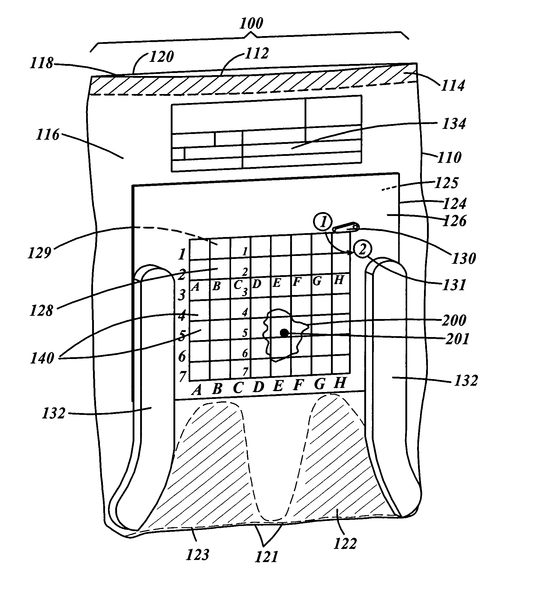
[0026]FIGS. 1-6 illustrate a first embodiment of a device for storing and / or for taking orthogonal images for radiographic examination of a tissue specimen. In FIG. 1, the device 100 comprises a flexible, transparent, sealable, liquid impervious bag 110 for receiving a tissue specimen 200 having an abnormality contained therein. In the preferred embodiment the bag has an opening 112 provided with a sealable adhesive strip 114. However, as should be obvious to one of ordinary skill in the art, opening 112 may be sealed by any suitable means for providing a fluid-tight seal to the opening 112 of the bag. In another embodiment the opening 112 is substantially resealable and liquid impervious, for example through the use of a Ziploc®-type device. The opening 112 provides access to a chamber or pocket 116 defined by walls 118 and 120 for receiving the specimen. In general, the pocket 116 is large enough to accommodate multiple tissue specimens from the same patient, each specimen measuri...


PUM


Abstract

A device is provided for transporting a tissue specimen without the risk of exposing health-care workers to potentially hazardous tissue fluids. The device includes a container, a flexible portion, a first support member received in the container, a second support member extending from the fist support member, a first locating indicia on a first side of the first supporting member, and a second substantially radiopaque locating indicia on a second side of the first support member, and an indicating member movably mounted on the first side of the first support member. The first support member has a first locating indicia on a first surface and a second locating indicia on a second surface, which generates a radiographic image when exposed to x-rays. The first and second indicia are substantially in registration such that when a tissue specimen is positioned on the first locating indicia and is then exposed to x-rays, a radiographic image of the specimen superimposed on the image of the second locating indicia is produced. Since both locating indicia are in registration, any tissue abnormality within the specimen can be precisely located with respect to both indicia. The device further includes a second support member, limiting contact of the container walls with the tissue specimen, and biasing the flexible portion away from the first support surface containing the tissue specimen. The device further includes an at least partially radiopaque indicating member for indicating the position of the tissue specimen on the radiographic image. A method for using the device to generate radiographic images of a tissue specimen is also disclosed.

Description

FIELD OF THE INVENTION [0001] The present invention relates generally to a method and apparatus for use in the storing and / or manipulation of tissue specimens, such as for storing and / or manipulating tissue specimens in the course of producing image radiographs of tissue specimens. BACKGROUND OF INVENTION [0002] Diagnostic radiology procedures often show a tissue abnormality which can only be identified as either malignant or benign by surgical biopsy and subsequent microscopic study. This is most often the case with mammography, the examination of the breast with radiography, where a suspected soft tissue tumor or an abnormal calcification seen on a mammogram frequently is not palpable due to its small size. [0003] There are roughly 750,000-1,000,000 surgical breast biopsies performed in the U.S. each year. Of these, roughly 55% are needle localized. In needle localization a hook located on the end of a wire is placed directly into the area of concern, and once the wire is in place...

Claims


Application Information

Patent Timeline
IPC IPC(8): A61B10/00C12M1/00
CPCA61B10/0096
Inventor ARCHAMBAULT, MAEGHANBROYLES, MARK
Owner BEEKLEY A CT
Why Eureka
Try Eureka