Method for detecting cancer cells and monitoring cancer therapy

a cancer cell and monitoring technology, applied in the field of cancer cell detection and monitoring cancer therapy, can solve the problems of not being able ct and mri are unable to detect microscopic tumors in vivo, and using pet for monitoring cancer therapy in these regions is not suitabl

Inactive Publication Date: 2005-07-14
NEW YORK UNIV
View PDF1 Cites 2 Cited by
  • Summary
  • Abstract
  • Description
  • Claims
  • Application Information

AI Technical Summary

Benefits of technology

[0007] In one aspect, the present invention provides a method for monitoring anti-cancer therapy in a mammal comprising administering to a mammal in need of such treatment a diagnostically effective amount of a Sindbis virus comprising a gene encoding a detectable label, and determining the amount of cancer cells in the body of said mammal, wherein the amount of cancer cells is proportional to the amount of label produced by said cancer cells.
[0008] In a further aspect, the present invention provides a method for identifying cancer cells in the body of a mammal comprising administering to a mammal in need of such treatment of diagnostically eff

Problems solved by technology

CT requires the use of x-rays and is not suitable for pregnant patients.
In addition, both CT and MRI are unable to detect microscopic tumors in vivo.
However, since

Method used


Image

  • Method for detecting cancer cells and monitoring cancer therapy
  • Method for detecting cancer cells and monitoring cancer therapy
  • Method for detecting cancer cells and monitoring cancer therapy

Examples


example 1

SinRep / Luc Viral Vector Specifically Infects Subcutaneous (s.c.) BHK Tumors and Suppresses Their Growth

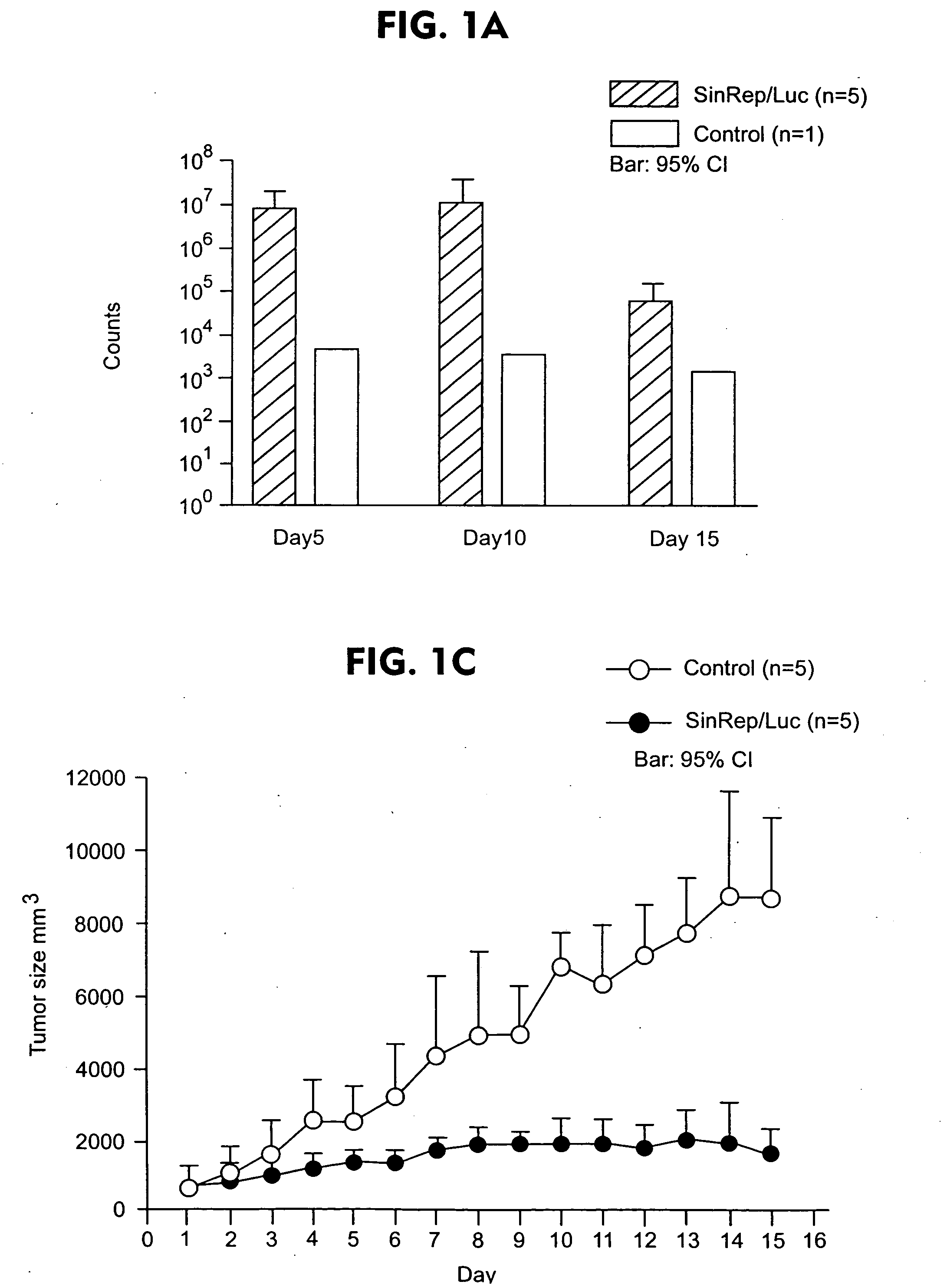
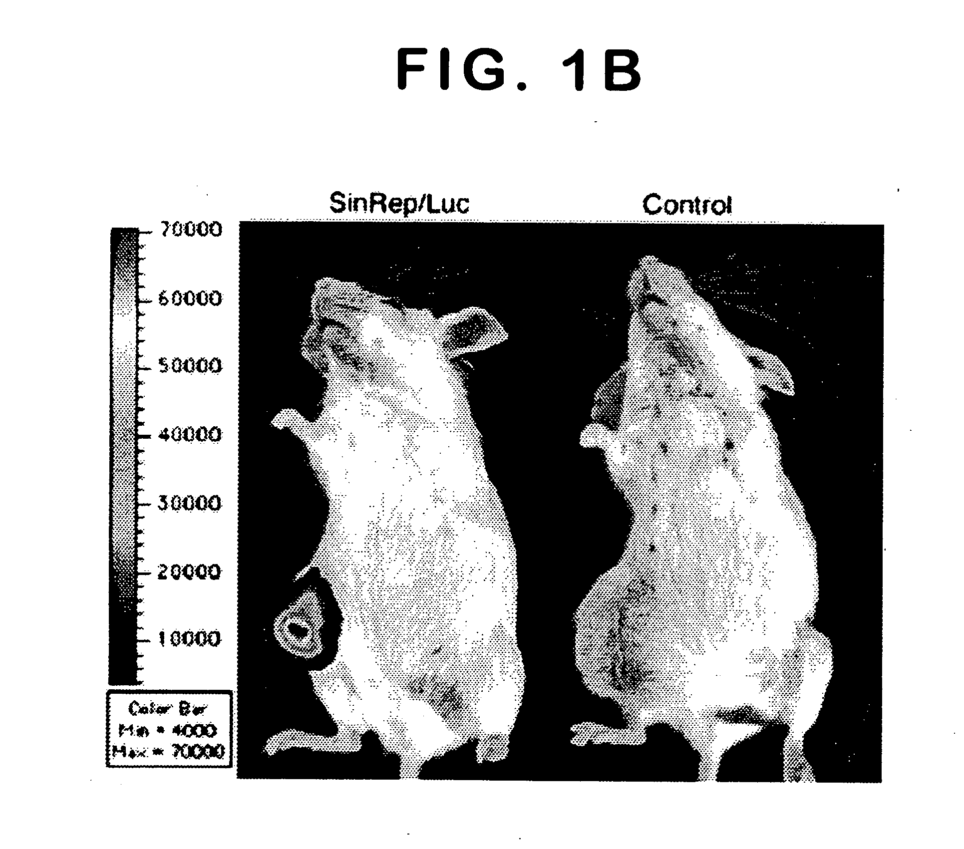
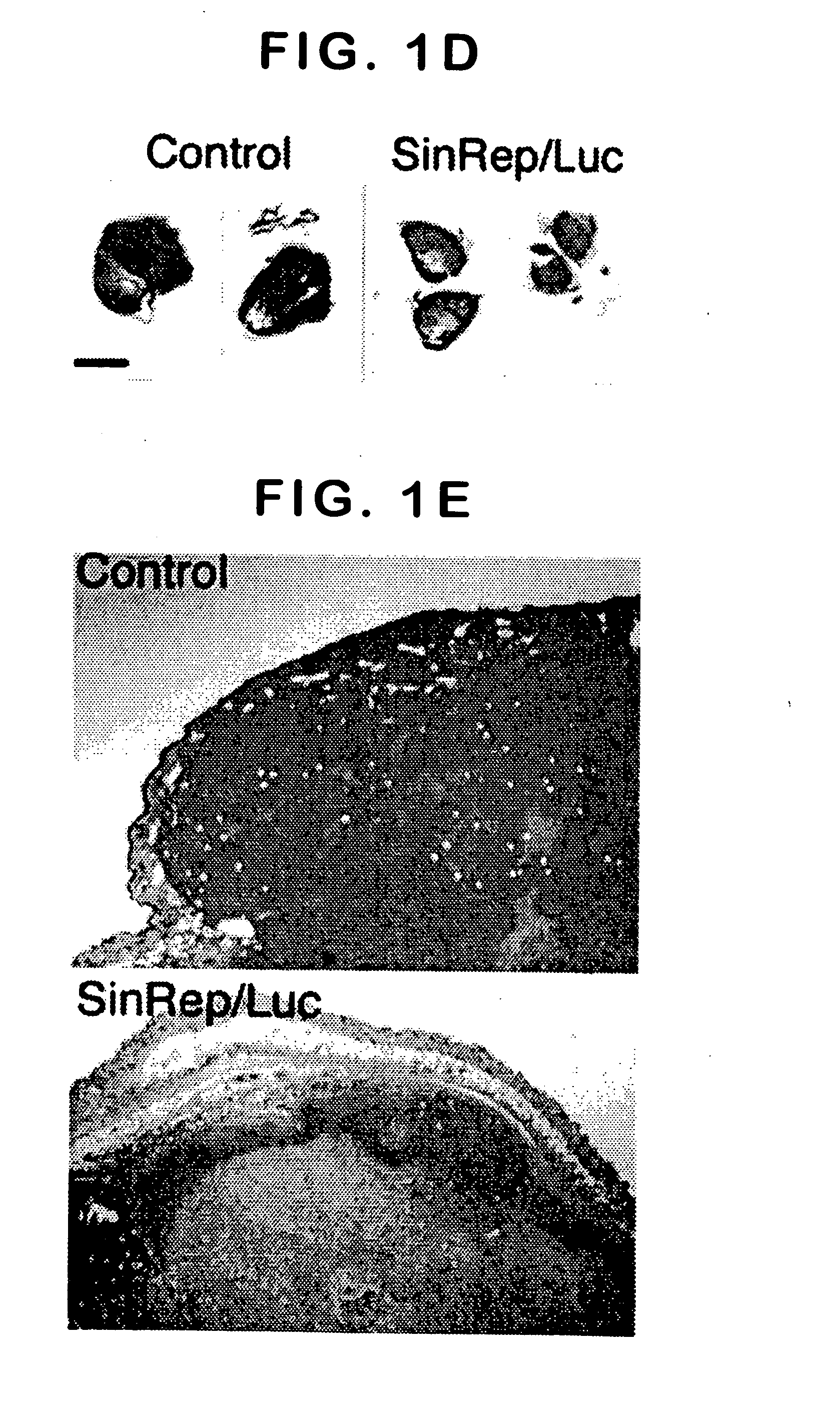
[0067] To test the potential of Sindbis viral vectors for systemic delivery and specific infection, a SinRep / Luc viral vector was injected daily, which carries a firefly luciferase gene, intraperitoneally (i.p.) to SCID mice bearing s.c. BHK tumors (FIG. 1). The daily i.p. injections of SinRep / Luc vectors was started when the tumors were approximately 500 mm3 (day 1) and the IVIS® system was used to monitor bioluminescence in the mice on days 5, 10, and 15. Control tumor-bearing mice received no SinRep / Luc treatment. On day 5, tumor specific bioluminescence that persisted until day 10 and dropped significantly on day 15 (P=0.0004, student t-test, FIG. 1a) was observed. S.c. BHK tumors on control mice generated very low background bioluminescence signals (˜103 photon counts) compared with SinRep / Luc treated tumors (˜107 photon counts). By day 10, a substantial therapeutic effect of...

example 2

Sindbis Viral Vectors Specifically Infect Intraperitoneal (i.p.) and Intrapancreatic BHK Tumors

[0069] To determine if Sindbis vectors can specifically infect BHK tumors growing at other locations, intrapancreatic tumors were established with a special BHK-derived line, BHKSINLuc2, which stably transcribes a defective Sindbis replicon RNA containing a firefly luciferase gene31. Since this cell line expresses luciferase activity in response to Sindbis infection, BHKSINLuc2 tumors as biological reporters of vector infection were used. Intrapancreatic inoculation of 1×10⁶ BHKSINLuc2 cells resulted in tumors mostly limited to the pancreas after eight days. A single i.p. injection of a Sindbis vector SinRep / LacZ that carries a bacerial β-galactosidase gene, led to specific infection of the intrapancrestic BHKSINLuc2 tumors and induction of luciferase activities (FIG. 2a, upper panels). Without SinRep / LacZ treatment, the control mice bearing intrapancreatic BHKSINLuc2 tumor showed no biol...

example 3

Sindbis Vectors Specifically Infected Intraperitoneal Metastasized Tumors

[0071] In another set of experiments, 1×10⁶ BHK cells were intrapancreatically inoculated in SCID mice. Intrapancreatic BHK tumors grew faster than BHKSINLuc2 tumors and resulted in metastasis throughout the peritoneal cavity (FIG. 3). Eight days after BHK cell inoculation, a single i.p. injection of SinRep / Luc vectors to mice induced specific and substantial bioluminescence signals (FIG. 3a) associated with tumor development on the diaphragm, pancreas, mesentery, and peritoneum (FIG. 3b). Background bioluminescence signals were minimal in the peritoneal cavity of tumor-free control mice that were given a single i.p. SinRep / Luc vector injection (FIG. 3a).

[0072] These results indicate that single i.p. delivery of Sindbis vectors can specifically infect the metastasized BHK tumors throughout the peritoneal cavity. Thus, Sindbis vectors, while delivered systemically, may serve as powerful tools for detecting mic...


PUM

PropertyMeasurementUnit
Fluorescenceaaaaaaaaaa

Abstract

Disclosed herein are methods for identifying cancer cells and monitoring anti-cancer therapy in the body of a mammal by systemically delivering Sindbis viral vectors. The vector can specifically target and identify tumor cells in mice growing subcutaneously, intraperitoneally, intrapancreatically, or in the lungs. These findings demonstrate the remarkable specificity of the Sindbis vector system that is relatively safe and can specifically target tumor cells throughout the body via the bloodstream.

Description

[0001] This application claims priority to U.S. Ser. No. 60 / 496,486, filed on Aug. 19, 2003. This prior application is incorporated herein by reference.[0002] The United States Government has certain rights to this invention by virtue of funding received from Public Health Service Grants CA22247 and CA68498 from the National Cancer Institute, National Institutes of Health, Department of Health and Human Services and U.S. Army grant OC000111.BACKGROUND OF THE INVENTION [0003] The likelihood of successful cancer gene therapy would be greatly enhanced by availability of a vector that could be delivered systemically and would have specific anti-tumor targeting capability along with the ability to induce death in primary and metastatic tumor cells. Vectors based on a prototype alphavirus, Sindbis virus, which were originally developed for efficient in vitro gene transfer to mammalian cells1, appear to have the desired properties2. Several factors contribute to the vectors' potential. Fir...

Claims


Application Information

Patent Timeline
IPC IPC(8): A61KA61K48/00A61K49/00A61K51/00A61M36/14C12N7/00C12N15/63C12Q1/70
CPCA61K35/76A61K47/48776A61K49/0013C12N2770/36143A61K49/0097C12N2770/32132A61K49/0047A61K47/6901
Inventor MERUELO, DANIELTSENG, JEN-CHIEH
Owner NEW YORK UNIV
Why Eureka
Try Eureka