Methods and devices for measuring diffusion by magnetic resonance imaging

a magnetic resonance imaging and measurement method technology, applied in the field of magnetic resonance imaging, can solve problems such as inhomogeneity of each of the three axes

Active Publication Date: 2005-07-28
CALIFORNIA INST OF TECH
View PDF1 Cites 5 Cited by
  • Summary
  • Abstract
  • Description
  • Claims
  • Application Information

AI Technical Summary

Benefits of technology

[0006] The diffusional permeability of a membrane is a key measure of how easily certain molecules, including water, pass through the membrane by diffusion. In biological tissues, the diffusional permeability may change due to disease, trauma or other failure, and is of significance in the study of many disease processes, including age-related macular degeneration, a leading cause of blindness in the older population. In this context, embodiments of the present invention provide methods and devices for the observing the diffusion of molecules across a permeable membrane using magnetic resonance techniques. Certain embodiments of the invention are useful for example for measuring changes in diffusional permeability and indices of diffusional permeability of Bruch's membrane (also known as the basal lamina of the choroid or vitreous membrane), one of the layers of the lining of the eyeball.

Problems solved by technology

Further, each of the three axes is not uniform but comprises a magnetic gradient, allowing each proton to be measured relative to the axis by its position within the gradient.

Method used


Image

  • Methods and devices for measuring diffusion by magnetic resonance imaging
  • Methods and devices for measuring diffusion by magnetic resonance imaging
  • Methods and devices for measuring diffusion by magnetic resonance imaging

Examples


example 1

Devices and Methods for Measuring Membrane Diffusional Permeability of Deuterium Oxide by Magnetic Resonance Imaging

example 1a

Membrane Cell Preparation

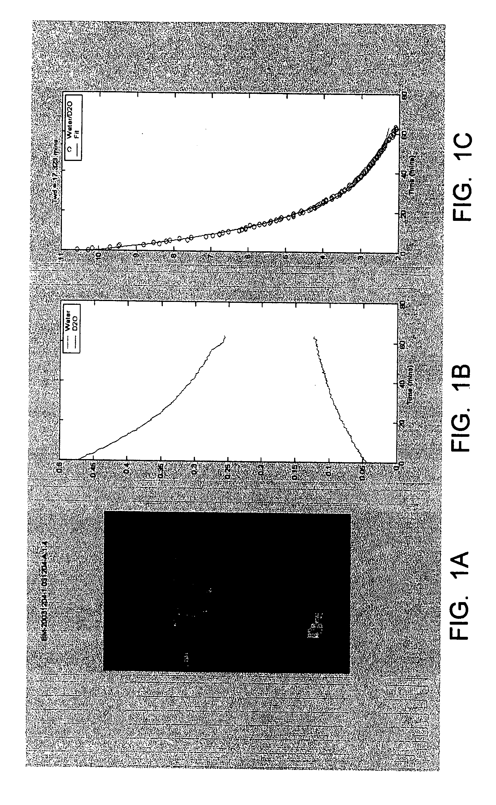
[0035] A 5 mm diameter tissue sample is mounted with tissue adhesive between two plastic disks with a central 3 mm hole in each disk to allow diffusion across the membrane. The central well of the membrane cell base is filled with either water or D2O (deuterium oxide). This sample assembly is then inserted in the upper circular recess of the membrane cell base. The membrane cell washer is then placed over the sample assembly and the membrane cell cap screwed onto the base, tightening the washer onto the sample assembly. The upper central well in the membrane cell cap is then filled with either D2O or water (the opposite of the contents of the lower well) and the sample placed in the magnetic resonance imaging system.

Magnetic Resonance Imaging

[0036] Sequential magnetic resonance images are acquired of a midline slice through the membrane cell, parallel to the long-axis of the cell. Sufficient images are acquired to record the change in signal intensity i...

example 1b

Membrane Cell Preparation

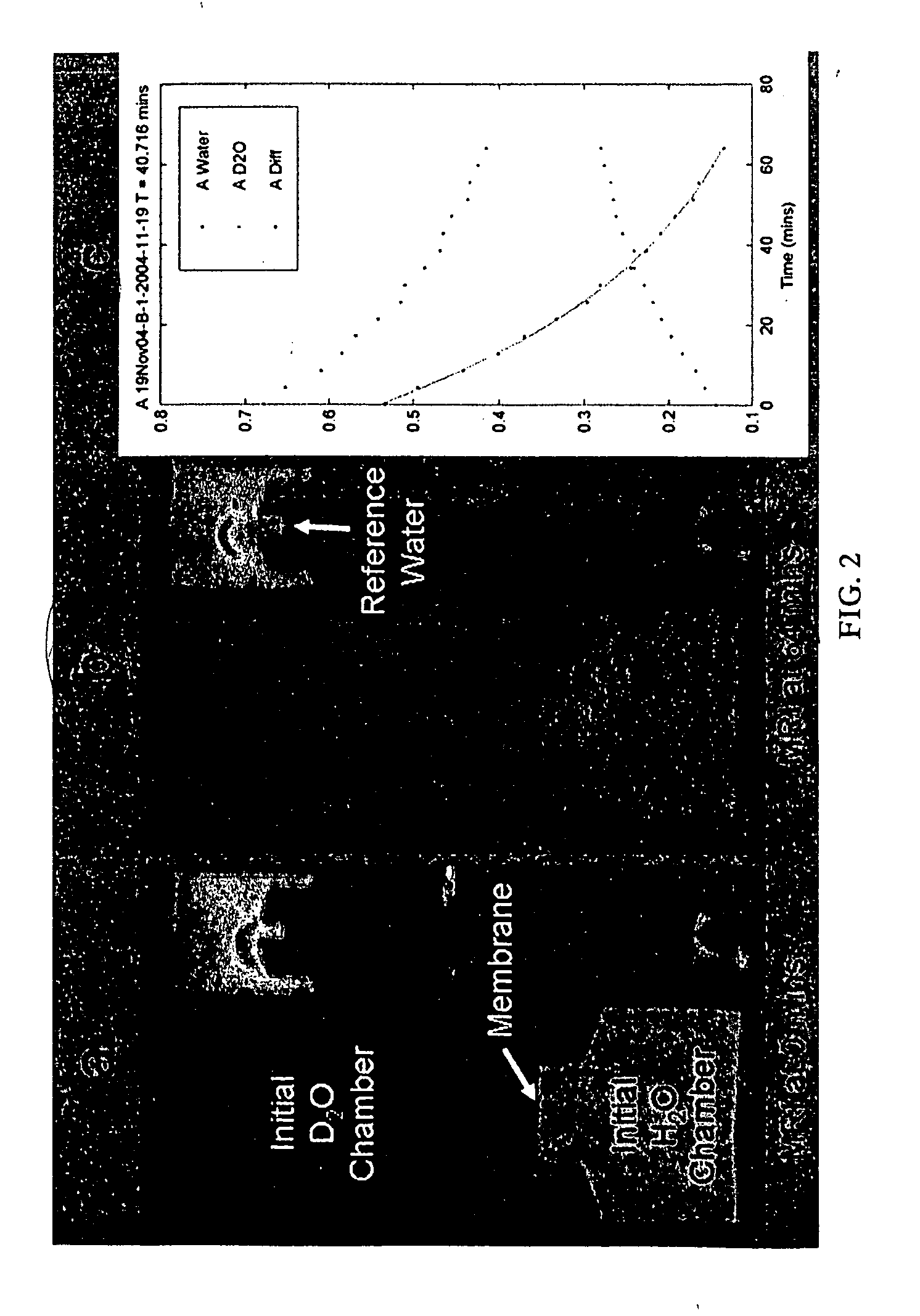
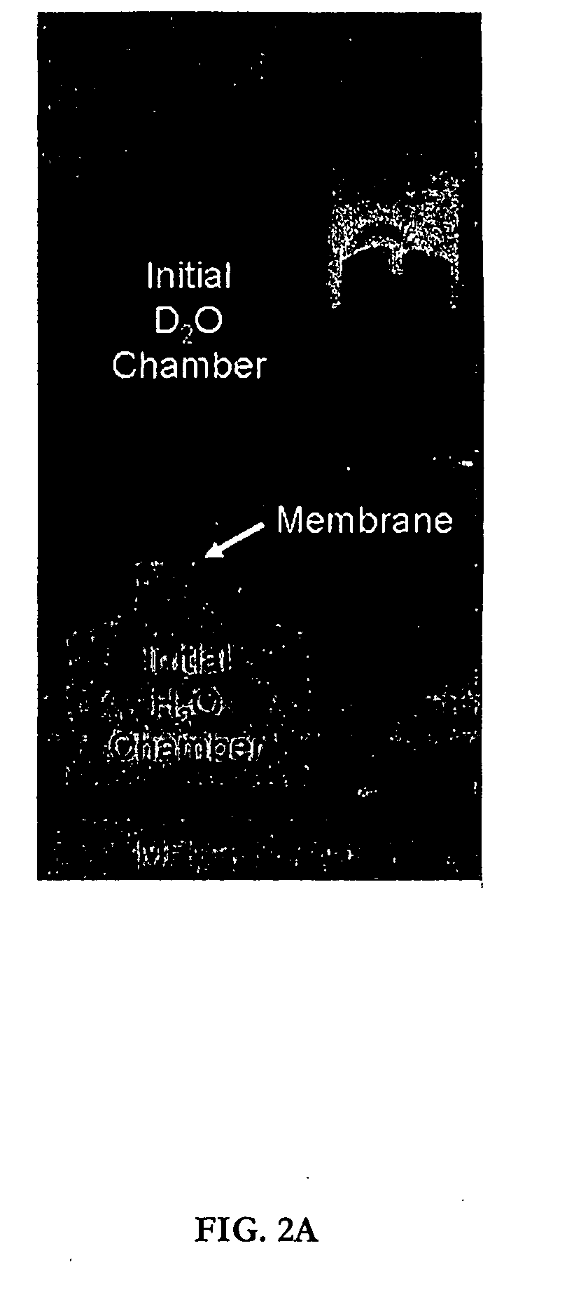
[0038] A 5 mm diameter membrane tissue sample is mounted between two layers of 125 micron pore-size nylon mesh. The tissue sample assembly is then mounted in the central recess of the cell base and the cell cap is tightened onto the base using three M2.5 nylon screws. O-rings mounted into the cap and base prevent fluid leakage between the upper and lower central chambers. The central well of the base is filled with water or saline water with the cell inverted. The base well is then sealed with impermeable adhesive tape. The central well of the upper chamber is filled with deuterium oxide (D2O) or D2O saline and sealed with impermeable adhesive tape as before.

Magnetic Resonance Imaging

[0039] Sequential magnetic resonance images are acquired of a midline slice through the membrane cell, parallel to the long-axis of the cell. Sufficient images are acquired to record the change in signal intensity in the upper and lower chambers as the D2O and water exchang...


PUM


Abstract

The present invention relates generally to the field of magnetic resonance imaging and, more particularly, to devices and methods used in the observation of the diffusion of molecules across a permeable membrane using magnetic resonance techniques. A typical embodiment of the invention is a method of observing the diffusion of a molecule in a container having a permeable membrane that is disposed between a first solution and a second solution in the container, by using magnetic resonance imaging to obtain an image of the diffusion of the molecule in the first solution across the membrane in to the second solution.

Description

CROSS REFERENCE TO RELATED APPLICATIONS [0001] This application claims the benefit of U.S. provisional patent application Ser. No. 60 / 528,535 filed Dec. 10, 2003 and U.S. provisional patent application Ser. No. 60 / 550,683 filed Mar. 5, 2003. The entire content of these provisional patent applications are incorporated herein by reference.FIELD OF THE INVENTION [0002] The present invention relates generally to the field of magnetic resonance imaging and, more particularly, to the observation of the diffusion of molecules across a permeable membrane using magnetic resonance imaging techniques. BACKGROUND OF THE INVENTION [0003] Magnetic resonance imaging (MRI) is a well established technique that is used, for example, to discriminate between normal and pathological tissue samples. MRI image contrast in this technology depends on differences in parameters such as the proton density, the longitudinal relaxation time T1, and the transverse relaxation time T2 for different media. MRI is al...

Claims


Application Information

Patent Timeline
Patent Type & Authority Applications(United States)
IPC IPC(8): A61K49/00G01R33/563
CPCG01R33/56341
Inventor TYSZKA, JULIAN MICHAELYU, CHANGJUNSCHWARTZ, DANIELSUGINO, ILENEFRASER, SCOTT E.
Owner CALIFORNIA INST OF TECH
Why Eureka
Try Eureka