High contrast optoacoustical imaging using nonoparticles

a non-oparticle, optoacoustic imaging technology, applied in the field of optoacoustic imaging, can solve the problems of inability to recognize tumor masses, low image resolution, and inability to achieve tumor masses, so as to accelerate the passage of nanoparticles, enhance the uptake of contrast agents, and slow the chemical degradation of components

Inactive Publication Date: 2005-08-11
SENO MEDICAL INSTR
View PDF40 Cites 121 Cited by
  • Summary
  • Abstract
  • Description
  • Claims
  • Application Information

AI Technical Summary

Benefits of technology

[0012] We have now discovered a way in which to optoacoustically detect the presence of objects as small as 1 mm and smaller in a body which can be penetrated by electromagnetic radiation. We have discovered that at least partially metallic nanoparticulates fabricated or manipulated to be non-spherical not only will shift the optical absorption spectrum into the near infrared range for deeper penetration into a body but also will both narrow the absorption band and simultaneously increase the effective absorbance, in certain instances by about an order of magnitude or more, thereby greatly increasing the optoacoustic efficacy of the nanoparticulate, making the manipulated nanoparticulate a very high contrast optoacoustic imaging agent. In accordance with our discoveries, effective acoustical response to electromagnetic radiation is increased by tailoring the choice of the composition, non-spherical shape and size of nanoparticulates to maximize both the optical absorbance of the particles at the irradiation wavelength(s) and the optoacoustocal pressure produced in response to absorbed electromagnetic energy.
[0020] The shape is engineered for longer length in one dimension in order to provide sufficient shift of an electromagnetic absorption maximum towards longer wavelengths compared with spherical (or otherwise fully symmetrical) nanoparticles, to yield desirable absorption at a selected wavelength or selected spread of wavelengths which are capable of penetrating the body.
[0064] In general, the particulate portion of the contrast agent will be dispersed in a sterile carrier liquid, which can be water, a water solution such as saline solution, an organic liquid, or an oil, including oils of animal, mineral or synthetic origin. Furthermore, the formulations used for practice of the invention can include a variety of excipients such as one or more neutral lipids, charged lipids, gases, gaseous precursors, liquids, oils, diagnostic agents, targeting ligands and / or bioactive agents. The formulation can also comprise stabilizers to slow the chemical degradation of the components.
[0065] In one embodiment of the invention, the contrast agents are administered in conjunction with the use of hyperthermia, that is, the artificial elevation of the local temperature of an organ or another body part. Kong, et al. (2000 and 2001) teach, that hyperthermia accelerates the passage of nanoparticles through the capillaries of the vascular system of growing tumors. Hyperthermia will also enhance the uptake of the contrast agent of other types of diseased tissue, sites of inflammation caused by infection or trauma.

Problems solved by technology

Optical imaging suffers from the severe disadvantage that resolution of the image is inherently low, because light, whether in the visible or near infrared spectrum, is strongly scattered in the body through interaction with local inhomogeneities.
However, they have not yet developed into recognizable tumor masses.
In particular, they have not yet developed the extensive vascular network that is characteristic of many larger solid tumors.
For detection of nascent cancerous tumors, it may no longer be sufficient to rely on differences in the blood content and normal tissue.
Currently, however, neither pure optical imaging can nor prior optoacoustical imaging could detect tumors as small as 1 mm.

Method used


Image

  • High contrast optoacoustical imaging using nonoparticles
  • High contrast optoacoustical imaging using nonoparticles
  • High contrast optoacoustical imaging using nonoparticles

Examples


example i

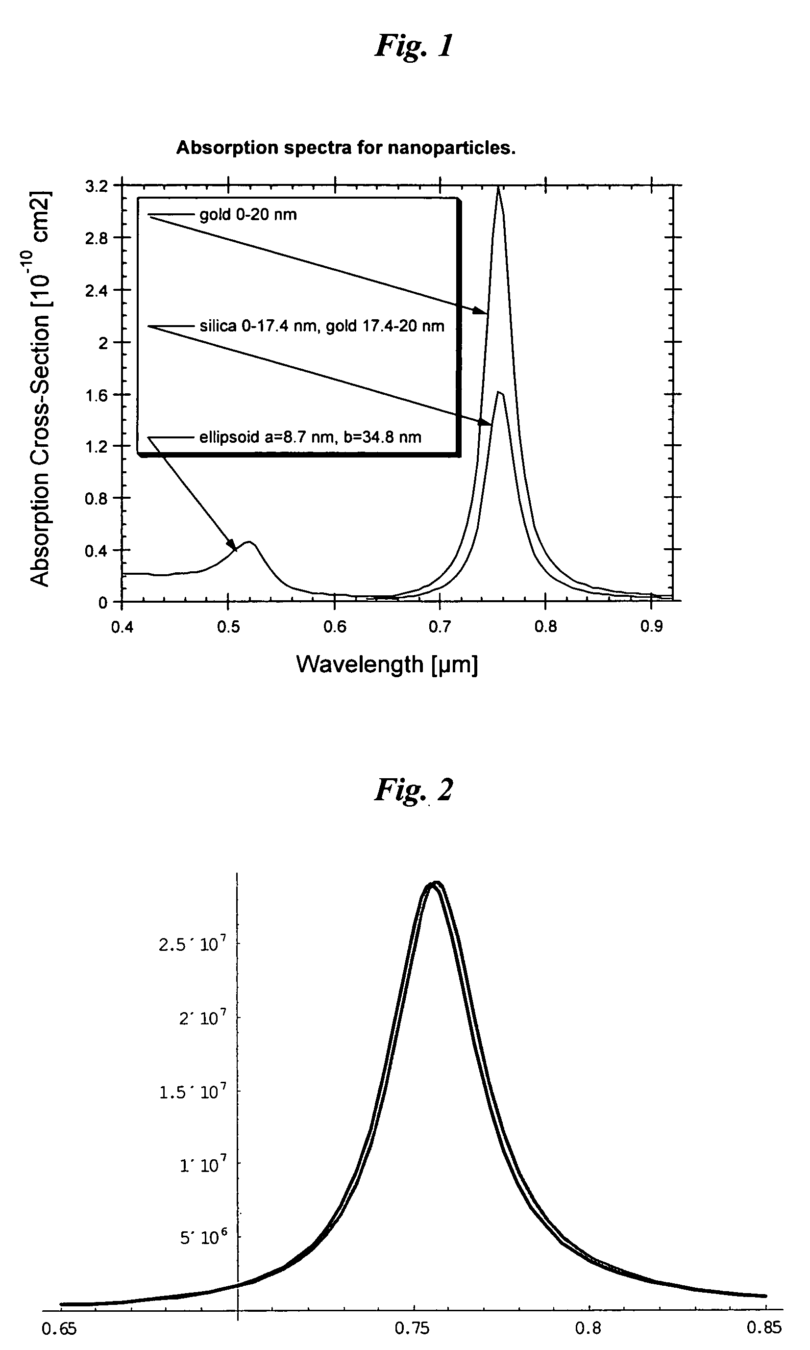
Optical Absorption Spectra of Metal Nanoparticles

[0214] To calculate the intensity and resonance absorption of an ellipsoid metal nanoparticles (NP) with radii, a and b and length, c, it is necessary to know the amplitude of an electromagnetic field inside the NP. When the wavelength is significantly longer than the linear dimensions of NP, the E-field components, Ek, inside the NP can be expressed in terms of the external field, E0k, incident upon metal NP as taught by Papavassilliou-1980, who provided details to a classical electromagnetic theory described by Stratton-1941: Emk=ɛ0(ɛm-ɛ0)⁢Pk+ɛ0⁢E0k(1)[0215] where εm=εm+iεm is the dielectric constant of the metal, from which NP is fabricated, ε0 is the dielectric constant of the surrounding medium (usually can be taken with properties of water). It is assumed that the axes x, y, z are directed along the NP axes. The quantity Pk {k=x,y,z} is called the depolarization factor along axes x, y, z is determined by the integral: Pk=12⁢...

example ii

Enhancement of Optoacoustic Signal at 532 nm with Contrast Agents Made of Metallic Nanoparticles.

[0237] If the optical fluence exceeds the threshold of phase transition for a substance covering the nanoparticles (let us for the purpose of this example assume that the substance surrounding nanoparticles is water) the amplitude of the optoacoustic signal can be increased one order of magnitude or more. Let us assume that the temperature of the nanoparticle and surrounding water comes to boiling point at some moment tev in a course of the laser pulse. A thin vapor layer may be generated around the particle heated above 100° C., or water may occur in the superheated steady state. Due to lower heat conductivity of vapor compared to that of water, the temperature of the particle may continue to increase in the course of laser pulse with fluence exceeding the threshold. After the end of the laser pulse the particle temperature relaxes to the initial value due to heat flux from the partic...

example iii

Optoacoustic Biosensor for Detection of Biological Warfare

[0251] Optoacoustics technique that employs laser pulses for spectrally selective excitation of multicomponent medium and detection of resulting acoustic waves is well known for its exceptional sensitivity in measuring small concentration of molecules in liquids and gases (Tam, 1986; Zharov and Letokhov, 1984). The method of optoacoustic tomography developed by Oraevsky et al (U.S. Pat. No. 5,840,023) permits sensitive detection, high contrast visualization and diagnostics of early malignant tumors in human organs (Oraevsky 1994, 1996, 2000, 2002). We teach here that resolution and sensitivity of this method can be further enhanced through selective administration of non-spherical gold nanoparticles into the volume of diagnostic interest. Gold nanoparticles help to dramatically enhance the optoacoustic contrast, since they can be designed to strongly absorb laser radiation in the near-infrared spectral range and, being heat...


PUM

PropertyMeasurementUnit
wavelengthaaaaaaaaaa
aspect ratioaaaaaaaaaa
sizeaaaaaaaaaa

Abstract

A method of enhancing detection for a specific object in a body. A nanoparticulate is administered to the body for location in an area to be explored for detection of the object, if present. The nanoparticulate is at least partially metallic, has a formed non-spherical shape having a minimal characteristic dimension in the range from about 1 to about 3000 nanometers, and has a formed composition capable of producing thermal pressure either in the nanoparticulate or in the object greater than the object could produce in the absence of the nanoparticulate. Electromagnetic radiation is directed into the body. The electromagnetic radiation has a specific wavelength or spectrum of wavelengths in the range from 300 nm to 300 mm selected so that the wavelength or wavelength spectrum is longer by a factor of at least 3 than the minimum characteristic dimension of the nanoparticulate. The nanoparticulate absorbs the electromagnetic radiation more than would one or more non-aggregated spherically shaped particles of the same total volume with a composition identical to the nanoparticulate. The nanoparticulate produces an enhanced optoacoustic signal resulting from the absorption that is received and converted into an electronic signal and presented for assessment of the at least one parameter by a human or a machine

Description

REFERENCE TO RELATED APPLICATION [0001] This application claims the benefit of 35 U.S.C. 111 (b) Provisional Patent Application Ser. No. 60 / 442,369, filed Jan. 25, 2003, and entitled, “Contrast Agents for Optoacoustic Imaging.”BACKGROUND OF THE INVENTION [0002] The present invention relates to optoacoustic imaging employing nanoparticles to enhance detection of objects in a body. The invention has applications that include imaging for medical reasons. [0003] Optoacoustic imaging is to be distinguished from optical imaging. In optical imaging a body is irradiated with light in the visible or infrared wavelength ranges. Transmitted or reflected light in the same ranges is detected to generate images. Optical imaging suffers from the severe disadvantage that resolution of the image is inherently low, because light, whether in the visible or near infrared spectrum, is strongly scattered in the body through interaction with local inhomogeneities. [0004] Optoacoustic methods of imaging re...

Claims


Application Information

Patent Timeline
Patent Type & Authority Applications(United States)
IPC IPC(8): A61B5/00A61K49/00A61K49/22G01H9/00G01N21/17G01N21/47G01N21/55
CPCA61B5/0059A61B5/0095A61B5/415A61B5/418G01N2021/1706B82Y15/00G01N21/1702G01N21/4795G01N21/554A61K49/225A61P35/00
Inventor ORAEVSKY, ALEXANDER A.HENRICHS, PAUL M.
Owner SENO MEDICAL INSTR
Why Eureka
Try Eureka