Non-invasive subject-information imaging method and apparatus

a subject-information imaging and non-invasive technology, applied in the field of non-invasive subject-information imaging methods and apparatuses, can solve the problems of affecting the subject's skin, affecting the operation, and causing a lot of pain to the subject, and causing the operator to be contaminated by biohazards

Active Publication Date: 2005-08-25
TOSHIBA MEDICAL SYST CORP
View PDF15 Cites 169 Cited by
  • Summary
  • Abstract
  • Description
  • Claims
  • Application Information

AI Technical Summary

Benefits of technology

[0028] According to a second aspect of the present invention, there is provided a non-invasive subject-information imaging method comprising irradiating a subject to be examined with light containing a specific wavelength component from a plurality of two-dimensionally arranged light irradiation positions, causing a plurality of two-dimensionally arranged electroacoustic transducer elements to receive acoustic waves generated in the subject upon the irradiation of light, driving the plurality of electroacoustic

Problems solved by technology

This method therefore damages the skin of the subject, causing much pain to the subject.
This also introduces the possibility of biohazard contamination to the subject and the operator.
Breast cancer is a major source of mortality in females.
These set of steps take considerable time before reaching a final conclusion.
These methods suffer from imprecision of image conversion and image distortions close to the edges of the body part, such as the breast.
Further, MRI and CAT scan require large expensive equipment that cannot be transformed easily.
They do not teach combination of photoacoustic and ultrasound images that are detected using positioned ultrasound transducers.
The method leads to the possibi

Method used


Image

  • Non-invasive subject-information imaging method and apparatus
  • Non-invasive subject-information imaging method and apparatus
  • Non-invasive subject-information imaging method and apparatus

Examples


Embodiment Construction

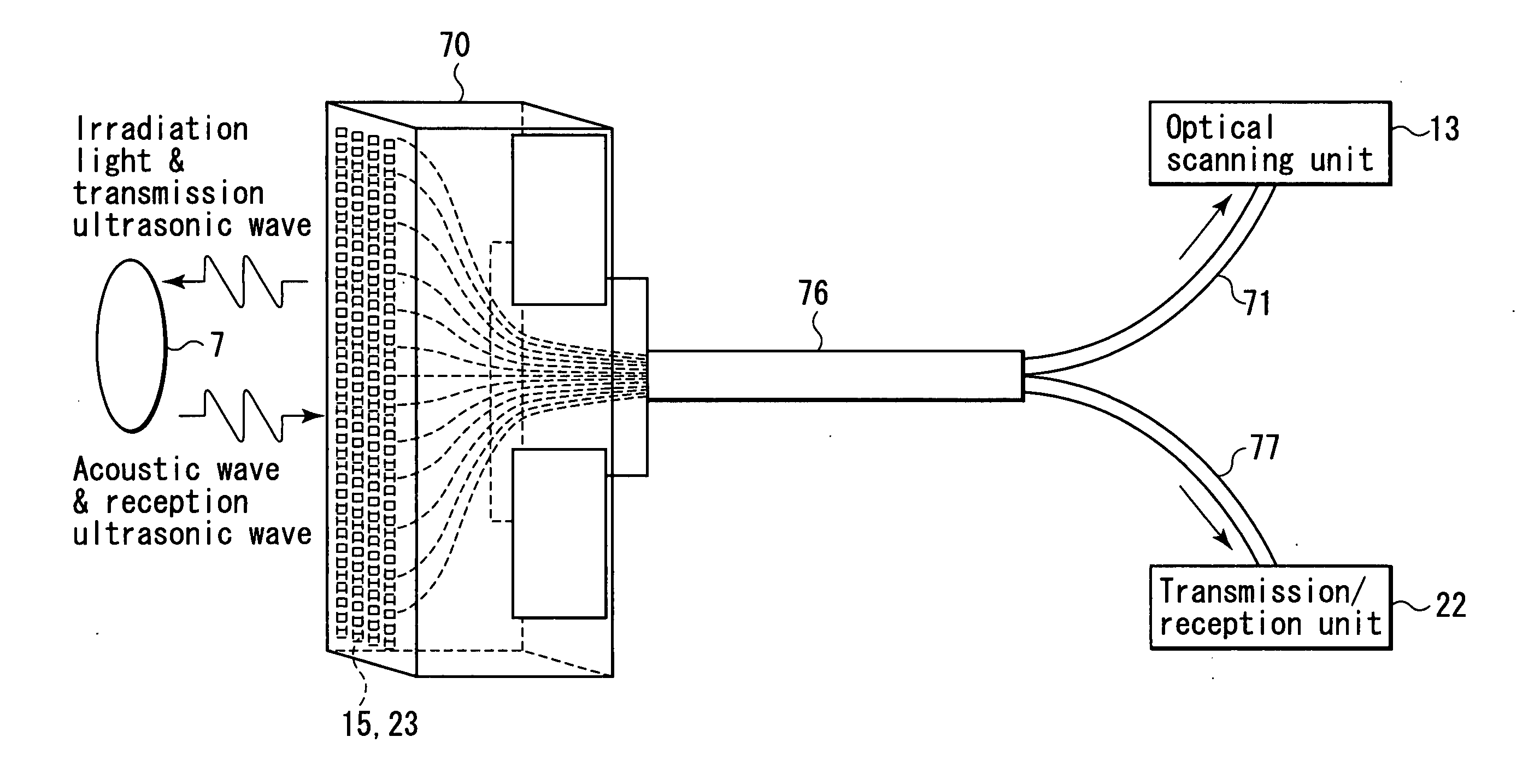
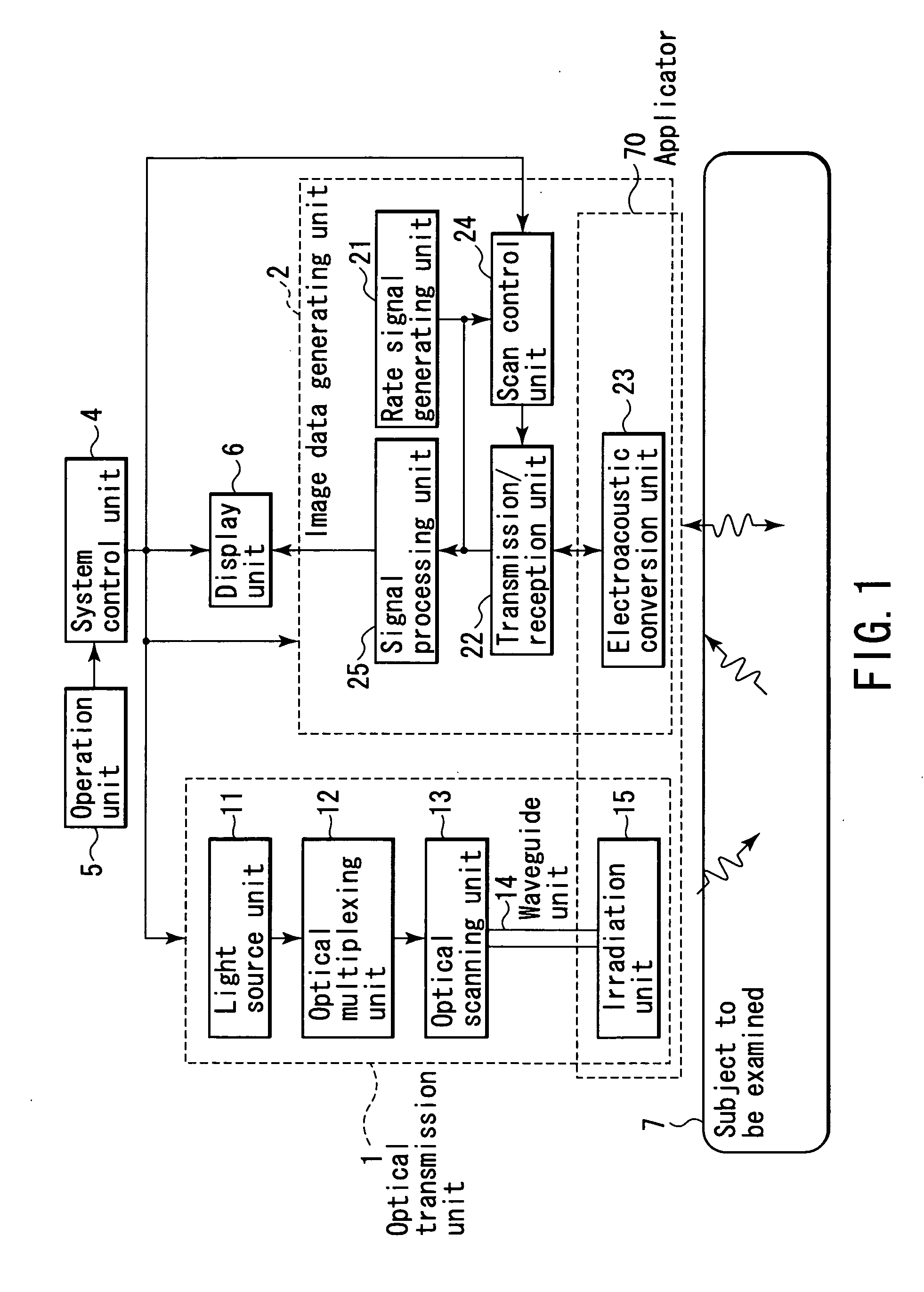
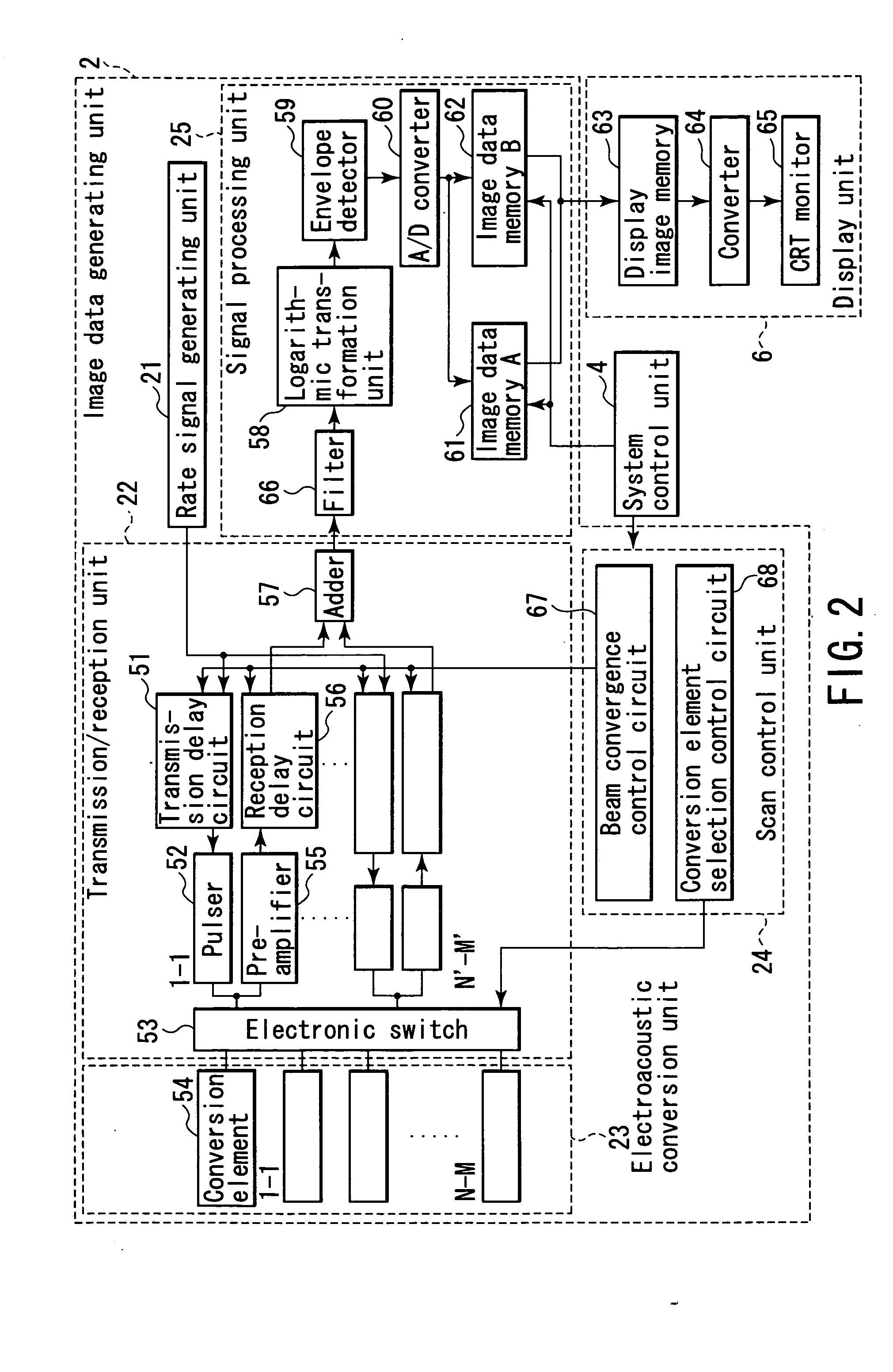
[0050] An embodiment of the present invention will be described below with reference to the views of the accompanying drawing. A subject-information imaging apparatus according to this embodiment can image a hemoglobin distribution in the subject, which is mainly directed to diagnosis of breast cancer. More specifically, a plurality of electroacoustic transducer elements are two-dimensionally arranged at predetermined intervals in the vertical and horizontal directions, and the output ends of a plurality of optical fibers for light irradiation are arranged in the gaps between the electroacoustic transducer elements, thereby forming an applicator in which an irradiation unit is integrated with an electroacoustic conversion unit. By using this arrangement, volume data corresponding to a three-dimensional region representing a living body function is acquired by two-dimensional electroacoustic scanning based on light irradiation from the irradiation unit and detection of the resultant ...


PUM


Abstract

A non-invasive subject-information imaging apparatus according to this invention includes a light generating unit which generates light containing a specific wavelength component, a light irradiation unit which radiates the generated light into a subject, a waveguide unit which guides the light from the light generating unit to the irradiation unit, a plurality of two-dimensionally arrayed electroacoustic transducer elements, a transmission/reception unit which transmits ultrasonic waves to the subject by driving the electroacoustic transducer elements, and generates a reception signal from electrical signals converted by electroacoustic transducer elements, and a signal processing unit which generates volume data about a living body function by processing a reception signal corresponding to acoustic waves generated in the subject by light irradiation, and generates volume data about a tissue morphology by processing a reception signal corresponding to echoes generated in the subject upon transmission of the ultrasonic waves.

Description

CROSS-REFERENCE TO RELATED APPLICATIONS [0001] This application is based upon and claims the benefit of priority from prior Japanese Patent Application No. 2004-30578, filed Feb. 6, 2004, the entire contents of which are incorporated herein by reference. BACKGROUND OF THE INVENTION [0002] 1. Field of the Invention [0003] The present invention relates to a non-invasive subject-information imaging method and apparatus for imaging living body anatomical, functional, and / or metabolic information of a subject to be examined by acquiring acoustic signals generated on the basis of the energy of light radiated into the subject and, more particularly, to a method and apparatus which acquire and superimpose two acoustic images, one generated from the energy of light radiated into a subject to be examined and the other is an ultrasound echo image generated from ultrasonic waves directed into the subject, and allow an operator to know the distribution of substance concentrations with respect to...

Claims


Application Information

Patent Timeline
IPC IPC(8): A61B5/00G01N29/00A61B8/08A61B8/14G01N21/00G01N29/06
CPCA61B5/0091A61B5/0095A61B5/145A61B5/14532A61B5/14546G01N2291/02809A61B8/14A61B8/5238A61B2562/046G01N29/0609A61B5/4312A61B5/0035A47G19/025A47G23/0225A47G23/0306A47G2400/08
Inventor KANAYAMA, SHOICHIITSUMI, KAZUHIRO
Owner TOSHIBA MEDICAL SYST CORP
Why Eureka
Try Eureka