Method for screening cervical cancer

a cervical cancer and reagent technology, applied in the field of reagents for diagnosing cervical cancer, can solve the problems of inability to obtain information for identifying the type of cancer, inability to accurately diagnose inability to identify the type of cancer, etc., and achieve the effect of high accuracy, effective diagnosis, and improved accuracy

Inactive Publication Date: 2005-10-06
SYSMEX CORP
View PDF3 Cites 31 Cited by
  • Summary
  • Abstract
  • Description
  • Claims
  • Application Information

AI Technical Summary

Benefits of technology

[0020] In consideration of individual features of gland cells and squamous cells and by paying attention to a marker specific to each cancer cell, the method for screening cervical cancer according to the present invention involves examining the presence or absence of the reactivity of a marker to each antibody thereby enabling not only discrimination of cancer cells from normal cells but also discrimination of adenocarcinoma cells from squamous carcinoma cells and further judging their precancerous state, and is thus utilizable effectively in diagnosis requiring judgment of the type of cervical cancer. Further, flow cytometry can be applied, whereby cervical cancer can be screened more efficiently.
[0021] The method for screening cervical cancer according to the present invention is capable of screening the presence or absence of cervical cancer and / or squamous cell carcinoma with high accuracy and high efficiency, and is thus useful in cellular diagnosis for cervical cancer wherein a large number of samples should be treated in medical examinations.

Problems solved by technology

Such accuracy and processing speed are not satisfactory for those who judge the presence or absence of cancer in medical examinations.
However, the markers used in U.S. Patent Application No. 2002 / 0106685 supra are markers for general cancer cells, and thus the presence or absence of cancer cells can be judged, but the type of cervical cancer cannot be identified.
The marker disclosed in U.S. Pat. No. 5,858,683 supra is a marker specific to cervical cancer, but it is a marker common to squamous cell carcinoma and adenocarcinoma, so information for identifying the type of cancer cannot be obtained.
NMP179 antibody is reported to react with a part of normal cells (Acta Cytologica, Volume 43, Number 6 / November-December 1999: 1015-1022), and its performance cannot be satisfactory.

Method used


Image

  • Method for screening cervical cancer
  • Method for screening cervical cancer
  • Method for screening cervical cancer

Examples


example 1

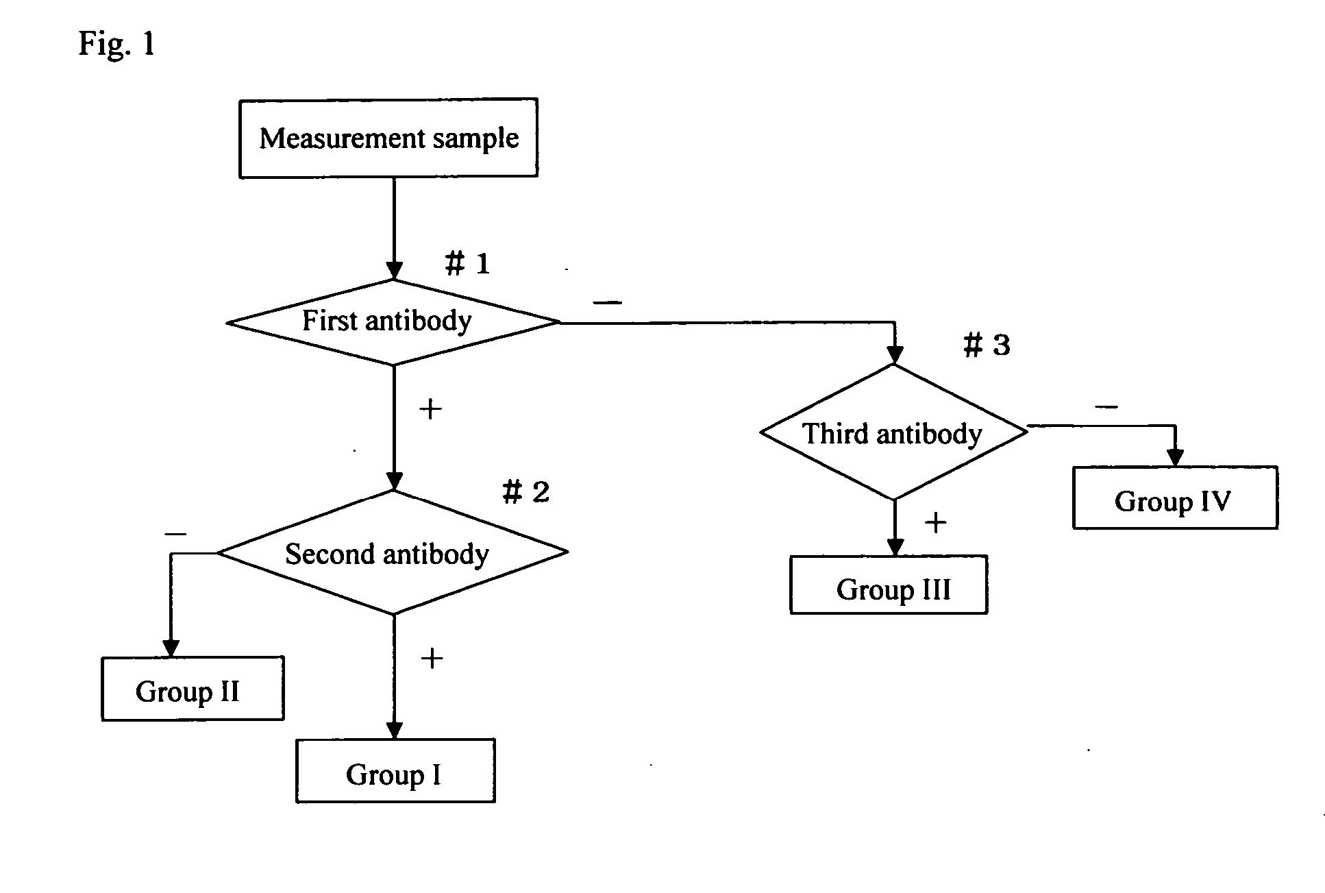
[0155] Four tubes were prepared, and HeLa cells, C33A cells and the clinical samples 1 and 2 were introduced at a density of about 1×10⁶ cells into the tubes respectively. The cells were fixed with PreservCyt solution from Cytyc Ltd. and then centrifuged at 10000 rpm for 1 minute to remove a supernatant. Then, 5% N-acetyl-L-cysteine was added thereto, and the cells were stirred and centrifuged at 10000 rpm for 1 minute to remove a supernatant. The cells were washed with PBS and centrifuged at 10000 rpm for 1 minute to remove a supernatant. One ml PBS or PBS-T (PBS containing 0.05% Tween 20) containing 1% goat serum was added thereto, and the cells were stirred and subjected to blocking reaction for 10 minutes.

[0156] After blocking, the cells were centrifuged at 10000 rpm for 1 minute to remove a supernatant. The first labeled antibody-containing reagent prepared above was added thereto, and the cells were reacted for 30 minutes at room temperature.

[0157] After the antibody reactio...

example 2

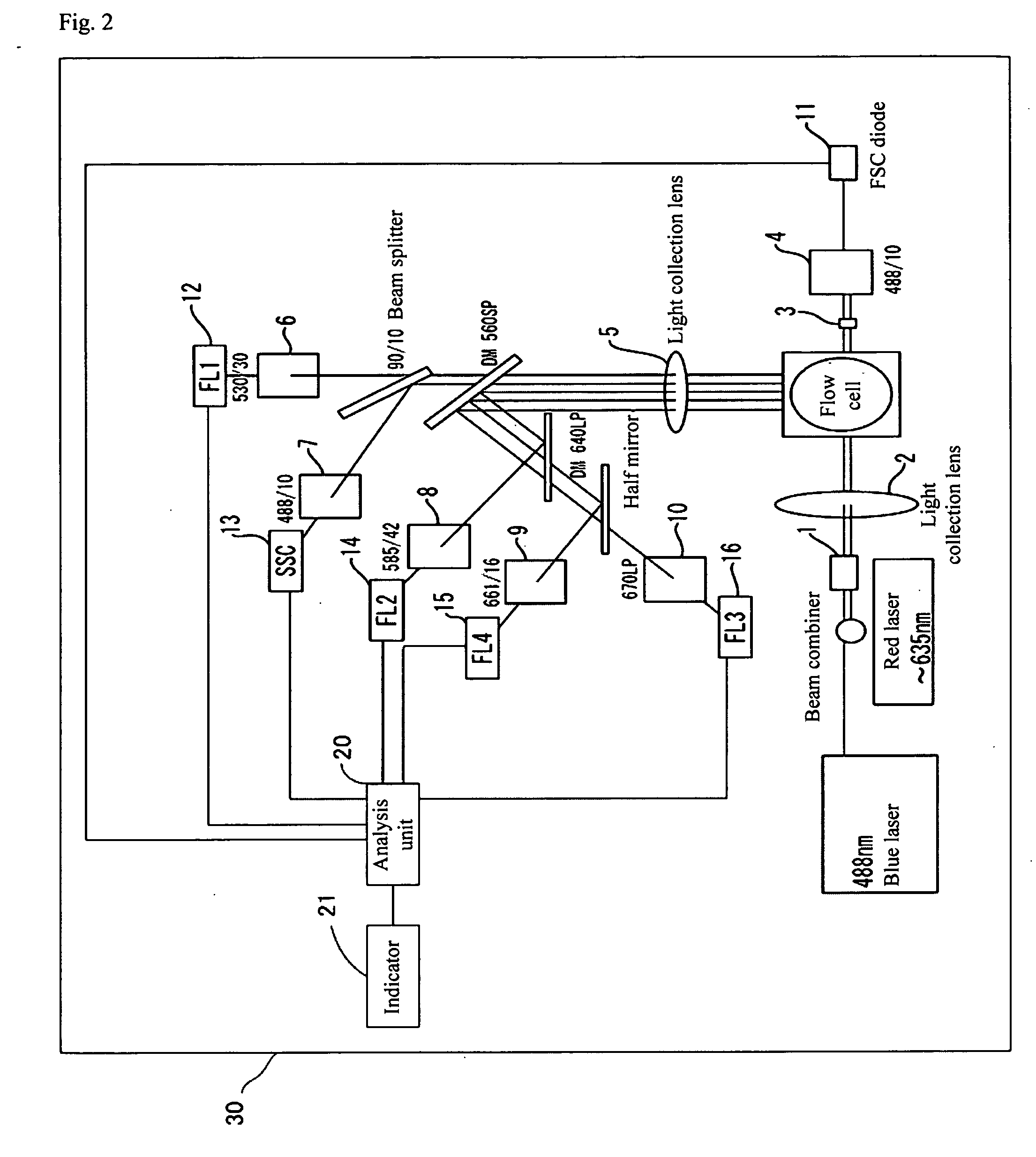
[0165] About 1×10⁶ epithelial cells in mouth cavity mucosa were introduced into a tube, and 100 μl of 300 μg / ml ribonuclease A (#R-4612 manufactured by Sigma) diluted with PBS was added to the cells to prevent non-specific staining of RNA. The cells were stirred for 5 minutes at room temperature and centrifuged at 10000 rpm for 1 minute to remove a supernatant. Then, 500 μl of 10 μM PI (propidium iodide) was added as a nucleus staining solution thereto, and the cells were stirred for 30 minutes at room temperature. The cells were centrifuged at 10000 rpm for 1 minute to remove a supernatant, and after 500 μl of 0.05% PBST was added thereto, the cells were centrifuged at 10000 rpm for 1 minute. The medium was replaced by a suitable amount of PBS, and the cells were subjected to a flow cytometer having the constitution shown in FIG. 6.

[0166] The sample was excited by an argon ion laser at 488 nm, and a fluorescence profile in orange derived from nuclear staining, a side scattered lig...

example 3

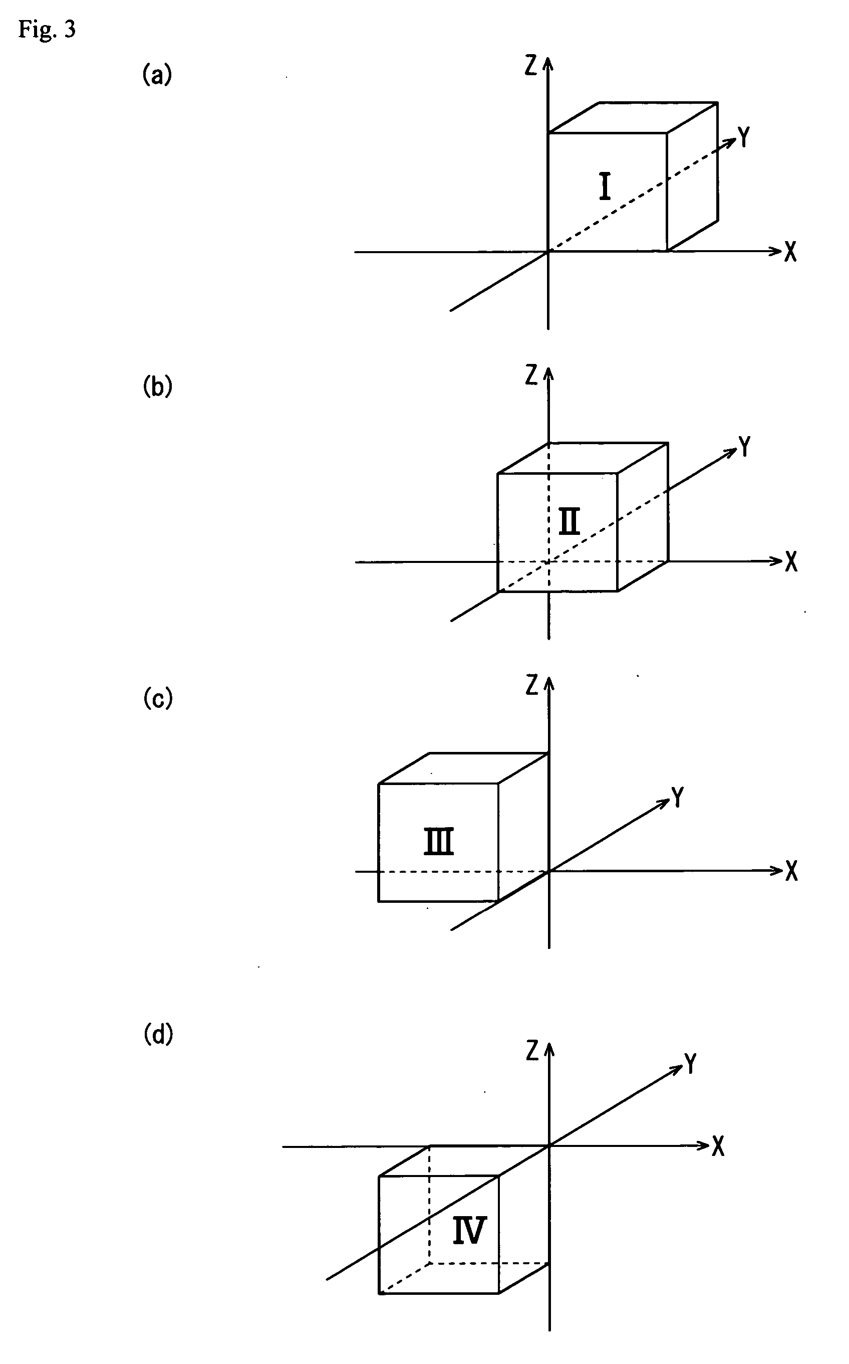
[0169] Oral mucosal epithelial cells as a substitute for the cervical normal squamous cells, HeLa cells as the cervical adenocarcinoma cells, and C33A cells as the cervical squamous carcinoma cells were used to prepare model samples, that is, a normal sample and 2 abnormal samples (cervical adenocarcinoma cells and cervical squamous carcinoma cells), and whether screening of cervical cancer in these samples was feasible or not was verified.

(Preparation of the Model Samples)

[0170] As the model sample (model sample of the normal sample) of cervical normal squamous cells, oral mucosal epithelial cells, about 2×10⁵ cells / tube, preserved in a preservative solution PreservCyt (Cytyc; Cat#0234004) were used.

[0171] As the model sample of cervical adenocarcinoma cells, about 1×10⁵ HeLa cells (from about 2×10⁵ cells / tube preserved in PreservCyt) were mixed with about 1×10⁵ cells of the above model sample of normal squamous cells, to prepare about 2×10⁵ cells in total per tube.

[0172] As t...


PUM

PropertyMeasurementUnit
oscillation wavelengthaaaaaaaaaa
pass wavelengthaaaaaaaaaa

Abstract

The invention provides a reagent for diagnosing cervical cancer, which can not only detect the presence or absence of cervical cancer but can also distinguish squamous cell carcinoma and adenocarcinoma from each other, a method for screening cervical cancer by using the reagent, particularly a screening method capable high speed processing by utilizing flow cytometry.
The reagent comprises a first labeled antibody reacting with gland cells, a second labeled antibody reacting with adenocarcinoma cells and a third labeled antibody reacting with atypical cervical squamous cells, the antibodies being labeled with mutually distinguishable labels respectively. Preferably, at least one selected from the group consisting of MUC1 antibody, cytokeratin 7 antibody, and cytokeratin 18 antibody is used as the first labeled antibody, at least one selected from the group consisting of cytokeratin 8 antibody and HIK1083 antibody is used as the second labeled antibody, and at least one member selected from the group consisting of NMP179 antibody, p16INK4A antibody, Ki-67 antibody, p53 antibody, p21 antibody, EMA antibody, CEA antibody and MIB-1 antibody is used as the third labeled antibody.

Description

BACKGROUND OF THE INVENTION [0001] 1. Field of the Invention [0002] The present invention relates to a reagent for diagnosing cervical cancer, which can detect the presence or absence of squamous cell carcinoma, its precancerous state, adenocarcinoma and its precancerous state in a sample of cervical cell group collected from the living body, a method for screening cervical cancer by using the diagnostic reagent, and an automatic diagnostic method of automatically judging adenocarcinoma and squamous cell carcinoma. [0003] 2. Description of the Related Art [0004] As a method of early finding of cervical cancer, cellular diagnosis is utilized effectively in medical examinations. [0005] Cellular diagnosis for cervical cancer is conduced by scrubbing a cervical surface with a cotton swab or a scrubber, immediately smearing the scrubbed cells on a slide glass to prepare a sample and observing the sample under a microscope or the like. The diagnosis of cells by observing the form of the c...

Claims


Application Information

Patent Timeline
Patent Type & Authority Applications(United States)
IPC IPC(8): G01N33/574G01N33/58
CPCG01N33/57411G01N33/582
Inventor NAKANO, KOICHIISHISAKA, MASAKIKISHI, KAZUKIWATANABE, MIHARUIMURA, YASUYUKI
Owner SYSMEX CORP
Why Eureka
Try Eureka