

Methods of inducing differentiation of stem cells
- Summary
- Abstract
- Description
- Claims
- Application Information
AI Technical Summary
Benefits of technology
Problems solved by technology
Method used
Image

Examples
example 1
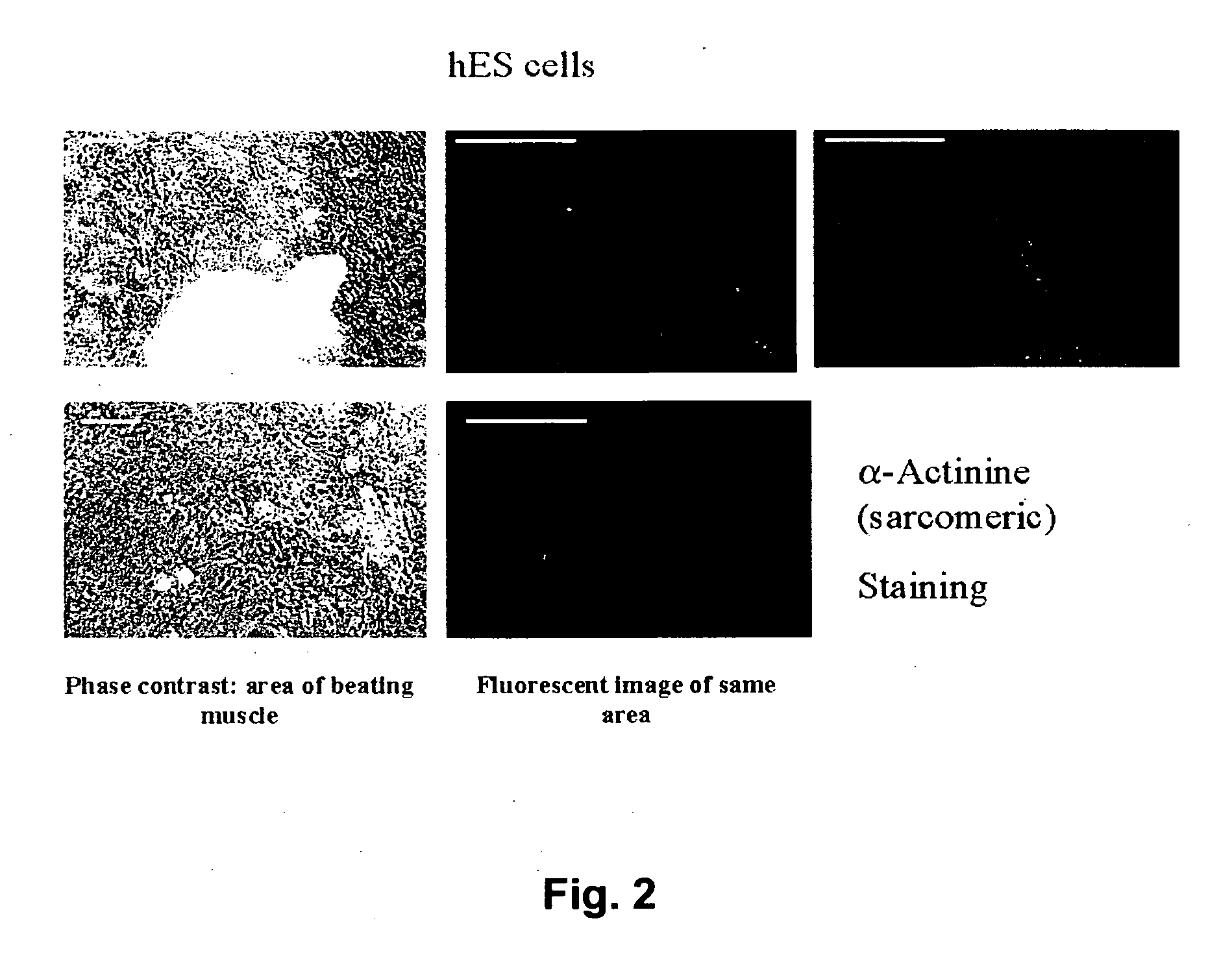
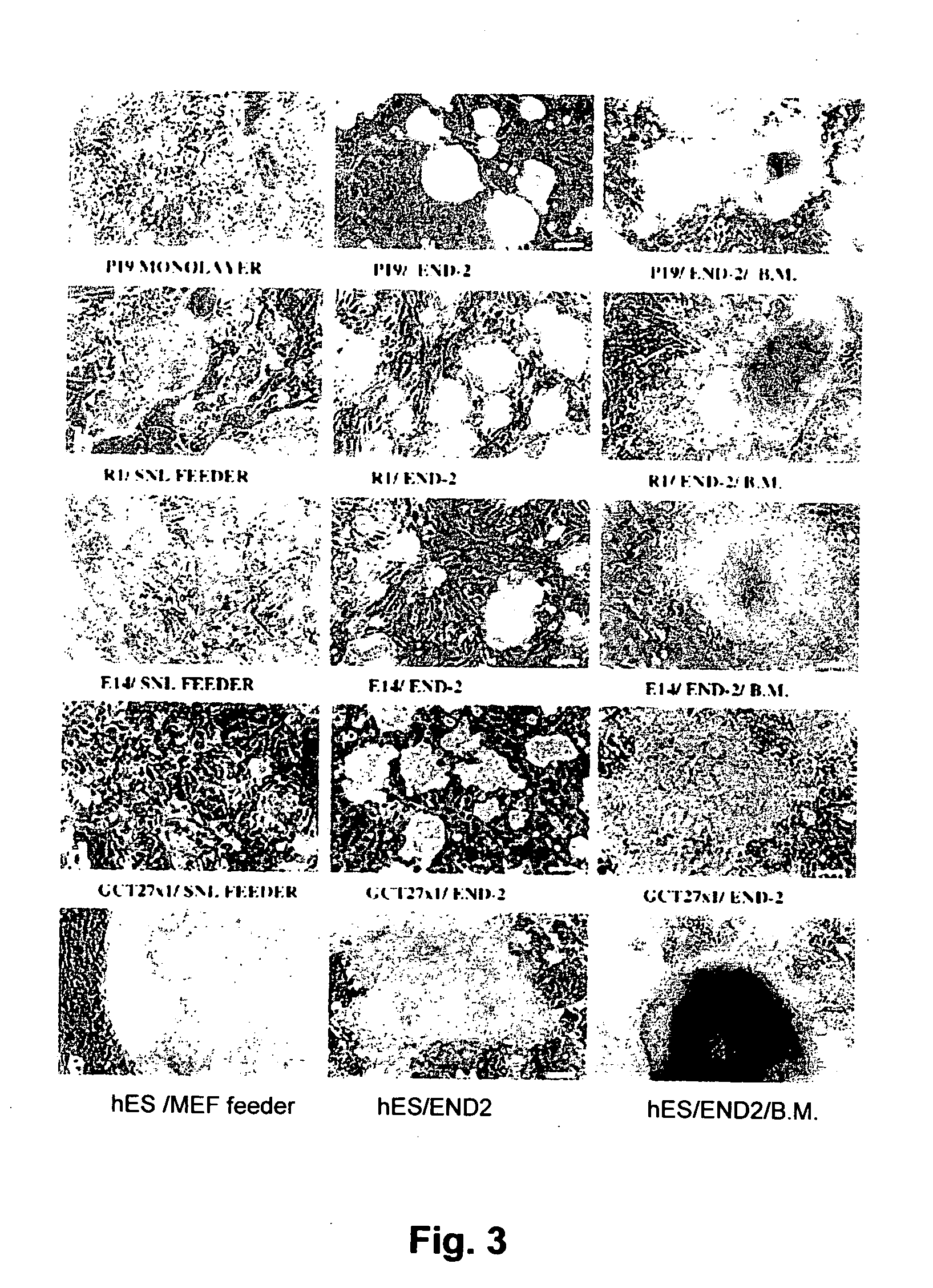
Differentiation of Human Embryonic Stem (hES) Cells into Cardiomyocytes
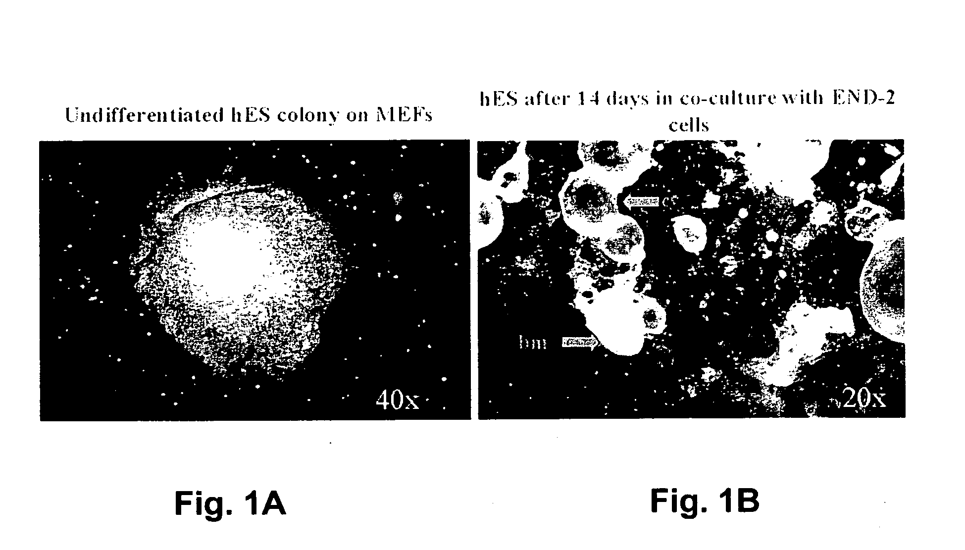
[0119] (a) Co-Culturing of hES Cells With END-2 Cells
[0120] Human embryonic stem cells (hES cells) Reubinoff et al, Nature Biotech. 16:399-404) were co-cultured with END-2 cells ((Mummery et al, 1985, Dev Biol. 109:402-410)). The END-2 cells are grown routinely in a 1:1 ratio of Dulbecco's minimum essential medium (DMEM) and Ham's F12 medium (DF) with 7.5% FCS. Cells were then passaged twice weekly, 1:5 using trypsin / EDTA (0.125%w / v; 50 mM resp). The hES cells were cultured in DMEM with 20% FCS, 0.1 mM β-mrcaptoethanol, 1% non-essential amino acids, 2 mM glutamine plus antibiotics (pen / strep) on mitomycin (10 μg / ml) treated embryonic feeder cells. HES were subcultured by treating with dispase and mechanical slicing of individual colonies into 6-10 pieces followed by transfer of the pieces to new feeder cells.
[0121] To initiate co-cultures, confluent cultures of END-2 cells were first passaged 1:10 on to gelati...
example 2
Differentiation of Human Embryonic Stem (hES) Cells into Skeletal Muscle Cells
[0133] Human stem cells (hES) as used in Example 1 were placed on ectoderm isolated from E7.5 day embryos (E0.5 is day of plug). With sharpened tweezers and tungsten needles, the embryos were prepared out of the decidua and kept on ice in HEPES-buffered DMEM containing 10% FCS. After removing Reichert's membrane, the embryonic and extraembryonic parts of the conceptus were separated with tungsten needles. The node and primitive streak were removed and the embryonic part incubated in 2.5% pancreatin and 0.5% trypsin in PBS on ice for 8 min. After incubation, the embryos were transferred to HEPES-buffered DMEM containing 10% FCS on ice. The ectoderm, endoderm and mesoderm could then be cleanly isolated using tungsten needles.
[0134] The hES cells initially resemble those on endoderm but by day 18 there are areas of highly elongated, twitching cells that resemble skeletal muscle. There are no areas reminisce...
example 3
Differentiation of Human Embryonic Stem (hES) Cells into Vascular Endothelial Cells
[0135] Human stem cells (hES) as used in Example 1 were placed on ectoderm and / or endoderm cells. Co-culture condition of hES with ectoderm or endoderm were as above. Vascular endothelial cells in networks accompanied differentiation to other somatic cell types.
PUM
| Property | Measurement | Unit |
|---|---|---|
| Fraction | aaaaa | aaaaa |
Abstract
Description
Claims
Application Information