Method for exploring and displaying tissues fo human or animal origin from a high frequency ultrasound probe

a high-frequency ultrasound and tissue technology, applied in ultrasonic/sonic/infrasonic diagnostics, instruments, applications, etc., can solve the problems of not allowing detailed analysis of the retina and the posterior wall of the eye, and achieve the effect of low spatial resolution and high spatial resolution

Inactive Publication Date: 2005-11-10
CENT NAT DE LA RECHERCHE SCI
View PDF9 Cites 20 Cited by
  • Summary
  • Abstract
  • Description
  • Claims
  • Application Information

AI Technical Summary

Benefits of technology

[0014] various signal processing operations are carried out on the data coming from the scanning, to improve the reproduction of the information and to facilitate the interpretation thereof by the practitioner.

Problems solved by technology

The major drawback of low-frequency echography is mainly the low resolution (600 to 700 μm) provided by these low frequencies, which do not allow detailed analysis of the retina and the other layers of the posterior wall of the eye (choroid and sclera) and more particularly in the macular region.

Method used


Image

  • Method for exploring and displaying tissues fo human or animal origin from a high frequency ultrasound probe
  • Method for exploring and displaying tissues fo human or animal origin from a high frequency ultrasound probe
  • Method for exploring and displaying tissues fo human or animal origin from a high frequency ultrasound probe

Examples


Embodiment Construction

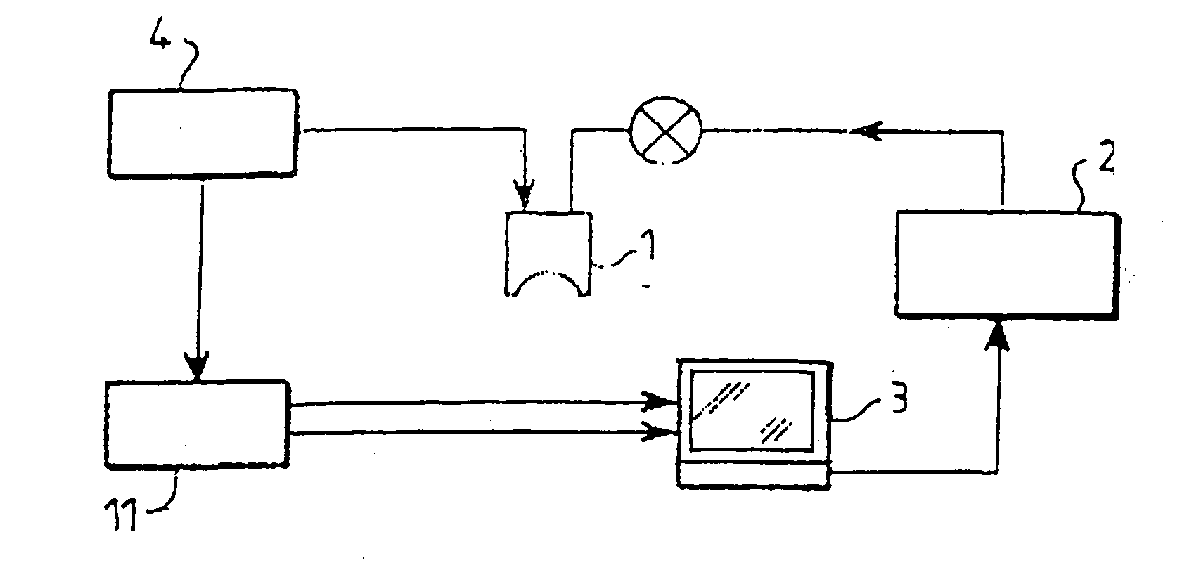
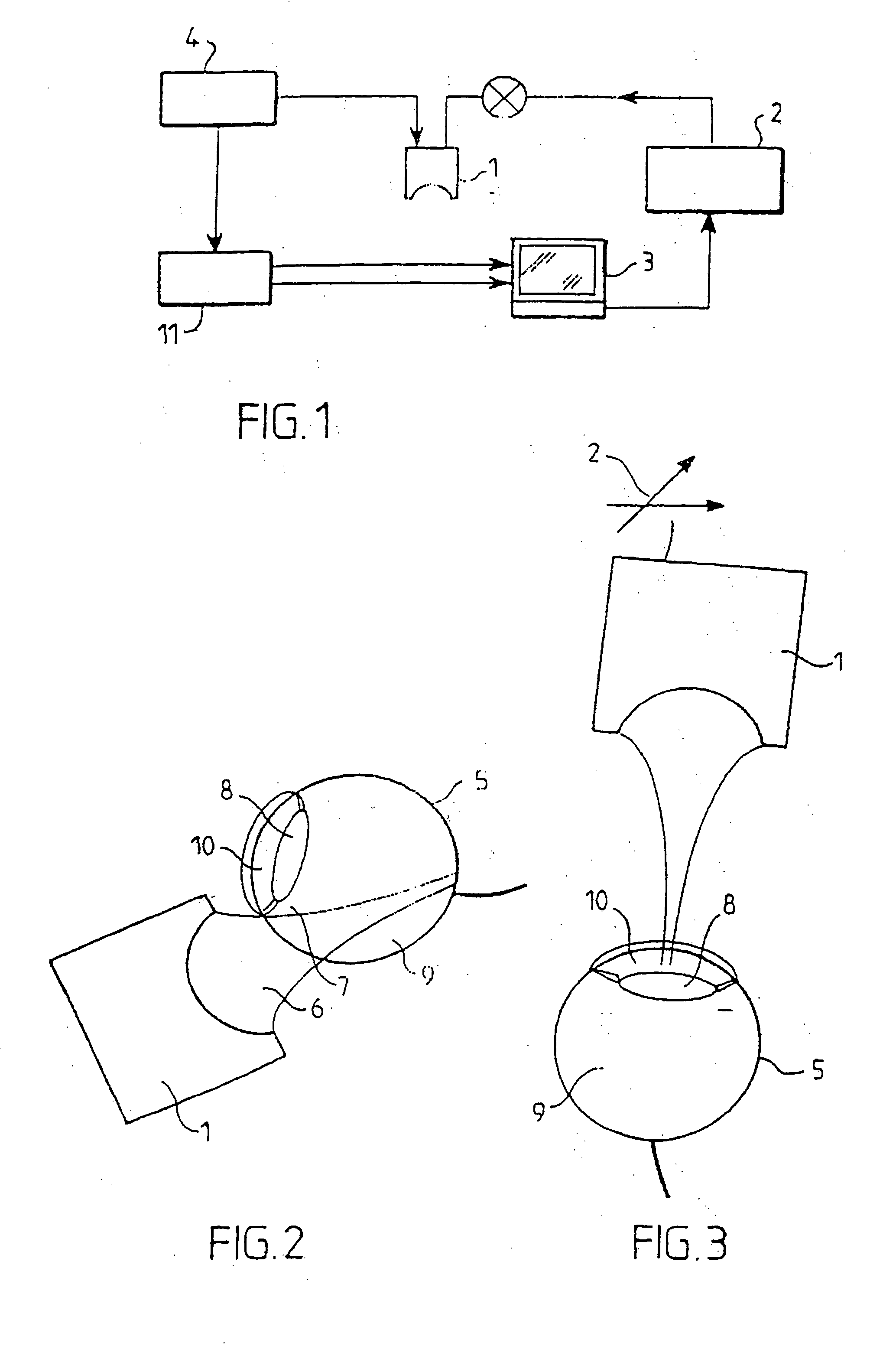
[0026] According to a preferred embodiment of the process forming the subject of the invention, of which one system enabling its implementation is shown schematically in FIG. 1, the process consists in positioning an ultrasound probe 1 mounted within a head articulated in three dimensions X, Y, Z, at least one direction of which can be fixed, this head being steered by a servo-controlled positioning system 2, controlled by a computer 3, in particular in a direction perpendicular to the medium to be investigated.

[0027] This ultrasound probe 1 consists mainly of a transducer, in particular one made of PVDF (polyvinylidene difluoride), controlled by a transmitter / receiver 4, in order to generate beams of convergent, broadband, ultrasonic waves, these waves being able to adopt a spherical or linear profile.

[0028] Next, FIG. 2 shows an investigation of the posterior segment of an ocular globe 5, previously inserted into a coupling medium 6 which does not impair the propagation of the w...


PUM


Abstract

A method for displaying scanned ultrasound images of tissue employs an apparatus including an ultrasound probe mounted to a mechanical head. A three-dimensional positioning system mounts the head for positioning the probe in proximate orthogonal relation to the tissue. A computer controls the three-dimensional positioning system thereby moving the probe during a scan. The probe transmits high frequency ultrasound waves whose nominal frequency is included within the range from 30 to 100 MHz and with a large pass band, adapted to frequencies reflected by the tissue. The beams of ultrasound transmission are focused in a given zone of the tissue over a vertical penetration distance of between 20 and 30 mm. Reflected signals are acquired and processed for display.

Description

CROSS REFERENCE INFORMATION [0001] This application is a Continuation of co-pending Application Ser. No. 09 / 600,073, filed on Feb. 5, 2001, which is the national phase of PCT International Application No. PCT / FR99 / 00040 filed on Jan. 12, 1999, which designated the United States, and on which priority is claimed under 35 U.S.C. § 120; and this application claims priority of Application No. 98-00209 filed in France on Jan. 12, 1998 under 35 U.S.C. § 119. The entire contents of all are hereby incorporated by reference.FIELD OF THE INVENTION [0002] The present invention relates to a process for the investigation and display, using ultrasound echography techniques, of tissue structures of human or animal origin such as in particular the ocular globes and more particularly of the posterior segment (the vitreous cavity, the posterior wall of the globe lined by the choroid and the retina, the macula), tissue structures of the anterior segment (the cornea, the anterior chamber, the iris and ...

Claims


Application Information

Patent Timeline
Patent Type & Authority Applications(United States)
IPC IPC(8): A61B3/10A61B8/10
CPCA61B8/4483A61B8/10G01S15/8993
Inventor SAIED, AMENABERGER, GENEVIEVELAUGIER, PASCALPUECH, MICHEL
Owner CENT NAT DE LA RECHERCHE SCI
Why Eureka
Try Eureka